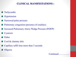
CARDIOGENIC
SHOCK
Presented By:
Mr. Nandish.S
Asso. Professor
Mandya Institute of Nursing Sciences
DEFINITION OF SHOCK:
It is a syndrome characterized by decreased tissue perfusion and
impaired cellular metabolism.
It is a failure of circulatory system to maintain adequate perfusion to
vital organs.
It is a state of cellular & tissue hypoxia due to either reduced oxygen
delivery, increased oxygen consumption, inadequate oxygen
utilization or a combination of these processes to vital organs.
It is a life threatening condition that occurs when the body is not
getting enough blood supply.
TYPES OF SHOCK :
1. Cardiogenic Shock
2. Hypovolemic shock
3. Neurogenic Shock
4. Septic Shock
5. Anaphylactic shock
CARDIOGENIC SHOCK :
It occurs when either systolic or diastolic dysfunction of the
myocardium results in compromised cardiac output.
It is also called as cardiac shock, happens when heart cannot pump
enough blood to meet body’s needs.
it occurs when the heart is unable to function adequately resulting in
insufficient blood flow to tissues and organs.
ETIOLOGY & RISK FACTORS :
Systolic Dysfunction : it occurs due to inability of the heart to pump
blood forward.
- Myocardial Infarction
- Cardiomyopathy
Diastolic Dysfunction : it occurs due to inability of the heart to fill
during diastole.
- Cardiac tamponade
Arrhythmias
- Tachycardia
- Bradycardia
Continued ………
Structural Factors / Problems
- Valvular abnormality (stenosis, regurgitation)
- Papillary muscle dysfunction
- Acute ventricular septal defect.
Other factors
- Endocarditis
- Myocarditis
- Sepsis
- Peripheral vascular disease
- Cerebro vascular disease
- Pneumothorax
- Acute aortic insufficiency
PATHOPHYSIOLOGY :
Structural problems
Systolic dysfunction : ineffective forward movement of blood
Decreased stroke volume
Decreased cardiac output
Decreased cellular oxygen supply
Decreased Tissue perfusion
Impaired cellular metabolism
Arrhythmias
Diastolic dysfunction : ineffective filling
Increased pulmonary pressure
Pulmonary Oedema
Decreased Oxygenation
Decreased tissue perfusion
Impaired cellular metabolism
CLINICAL MANIFESTATIONS :
Tachycardia
Hypotension
Narrowed pulse pressure
Pulmonary congestion (presence of crackles)
Increased Pulmonary Artery Wedge Pressure (PAWP)
Cyanosis
Pallor
Cool & clammy skin
Capillary refill time more than 3 seconds
Oliguria
Continued …………..
Anxiety
Delirium / confusion
Chest pain
Nausea & vomiting
Palpitation
Syncope
Restlessness
Dyspnoea
DIAGNOSTIC STUDIES :
History collection & Physical Examination
Blood examination
• Complete blood count
• ABG Analysis
• Cardiac Enzymes
ECG
Chest X - Ray
Echocardiogram
Angiogram
MANAGEMENT :
The goal of management is :
- To restore the blood flow to myocardium by restoring the balance
between oxygen supply and demand.
MAINTAINING OXYGEN SATURATION / LEVEL
• Improve oxygen delivery by decreasing demand.
• Provide analgesics & anxiolytics to relax muscles.
• Maintain arterial Oxygen saturation by providing supplemental
oxygen.
• Maintain Hemoglobin level more than 10g/dl.
• Monitor central venous oxygen saturation to rule out tissue oxygen
extraction.
OPTIMIZING CIRCULATION :
• Initiate isotonic crystalloid fluids.
• Maintain central venous pressure (CVP) between 8 – 12 mm of Hg.
• Patients may require 4 to 6 litres of fluid per day.
• Measure the urine output and report if output is less than 30 ml / hour.
• Improve heart rate and it should not fall below 60.
• Place the patient in supine position with leg end elevation (45
degree).
PHARMACOLOGIC THERAPY :
• Dilate coronary arteries – Nitrates
• Improve contractility – inotropes (Epinephrine)
• Reduce preload – ACE Inhibitors, Diuretics, Morphine
• Reduce afterload – phospho-diesterase inhibitors, vasodilators
• Reduce heart rate – β Adrenergic blockers, calcium channel blockers
(Amlong)
• Reduce contractility - β Adrenergic blockers (Aten)
OTHER TREATMENT:
• Intra Aortic Balloon Pump (IABP)
• Ventricular Assist Device (VAD)
• Angioplasty & Stent
• Coronary Artery Bypass Graft (CABG)
• Percutaneous Transluminal Coronary Angioplasty (PTCA)
• Thrombolysis
• Cardiac Transplantation.
NURSING DIAGNOSIS :
• Decreased cardiac output related to impaired cardiac contractility as
manifested by hypotension.
• Impaired gas exchange related to pulmonary congestion as manifested
by decreased oxygen saturation levels.
• Impaired tissue perfusion related to decreased cardiac contractility &
blood flow as manifested by increased capillary refill time.
• Fear & Anxiety related to intensive care environment as evidenced by
fearful facial expression.
• Activity intolerance related to decreased cardiac activity & laboured
respirations as evidenced by difficulty in performing ADL.
Cardiogenic Shock.pptx

Cardiogenic Shock.pptx

  • 1.
    CARDIOGENIC SHOCK Presented By: Mr. Nandish.S Asso.Professor Mandya Institute of Nursing Sciences
  • 3.
    DEFINITION OF SHOCK: Itis a syndrome characterized by decreased tissue perfusion and impaired cellular metabolism. It is a failure of circulatory system to maintain adequate perfusion to vital organs. It is a state of cellular & tissue hypoxia due to either reduced oxygen delivery, increased oxygen consumption, inadequate oxygen utilization or a combination of these processes to vital organs. It is a life threatening condition that occurs when the body is not getting enough blood supply.
  • 4.
    TYPES OF SHOCK: 1. Cardiogenic Shock 2. Hypovolemic shock 3. Neurogenic Shock 4. Septic Shock 5. Anaphylactic shock
  • 5.
    CARDIOGENIC SHOCK : Itoccurs when either systolic or diastolic dysfunction of the myocardium results in compromised cardiac output. It is also called as cardiac shock, happens when heart cannot pump enough blood to meet body’s needs. it occurs when the heart is unable to function adequately resulting in insufficient blood flow to tissues and organs.
  • 7.
    ETIOLOGY & RISKFACTORS : Systolic Dysfunction : it occurs due to inability of the heart to pump blood forward. - Myocardial Infarction - Cardiomyopathy Diastolic Dysfunction : it occurs due to inability of the heart to fill during diastole. - Cardiac tamponade Arrhythmias - Tachycardia - Bradycardia Continued ………
  • 8.
    Structural Factors /Problems - Valvular abnormality (stenosis, regurgitation) - Papillary muscle dysfunction - Acute ventricular septal defect. Other factors - Endocarditis - Myocarditis - Sepsis - Peripheral vascular disease - Cerebro vascular disease - Pneumothorax - Acute aortic insufficiency
  • 9.
    PATHOPHYSIOLOGY : Structural problems Systolicdysfunction : ineffective forward movement of blood Decreased stroke volume Decreased cardiac output Decreased cellular oxygen supply Decreased Tissue perfusion Impaired cellular metabolism
  • 10.
    Arrhythmias Diastolic dysfunction :ineffective filling Increased pulmonary pressure Pulmonary Oedema Decreased Oxygenation Decreased tissue perfusion Impaired cellular metabolism
  • 11.
    CLINICAL MANIFESTATIONS : Tachycardia Hypotension Narrowedpulse pressure Pulmonary congestion (presence of crackles) Increased Pulmonary Artery Wedge Pressure (PAWP) Cyanosis Pallor Cool & clammy skin Capillary refill time more than 3 seconds Oliguria Continued …………..
  • 12.
    Anxiety Delirium / confusion Chestpain Nausea & vomiting Palpitation Syncope Restlessness Dyspnoea
  • 13.
    DIAGNOSTIC STUDIES : Historycollection & Physical Examination Blood examination • Complete blood count • ABG Analysis • Cardiac Enzymes ECG Chest X - Ray Echocardiogram Angiogram
  • 14.
    MANAGEMENT : The goalof management is : - To restore the blood flow to myocardium by restoring the balance between oxygen supply and demand.
  • 15.
    MAINTAINING OXYGEN SATURATION/ LEVEL • Improve oxygen delivery by decreasing demand. • Provide analgesics & anxiolytics to relax muscles. • Maintain arterial Oxygen saturation by providing supplemental oxygen. • Maintain Hemoglobin level more than 10g/dl. • Monitor central venous oxygen saturation to rule out tissue oxygen extraction.
  • 16.
    OPTIMIZING CIRCULATION : •Initiate isotonic crystalloid fluids. • Maintain central venous pressure (CVP) between 8 – 12 mm of Hg. • Patients may require 4 to 6 litres of fluid per day. • Measure the urine output and report if output is less than 30 ml / hour. • Improve heart rate and it should not fall below 60. • Place the patient in supine position with leg end elevation (45 degree).
  • 17.
    PHARMACOLOGIC THERAPY : •Dilate coronary arteries – Nitrates • Improve contractility – inotropes (Epinephrine) • Reduce preload – ACE Inhibitors, Diuretics, Morphine • Reduce afterload – phospho-diesterase inhibitors, vasodilators • Reduce heart rate – β Adrenergic blockers, calcium channel blockers (Amlong) • Reduce contractility - β Adrenergic blockers (Aten)
  • 18.
    OTHER TREATMENT: • IntraAortic Balloon Pump (IABP) • Ventricular Assist Device (VAD) • Angioplasty & Stent • Coronary Artery Bypass Graft (CABG) • Percutaneous Transluminal Coronary Angioplasty (PTCA) • Thrombolysis • Cardiac Transplantation.
  • 19.
    NURSING DIAGNOSIS : •Decreased cardiac output related to impaired cardiac contractility as manifested by hypotension. • Impaired gas exchange related to pulmonary congestion as manifested by decreased oxygen saturation levels. • Impaired tissue perfusion related to decreased cardiac contractility & blood flow as manifested by increased capillary refill time. • Fear & Anxiety related to intensive care environment as evidenced by fearful facial expression. • Activity intolerance related to decreased cardiac activity & laboured respirations as evidenced by difficulty in performing ADL.