SlideShare a Scribd company logo
1 of 86
Download to read offline
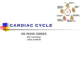
CARDIAC CYCLE
DR MOHD IDREES
SR2 Cardiology
LPSIC KANPUR
Cardiac Cycle
■ Def: The cardiac events that occur from
beginning of one heart beat to the beginning of
the next.
■ first assembled by Lewis in 1920 but first
conceived by Wiggers in 1915
■ Atria act as PRIMER PUMPS for
ventricles & ventricles provide major
source of power for moving the blood
through the vascular system.
■ Initiated by spontaneous generation of
AP in SA node (located in the superior lateral wall of
the right atrium near the opening of the superior vena cava)
Electrical System: Brief
Action potentials originating
in the sinus node travel to
AV node (1m/s) in 0.03 sec.
1. AV nodal delay of 0.09 sec before the impulse
enters the penetrating portion of the A-V bundle
2. A final delay of another 0.04 sec occurs mainly in
this penetrating A-V bundle
total delay in the A-V nodal and A-V bundle
system is about 0.13 sec
A total delay of 0.16 sec occurs before the excitatory
signal finally reaches the contracting muscle of the
ventricles from its origin in sinus node.
Delay in AV node (0.13sec)
■ Why delay?
Diminished numbers of gap junctions Between
successive cells in the conducting pathways.
■ Significance?
Delay allows time for the atria to empty their blood
into the ventricles before ventricular contraction
begins
■ Rapid Transmission in the Purkinje
System (1.5 to 4.0 m/sec)
allowing almost instantaneous
transmission of the cardiac impulse
throughout the ventricular muscle
■ (B/c of very high level of permeability of the gap
junctions)
Summary of Cardiac Impulse
Transmission
Mechanical Phase
Cardiac cycle – basically
describes…
1. Pressure
2. Volume, and
3. Flow phenomenon
in ventricles as a function of time
Basics
■ 1 Beat = 0.8 sec (800 msec)
■ Systole = 0.3 sec
■ Diastole = 0.5 sec
In tachycardia, Diastolic phase decreases more
than systolic phase
Phases of cardiac cycle
LV Contraction
Isovolumic contraction (b)
Maximal ejection (c)
LV Relaxation
Start of relaxation and reduced ejection (d)
Isovolumic relaxation (e)
LV Filling
Rapid phase (f)
Slow filling (diastasis) (g)
Atrial systole or booster (a)
Time Intervals
Total ventricular systole 0.3 sec
■ Isovolumic contraction (b) 0.05 sec
■ Maximal ejection (c) 0.1 sec
■ Reduced ejection (d) 0.15 sec
Total ventricular diastole 0.5 sec
■ Isovolumic relaxation (e) 0.1 sec
■ Rapid filling phase (f) 0.1 sec
■ Slow filling (diastasis) (g) 0.2 sec
■ Atrial systole or booster (a) 0.1 sec
GRAND TOTAL (Syst+Diast) = 0.8 sec
Physiologic Versus Cardiologic Systole
and Diastole
PHYSIOLOGIC
SYSTOLE
CARDIOLOGIC
SYSTOLE
Isovolumic
contraction
Maximal
ejection
From M1 to A2,
including:
Major part of isovolumic
contraction
Maximal ejection
Reduced ejection
PHYSIOLOGIC
DIASTOLE
CARDIOLOGIC
DIASTOLE
Reduced
ejection
Isovolumic
relaxation
Filling phases
A2-M1 interval
(filling phases included)
20msec
Physiological systole
cardiologic systole, demarcated by
heart sounds rather than by physiologic
events, starts fractionally later than
physiologic systole and ends significantly
later.
Cardiologic systole> physiologic systole
Description of Cardiac cycle
phases
1. Pressure & Volume events
2. ECG correlation
3. Heart sounds
4. Clinical significance
Atrial Systole
A-V Valves Open; Semilunar Valves Closed
➢ Blood normally flows
continually from great
veins into atria
➢ 80% flows directly
through atria into
ventricle before the atria
contracts.
➢ 20% of filling of
ventricles – atrial
contraction
➢ Atrial contraction is
completed before the
ventricle begins to
contract.
■ Atrial contraction normally accounts for
about 10%-15% of LV filling at rest,
however, At higher heart rates, atrial
contraction may account for up to 40% of
LV filling referred to as the "atrial kick”
■ The atrial contribution to ventricular
filling varies inversely with duration of
ventricular diastole and directly with
atrial contractility
Atrial Systole
Pressures & Volumes
■ ‘ a ‘ wave – atrial
contraction, when atrial
pressure rises.
■ Atrial pressure drops
when the atria stop
contracting.
■ After atrial contraction is complete
LVEDV typically about 120 ml (preload)
End-diastolic pressures of
LV = 8-12 mmHg and
RV = 3-6 mmHg
■ AV valves floats upward (pre-
position)
Abnormalities of “a” wave
■ Elevated a wave
Tricuspid stenosis
Decreased ventricular compliance (ventricular failure, pulmonic
valve stenosis, or pulmonary hypertension)
■ Cannon a wave
Atrial-ventricular asynchrony (atria contract against a closed tricuspid valve)
complete heart block, following premature ventricular contraction,
during ventricular tachycardia, with ventricular pacemaker
■ Absent a wave
Atrial fibrillation or atrial standstill
Atrial flutter
Why blood does not flow back in to SVC/PV
while atria contracting, even though no valve
in between?
■ Wave of contraction through the atria
moves toward the AV valve thereby
having a "milking effect."
■ Inertial effects of the venous return.
Atrial Systole
ECG
■ p wave – atrial depolarization
■ impulse from SA node results in depolarization
& contraction of atria ( Rt before Lt )
■ PR segment – isoelectric line as depolarization
proceeds to AV node.
■ This brief pause before contraction allows the
ventricles to fill completely with blood.
Atrial Systole
Heart Sounds
■ S4 (atrial or presystolic gallop) - atrial emptying after
forcible atrial contraction.
■ appears at 0.04 s after the P wave (late diastolic)
■ lasts 0.04-0.10 s
■ Caused by vibration of ventricular wall during
rapid atrium emptying into non compliant
ventricle
Causes of S4
■ Physiological;
>60yrs (Recordable, not audible)
■ Pathological;
All causes of concentric LV/RV hypertrophy
Coronary artery disease
Acute regurgitant lesions
An easily audible S4 at any age is generally
abnormal.
JVP: x descent
■ Prominent x descent
1 Cardiac tamponade
2 Constrictive pericarditis
3 Right ventricular ischemia with preservation of atrial
contractility
■ Blunted x descent
1 Atrial fibrillation
2 Right atrial ischemia
Beginning of VenTRICULAR Systole
Isovolumetric Contraction
All Valves Closed
Isovolumetric Contraction
Pressure & Volume Changes
■ The AV valves close when the
pressure in the ventricles (red)
exceeds the pressure in the
atria (yellow).
■ As the ventricles contract
isovolumetrically -- their volume
does not change (white) -- the
pressure inside increases,
approaching the pressure in the
aorta and pulmonary arteries
(green).
■ JVP: c wave- d/t Right
ventricular contraction pushes
the tricuspid valve into the
atrium and increases atrial
pressure, creating a small wave
into the jugular vein. It is
normally simultaneous with the
carotid pulse.
■ Ventricular chamber geometry changes considerably as the
heart becomes more spheroid in shape; circumference
increases and atrial base-to-apex length decreases.
■ Early in this phase, the rate of pressure development
becomes maximal. This is referred to as maximal dP/dt.
■ Ventricular pressure increases rapidly
LV ~10mmHg to ~ 80mmHg (~Aortic pressure)
RV ~4 mmHg to ~15mmHg (~Pulmonary A pressure)
At this point, semilunar (aortic and pulmonary) valves open
against the pressures in the aorta and pulmonary artery
Isovolumetric Contraction
ECG
■ The QRS complex is due to ventricular
depolarization, and it marks the
beginning of ventricular systole.
Isovolumetric Contraction
Heart Sounds
■ S1 is d/t closure and after
vibrations of AV Valves. (M1
occurs with a definite albeit
20 msec delay after the LV-
LA pressure crossover.)
■ S1 is normally split (~0.04
sec) because mitral valve
closure precedes tricuspid
closure.
(Heard in only 40% of normal
individuals)
S1 heart sound
■ low pitch and relatively long-lasting
■ lasts ~ 0.12-0.15 sec
■ frequency ~ 30-100 Hz
■ appears 0.02 – 0.04 sec after the
beginning of the QRS complex
Some Clinical facts about S1
■ S1 is a relatively prolonged, low
frequency sound, best heard at apex.
■ Normally split of S1 (~40%)is heard
only at tricuspid area.(As tricuspid
component is heard only here.)
■ If S1 is equal to or higher in intensity
than S2 at base, S1 is considered
accentuated.
■ Variable intensity of S1 and jugular venous
pulse are highly specific and sensitive in the
diagnosis of ventriculoatrial dissociation during
VT, and is helpful in distinguishing it from
supraventricular tachycardia with aberration.
Value of physical signs in the diagnosis of ventricular tachycardia. C J
Garratt, M J Griffith, G Young, N Curzen, S Brecker, A F Rickards and A J Camm,
Circulation. 1994;90:3103-3107
Ejection
Aortic and Pulmonic Valves Open; AV Valves Remain
Closed
■ The Semilunar valves ( aortic ,
pulmonary ) open at the
beginning of this phase.
■ Two Phases
• Rapid ejection - 70% of the blood
ejected during the first 1/3 of
ejection
• Slow ejection - remaining 30% of
the blood emptying occurs during
the latter 2/3 of ejection
Rapid Ejection
Pressure & Volume Changes
■ When ventricles
continue to contract ,
pressure in ventricles
exceed that of in aorta
& pul arteries & then
semilunar valves open,
blood is pumped out of
ventricles & Ventricular
vol decreases rapidly.
Rapid Ejection
ECG & Heart Sounds
■ In rapid ejection part of
the ejection phase there
no specific ECG changes /
heart sounds heard.
Slow Ejection
Aortic and Pulmonic Valves Open; AV Valves
Remain Closed
■ Approx. 200msec after the QRS
vent. repolarisation occurs as
shown by T wave, which leads to
decline in ventricular active
tension & pressure generation, so
rate of ejection falls. Ventricular
pressure falls slightly below
outflow tract pressure, however
outflow still occurs due to kinetic
energy of blood and elastic recoil
of aorta(Windkessel effect)
■ At the end of ejection, the
semilunar valves close. This marks
the end of ventricular systole
mechanically.
Slow Ejection
ECG & Heart Sounds
■ T wave – slightly
before the end of
ventricular
contraction
■ heart sounds :
none
Beginning of Diastole
■ At the end of systole, ventricular relaxation
begins, allowing intraventricular pressures to
decrease rapidly (LV from 100mmHg to
20mmHg & RV from 15mmHg to 0mmHg),
aortic and pulmonic valves abruptly close
(aortic precedes pulmonic) causing the
second heart sound (S2)
■ Valve closure is associated with a small
backflow of blood into the ventricles and a
characteristic notch (incisura or dicrotic
notch) in the aortic and pulmonary artery
pressure tracings
■ After valve closure, the aortic and pulmonary
artery pressures rise slightly (dicrotic wave)
following by a slow decline in pressure
Isovolumetric relaxation
■ Volumes remain constant because all
valves are closed
■ volume of blood that remains in a
ventricle is called the end-systolic
volume (LV ~50ml).
■ pressure & volume of ventricle are low
in this phase .
Isovolumetric relaxation
■ Throughout this and the
previous two phases, the
atrium in diastole has
been filling with blood on
top of the closed AV
valve, causing atrial
pressure to rise gradually
■ JVP - "v" wave occurs
toward end of ventricular
contraction – results from
slow flow of blood into
atria from veins while AV
valves are closed .
Isovolumetric relaxation
ECG & Heart Sounds
■ ECG : no deflections
■ Heart Sounds : S2
is heard when the
semilunar vlaves
close.
■ A2 is heard prior to
P2 as Aortic valve
closes prior to
pulmonary valve.
Why A2 occurs prior to P2 ?
■ “Hangout interval” is longer for pulmonary
side (~80msec),compared to aortic side
(~30msec).
Hangout interval is the time interval from crossover
of pressures (ventricle with their respective vessel) to
the actual occurrence of sound.
■ Due to lower pressure and higher
distensibility, pulmonary artery having longer
hangout interval causing delayed PV closure
and P2.
S2 heart sound
■ Appears in the terminal period of
the T wave
■ lasts 0.08 – 0.12s
Some clinical facts about S2
■ Normal split: Two components heard during
inspiration and is single sound during expiration.
(A2-P2 ~20- 50 msec in inspiration)
■ Clinically split is defined as wide, if it is heard
well in standing position, in expiration (normally
not heard as the split is 15 msec, which can not be heard
by human ears)
■ Single S2: absence of audible split in either
phase of respiration.
Common causes of wide split S2
■ RBBB
■ Sev PAH
■ ASD
■ Idiopathic dilatation of pul artery
■ Sev right heart failure
■ Moderate to severe PS
■ Severe MR
■ Normal variant
Common causes of wide fixed split
S2
■ ASD
■ All causes of wide split with
associated severe right ventricular
failure.
JVP: V wave
■ Elevated v wave
1 Tricuspid regurgitation
2 Right ventricular heart failure
3 Reduced atrial compliance (restrictive myopathy)
■ a wave equal to v wave
1 Tamponade
2 Constrictive pericardial disease
3 Hypervolemia
Rapid Inflow ( Rapid Ven. Filling)
A-V Valves Open
■ Once AV valves are open
the blood that has
accumulated in atria flows
into the ventricle.
Rapid Inflow
Volume changes
■ Despite the inflow of blood
from the atria,
intraventricular pressure
continues to briefly fall
because the ventricles are
still undergoing relaxation
■ JVP: Seen as y-descent.
Rapid Inflow ( Rapid Ven. Filling)
ECG & Heart Sounds
■ ECG : no deflections
■ Heart sounds : S3 is heard,
lasts 0.02-0.04 sec
(represent tensing of chordae
tendineae and AV ring during
ventricular relaxation and filling)
Causes of S3
■ Physiological: Childrens & young adults <40 yrs
(nearly 25%)
(Not heard in normal infants & adult
>40 yrs.)
■ Pathological:
Ventricular failure
Hyperkinetic state (anemia, thyrotoxicosis, beri-beri)
MR, TR
AR, PR
Systemic AV fistula
JVP: y descent
■ Prominent y descent
1 Constrictive pericarditis
2 Restrictive myopathies
3 Tricuspid regurgitation
■ Blunted y descent
1 Tamponade
2 Right ventricular ischemia
3 Tricuspid stenosis
Diastasis OR REDUCED FILLING
A-V Valves Open
■ remaining blood
which has
accumulated in
atria slowly flows
into the ventricle.
Diastasis
Volume changes
■ Ventricular volume
increases more slowly
now. The ventricles
continue to fill with blood
until they are nearly full.
Diastasis
ECG & Heart Sounds
■ ECG : no deflections
■ Heart Sounds : none
The Lewis or wiggers cycle
Volumes
■ End diastolic vol : During diastole, filling of
ventricle increases vol of each ventricle to
~ 110 -120 ml
■ Stroke Vol : amount of blood pumped out
of ventricle during systole. ~ 70 ml
■ End systolic vol : the remaining amount of
blood in ventricle after the systole. ~40
-50 ml
RV v/s LV
Rt Ventricular
• Pressure wave 1/5th
• dp/dt is less
• Isovolumic
contraction &
relaxation phases
are short.
Timing of Cardiac EVENTS
1. RA start contracting before
LA
2. LV start contracting before
RV
3. TV open before MV,
so RV filling start before
LV.
4. RV peak pressure 1/5th of
LV.
5. RV outflow velocity smooth
rise & fall, while Lt side
initial
peak followed by quick
fall.
PRESSURE VOLUME LOOP
Pressure-Volume Loop
Pressure-volume loop of RV
is same as that of LV,
however the area is only
1/5th
of LV because pressures
are so much lower on right
■ Maximal pressure that can be developed by
LV at any given LV volume is defined by end
systolic PV relationship (ESPVR)
■ ESPVR line is representative of contractility of
heart
■ When contractility ↑ slope of ESPVR is higher
■ ↓ slope of ESPVR is s/o ↓ contractility
■ Slope of EDPVR is reciprocal of vent.
compliance
PRELOAD, AFTERLOAD & STROKE VOLUME
ABNORMAL PRESSURE VOLUME LOOP
CHANGES IN PRELOAD AND STROKE
VOLUME
CHANGES IN AFTERLOAD AND STROKE VOLUME
SYSTOLIC DYSFUNCTION
DIASTOLIC DYSFUNCTION
CARDIAC TAMPONADE
■ Increased
stroke volume
■ Increased EF
■ Decreased
ESV
AORTIC STENOSIS
PV Loop in MS
PV loop in MR
■ EDV will ↑↑, ESV↓
■ Total SV↑(apparent
SV↓)
■ No true
isovolumetric
contraction or isovol.
relaxation
■ Afterload ↓-height↓
PV Loop in AR
■ No true isovol.
Contraction or
isovol. Relaxation
■ EDV ↑↑, ESV↑
■ Total SV ↑
■ Vol.↑, afterload↑,
height↑
Uses of PV loop
■ Work done by heart(stroke work)= total
area under curve
■ Width indicated SV=EDV-ESV
■ Height indicates afterload, ventricular
Activity
■ EF= SV/EDV X100
SYSTOLIC TIME
INTERVALS
■ Systole has 2 distinct time periods-pre-
ejection period(PEP) & left ventricular
ejection time(LVET)
■ PEP is defined as time between the onset
of electrocardiographic systole & the
opening of aortic valve
■ LVET begins with the AoV opening &
terminates at its closure-marked by the
onset of S2
■ QS2(Electromechanical
systole)=PEP+LVET
■ QS2 is constant over a wide variety
of cardiac disorders
■ Increased inotropic state is reflected
by ↓ in QS2 & a decreased
inotropic state prolongs QS2.
THANK YOU
ATRIAL PV LOOP

More Related Content

Similar to CARDIAC CYCLE phases heart sound jvp3.pdf

Cardiac cycle,e.c.g,heart sounds
Cardiac cycle,e.c.g,heart soundsCardiac cycle,e.c.g,heart sounds
Cardiac cycle,e.c.g,heart soundsDr. Jaishree Tiwari
 
Physiology presntation Cardaic cycle
Physiology presntation  Cardaic cyclePhysiology presntation  Cardaic cycle
Physiology presntation Cardaic cycleQurba Kiran
 
Cardiac cycle and jvp
Cardiac cycle and jvpCardiac cycle and jvp
Cardiac cycle and jvpRamesh Babu
 
Cardiovascular System3
Cardiovascular System3Cardiovascular System3
Cardiovascular System3msu
 
Cardiovascular System3
Cardiovascular System3Cardiovascular System3
Cardiovascular System3msu
 
Matters of the heart: cardiac physiology
Matters of the heart: cardiac physiologyMatters of the heart: cardiac physiology
Matters of the heart: cardiac physiologyBien Eli Nillos
 
Cardiac cycle made easy
Cardiac cycle made easy Cardiac cycle made easy
Cardiac cycle made easy Md Rahman
 
CARDIAC CYCLE Rajesh.pptx
CARDIAC CYCLE Rajesh.pptxCARDIAC CYCLE Rajesh.pptx
CARDIAC CYCLE Rajesh.pptxRajeshPonnada3
 
Cardiac cycle DR NIKUNJ R SHEKHADA (MBBS,MS GEN SURG ,DNB CTS SR)
Cardiac cycle DR  NIKUNJ R SHEKHADA (MBBS,MS GEN SURG ,DNB CTS SR)Cardiac cycle DR  NIKUNJ R SHEKHADA (MBBS,MS GEN SURG ,DNB CTS SR)
Cardiac cycle DR NIKUNJ R SHEKHADA (MBBS,MS GEN SURG ,DNB CTS SR)DR NIKUNJ SHEKHADA
 
Cardiac cycle.full
Cardiac  cycle.fullCardiac  cycle.full
Cardiac cycle.fullSidra Nawaz
 
Arterial and venous pulse
Arterial and venous  pulseArterial and venous  pulse
Arterial and venous pulseAnu Priya
 

Similar to CARDIAC CYCLE phases heart sound jvp3.pdf (20)

Cardiac cycle,e.c.g,heart sounds
Cardiac cycle,e.c.g,heart soundsCardiac cycle,e.c.g,heart sounds
Cardiac cycle,e.c.g,heart sounds
 
Physiology presntation Cardaic cycle
Physiology presntation  Cardaic cyclePhysiology presntation  Cardaic cycle
Physiology presntation Cardaic cycle
 
Cardiac cycle and jvp
Cardiac cycle and jvpCardiac cycle and jvp
Cardiac cycle and jvp
 
Cardiac cycle ii
Cardiac   cycle     iiCardiac   cycle     ii
Cardiac cycle ii
 
Cardiovascular System3
Cardiovascular System3Cardiovascular System3
Cardiovascular System3
 
Cardiovascular System3
Cardiovascular System3Cardiovascular System3
Cardiovascular System3
 
CVS Item 2.pdf
CVS Item 2.pdfCVS Item 2.pdf
CVS Item 2.pdf
 
Matters of the heart: cardiac physiology
Matters of the heart: cardiac physiologyMatters of the heart: cardiac physiology
Matters of the heart: cardiac physiology
 
Cardiac cycle made easy
Cardiac cycle made easy Cardiac cycle made easy
Cardiac cycle made easy
 
Cardiac cyclen
Cardiac cyclenCardiac cyclen
Cardiac cyclen
 
CARDIAC CYCLE Rajesh.pptx
CARDIAC CYCLE Rajesh.pptxCARDIAC CYCLE Rajesh.pptx
CARDIAC CYCLE Rajesh.pptx
 
Cardiac cycle
Cardiac cycleCardiac cycle
Cardiac cycle
 
Cardiac cycle DR NIKUNJ R SHEKHADA (MBBS,MS GEN SURG ,DNB CTS SR)
Cardiac cycle DR  NIKUNJ R SHEKHADA (MBBS,MS GEN SURG ,DNB CTS SR)Cardiac cycle DR  NIKUNJ R SHEKHADA (MBBS,MS GEN SURG ,DNB CTS SR)
Cardiac cycle DR NIKUNJ R SHEKHADA (MBBS,MS GEN SURG ,DNB CTS SR)
 
Cardiac cycle
Cardiac cycleCardiac cycle
Cardiac cycle
 
Cardiac cycle.full
Cardiac  cycle.fullCardiac  cycle.full
Cardiac cycle.full
 
Cardiac cycle
Cardiac cycleCardiac cycle
Cardiac cycle
 
cath Lab Hemoduhynamic
cath Lab Hemoduhynamiccath Lab Hemoduhynamic
cath Lab Hemoduhynamic
 
Cardiac cycle
Cardiac cycleCardiac cycle
Cardiac cycle
 
Cardiac cycle made easy
Cardiac cycle made easy Cardiac cycle made easy
Cardiac cycle made easy
 
Arterial and venous pulse
Arterial and venous  pulseArterial and venous  pulse
Arterial and venous pulse
 

More from ddocofdera

Intravascular Ultrasound Versus Angiography-Guided Drug-Eluting Stent Implant...
Intravascular Ultrasound Versus Angiography-Guided Drug-Eluting Stent Implant...Intravascular Ultrasound Versus Angiography-Guided Drug-Eluting Stent Implant...
Intravascular Ultrasound Versus Angiography-Guided Drug-Eluting Stent Implant...ddocofdera
 
The DELIVER Trial: Dapagliflozin in Heart Failure with Mildly Reduced or Pres...
The DELIVER Trial: Dapagliflozin in Heart Failure with Mildly Reduced or Pres...The DELIVER Trial: Dapagliflozin in Heart Failure with Mildly Reduced or Pres...
The DELIVER Trial: Dapagliflozin in Heart Failure with Mildly Reduced or Pres...ddocofdera
 
HEMODYNAMICS PRINCIPLES 
-PRESSURE MEASUREMENT
-MEASUREMENT OF CARDIAC OUTPUT
HEMODYNAMICS PRINCIPLES 
-PRESSURE MEASUREMENT
-MEASUREMENT OF CARDIAC OUTPUTHEMODYNAMICS PRINCIPLES 
-PRESSURE MEASUREMENT
-MEASUREMENT OF CARDIAC OUTPUT
HEMODYNAMICS PRINCIPLES 
-PRESSURE MEASUREMENT
-MEASUREMENT OF CARDIAC OUTPUTddocofdera
 
JVP SURAJIT.pptx
JVP SURAJIT.pptxJVP SURAJIT.pptx
JVP SURAJIT.pptxddocofdera
 
LONG­-TERM OUTCOMES OF PATENT FORAMEN OVALE 1.pptx
LONG­-TERM OUTCOMES OF PATENT FORAMEN OVALE 1.pptxLONG­-TERM OUTCOMES OF PATENT FORAMEN OVALE 1.pptx
LONG­-TERM OUTCOMES OF PATENT FORAMEN OVALE 1.pptxddocofdera
 
INTRACARDIAC DEVICES AND ITS COMPLICATION seminar..pptx
INTRACARDIAC DEVICES AND ITS COMPLICATION seminar..pptxINTRACARDIAC DEVICES AND ITS COMPLICATION seminar..pptx
INTRACARDIAC DEVICES AND ITS COMPLICATION seminar..pptxddocofdera
 
THE EMPEROR-PRESERVED TRIAL ppt.pptx
THE EMPEROR-PRESERVED TRIAL ppt.pptxTHE EMPEROR-PRESERVED TRIAL ppt.pptx
THE EMPEROR-PRESERVED TRIAL ppt.pptxddocofdera
 
Nonculprit Lesion Myocardial Infarction Following Percutaneous Coronary Inter...
Nonculprit Lesion Myocardial Infarction Following Percutaneous Coronary Inter...Nonculprit Lesion Myocardial Infarction Following Percutaneous Coronary Inter...
Nonculprit Lesion Myocardial Infarction Following Percutaneous Coronary Inter...ddocofdera
 

More from ddocofdera (10)

Intravascular Ultrasound Versus Angiography-Guided Drug-Eluting Stent Implant...
Intravascular Ultrasound Versus Angiography-Guided Drug-Eluting Stent Implant...Intravascular Ultrasound Versus Angiography-Guided Drug-Eluting Stent Implant...
Intravascular Ultrasound Versus Angiography-Guided Drug-Eluting Stent Implant...
 
The DELIVER Trial: Dapagliflozin in Heart Failure with Mildly Reduced or Pres...
The DELIVER Trial: Dapagliflozin in Heart Failure with Mildly Reduced or Pres...The DELIVER Trial: Dapagliflozin in Heart Failure with Mildly Reduced or Pres...
The DELIVER Trial: Dapagliflozin in Heart Failure with Mildly Reduced or Pres...
 
HEMODYNAMICS PRINCIPLES 
-PRESSURE MEASUREMENT
-MEASUREMENT OF CARDIAC OUTPUT
HEMODYNAMICS PRINCIPLES 
-PRESSURE MEASUREMENT
-MEASUREMENT OF CARDIAC OUTPUTHEMODYNAMICS PRINCIPLES 
-PRESSURE MEASUREMENT
-MEASUREMENT OF CARDIAC OUTPUT
HEMODYNAMICS PRINCIPLES 
-PRESSURE MEASUREMENT
-MEASUREMENT OF CARDIAC OUTPUT
 
JVP SURAJIT.pptx
JVP SURAJIT.pptxJVP SURAJIT.pptx
JVP SURAJIT.pptx
 
PHV.pptx
PHV.pptxPHV.pptx
PHV.pptx
 
LONG­-TERM OUTCOMES OF PATENT FORAMEN OVALE 1.pptx
LONG­-TERM OUTCOMES OF PATENT FORAMEN OVALE 1.pptxLONG­-TERM OUTCOMES OF PATENT FORAMEN OVALE 1.pptx
LONG­-TERM OUTCOMES OF PATENT FORAMEN OVALE 1.pptx
 
MURMUR.pptx
MURMUR.pptxMURMUR.pptx
MURMUR.pptx
 
INTRACARDIAC DEVICES AND ITS COMPLICATION seminar..pptx
INTRACARDIAC DEVICES AND ITS COMPLICATION seminar..pptxINTRACARDIAC DEVICES AND ITS COMPLICATION seminar..pptx
INTRACARDIAC DEVICES AND ITS COMPLICATION seminar..pptx
 
THE EMPEROR-PRESERVED TRIAL ppt.pptx
THE EMPEROR-PRESERVED TRIAL ppt.pptxTHE EMPEROR-PRESERVED TRIAL ppt.pptx
THE EMPEROR-PRESERVED TRIAL ppt.pptx
 
Nonculprit Lesion Myocardial Infarction Following Percutaneous Coronary Inter...
Nonculprit Lesion Myocardial Infarction Following Percutaneous Coronary Inter...Nonculprit Lesion Myocardial Infarction Following Percutaneous Coronary Inter...
Nonculprit Lesion Myocardial Infarction Following Percutaneous Coronary Inter...
 

Recently uploaded

VIP Call Girls Tirunelveli Aaradhya 8250192130 Independent Escort Service Tir...
VIP Call Girls Tirunelveli Aaradhya 8250192130 Independent Escort Service Tir...VIP Call Girls Tirunelveli Aaradhya 8250192130 Independent Escort Service Tir...
VIP Call Girls Tirunelveli Aaradhya 8250192130 Independent Escort Service Tir...narwatsonia7
 
Call Girl Coimbatore Prisha☎️ 8250192130 Independent Escort Service Coimbatore
Call Girl Coimbatore Prisha☎️  8250192130 Independent Escort Service CoimbatoreCall Girl Coimbatore Prisha☎️  8250192130 Independent Escort Service Coimbatore
Call Girl Coimbatore Prisha☎️ 8250192130 Independent Escort Service Coimbatorenarwatsonia7
 
Call Girl Number in Panvel Mumbai📲 9833363713 💞 Full Night Enjoy
Call Girl Number in Panvel Mumbai📲 9833363713 💞 Full Night EnjoyCall Girl Number in Panvel Mumbai📲 9833363713 💞 Full Night Enjoy
Call Girl Number in Panvel Mumbai📲 9833363713 💞 Full Night Enjoybabeytanya
 
VIP Call Girls Pune Vrinda 9907093804 Short 1500 Night 6000 Best call girls S...
VIP Call Girls Pune Vrinda 9907093804 Short 1500 Night 6000 Best call girls S...VIP Call Girls Pune Vrinda 9907093804 Short 1500 Night 6000 Best call girls S...
VIP Call Girls Pune Vrinda 9907093804 Short 1500 Night 6000 Best call girls S...Miss joya
 
Call Girls Service Pune Vaishnavi 9907093804 Short 1500 Night 6000 Best call ...
Call Girls Service Pune Vaishnavi 9907093804 Short 1500 Night 6000 Best call ...Call Girls Service Pune Vaishnavi 9907093804 Short 1500 Night 6000 Best call ...
Call Girls Service Pune Vaishnavi 9907093804 Short 1500 Night 6000 Best call ...Miss joya
 
Russian Call Girls in Pune Riya 9907093804 Short 1500 Night 6000 Best call gi...
Russian Call Girls in Pune Riya 9907093804 Short 1500 Night 6000 Best call gi...Russian Call Girls in Pune Riya 9907093804 Short 1500 Night 6000 Best call gi...
Russian Call Girls in Pune Riya 9907093804 Short 1500 Night 6000 Best call gi...Miss joya
 
(Rocky) Jaipur Call Girl - 9521753030 Escorts Service 50% Off with Cash ON De...
(Rocky) Jaipur Call Girl - 9521753030 Escorts Service 50% Off with Cash ON De...(Rocky) Jaipur Call Girl - 9521753030 Escorts Service 50% Off with Cash ON De...
(Rocky) Jaipur Call Girl - 9521753030 Escorts Service 50% Off with Cash ON De...indiancallgirl4rent
 
Bangalore Call Girls Nelamangala Number 7001035870 Meetin With Bangalore Esc...
Bangalore Call Girls Nelamangala Number 7001035870  Meetin With Bangalore Esc...Bangalore Call Girls Nelamangala Number 7001035870  Meetin With Bangalore Esc...
Bangalore Call Girls Nelamangala Number 7001035870 Meetin With Bangalore Esc...narwatsonia7
 
Call Girls Colaba Mumbai ❤️ 9920874524 👈 Cash on Delivery
Call Girls Colaba Mumbai ❤️ 9920874524 👈 Cash on DeliveryCall Girls Colaba Mumbai ❤️ 9920874524 👈 Cash on Delivery
Call Girls Colaba Mumbai ❤️ 9920874524 👈 Cash on Deliverynehamumbai
 
💎VVIP Kolkata Call Girls Parganas🩱7001035870🩱Independent Girl ( Ac Rooms Avai...
💎VVIP Kolkata Call Girls Parganas🩱7001035870🩱Independent Girl ( Ac Rooms Avai...💎VVIP Kolkata Call Girls Parganas🩱7001035870🩱Independent Girl ( Ac Rooms Avai...
💎VVIP Kolkata Call Girls Parganas🩱7001035870🩱Independent Girl ( Ac Rooms Avai...Taniya Sharma
 
Low Rate Call Girls Pune Esha 9907093804 Short 1500 Night 6000 Best call girl...
Low Rate Call Girls Pune Esha 9907093804 Short 1500 Night 6000 Best call girl...Low Rate Call Girls Pune Esha 9907093804 Short 1500 Night 6000 Best call girl...
Low Rate Call Girls Pune Esha 9907093804 Short 1500 Night 6000 Best call girl...Miss joya
 
Russian Call Girls in Bangalore Manisha 7001305949 Independent Escort Service...
Russian Call Girls in Bangalore Manisha 7001305949 Independent Escort Service...Russian Call Girls in Bangalore Manisha 7001305949 Independent Escort Service...
Russian Call Girls in Bangalore Manisha 7001305949 Independent Escort Service...narwatsonia7
 
Premium Call Girls Cottonpet Whatsapp 7001035870 Independent Escort Service
Premium Call Girls Cottonpet Whatsapp 7001035870 Independent Escort ServicePremium Call Girls Cottonpet Whatsapp 7001035870 Independent Escort Service
Premium Call Girls Cottonpet Whatsapp 7001035870 Independent Escort Servicevidya singh
 
VIP Call Girls Pune Sanjana 9907093804 Short 1500 Night 6000 Best call girls ...
VIP Call Girls Pune Sanjana 9907093804 Short 1500 Night 6000 Best call girls ...VIP Call Girls Pune Sanjana 9907093804 Short 1500 Night 6000 Best call girls ...
VIP Call Girls Pune Sanjana 9907093804 Short 1500 Night 6000 Best call girls ...Miss joya
 
Bangalore Call Girls Hebbal Kempapura Number 7001035870 Meetin With Bangalor...
Bangalore Call Girls Hebbal Kempapura Number 7001035870  Meetin With Bangalor...Bangalore Call Girls Hebbal Kempapura Number 7001035870  Meetin With Bangalor...
Bangalore Call Girls Hebbal Kempapura Number 7001035870 Meetin With Bangalor...narwatsonia7
 
Bangalore Call Girls Majestic 📞 9907093804 High Profile Service 100% Safe
Bangalore Call Girls Majestic 📞 9907093804 High Profile Service 100% SafeBangalore Call Girls Majestic 📞 9907093804 High Profile Service 100% Safe
Bangalore Call Girls Majestic 📞 9907093804 High Profile Service 100% Safenarwatsonia7
 
Call Girls Service Surat Samaira ❤️🍑 8250192130 👄 Independent Escort Service ...
Call Girls Service Surat Samaira ❤️🍑 8250192130 👄 Independent Escort Service ...Call Girls Service Surat Samaira ❤️🍑 8250192130 👄 Independent Escort Service ...
Call Girls Service Surat Samaira ❤️🍑 8250192130 👄 Independent Escort Service ...CALL GIRLS
 
Artifacts in Nuclear Medicine with Identifying and resolving artifacts.
Artifacts in Nuclear Medicine with Identifying and resolving artifacts.Artifacts in Nuclear Medicine with Identifying and resolving artifacts.
Artifacts in Nuclear Medicine with Identifying and resolving artifacts.MiadAlsulami
 
Aspirin presentation slides by Dr. Rewas Ali
Aspirin presentation slides by Dr. Rewas AliAspirin presentation slides by Dr. Rewas Ali
Aspirin presentation slides by Dr. Rewas AliRewAs ALI
 
Bangalore Call Girls Marathahalli 📞 9907093804 High Profile Service 100% Safe
Bangalore Call Girls Marathahalli 📞 9907093804 High Profile Service 100% SafeBangalore Call Girls Marathahalli 📞 9907093804 High Profile Service 100% Safe
Bangalore Call Girls Marathahalli 📞 9907093804 High Profile Service 100% Safenarwatsonia7
 

Recently uploaded (20)

VIP Call Girls Tirunelveli Aaradhya 8250192130 Independent Escort Service Tir...
VIP Call Girls Tirunelveli Aaradhya 8250192130 Independent Escort Service Tir...VIP Call Girls Tirunelveli Aaradhya 8250192130 Independent Escort Service Tir...
VIP Call Girls Tirunelveli Aaradhya 8250192130 Independent Escort Service Tir...
 
Call Girl Coimbatore Prisha☎️ 8250192130 Independent Escort Service Coimbatore
Call Girl Coimbatore Prisha☎️  8250192130 Independent Escort Service CoimbatoreCall Girl Coimbatore Prisha☎️  8250192130 Independent Escort Service Coimbatore
Call Girl Coimbatore Prisha☎️ 8250192130 Independent Escort Service Coimbatore
 
Call Girl Number in Panvel Mumbai📲 9833363713 💞 Full Night Enjoy
Call Girl Number in Panvel Mumbai📲 9833363713 💞 Full Night EnjoyCall Girl Number in Panvel Mumbai📲 9833363713 💞 Full Night Enjoy
Call Girl Number in Panvel Mumbai📲 9833363713 💞 Full Night Enjoy
 
VIP Call Girls Pune Vrinda 9907093804 Short 1500 Night 6000 Best call girls S...
VIP Call Girls Pune Vrinda 9907093804 Short 1500 Night 6000 Best call girls S...VIP Call Girls Pune Vrinda 9907093804 Short 1500 Night 6000 Best call girls S...
VIP Call Girls Pune Vrinda 9907093804 Short 1500 Night 6000 Best call girls S...
 
Call Girls Service Pune Vaishnavi 9907093804 Short 1500 Night 6000 Best call ...
Call Girls Service Pune Vaishnavi 9907093804 Short 1500 Night 6000 Best call ...Call Girls Service Pune Vaishnavi 9907093804 Short 1500 Night 6000 Best call ...
Call Girls Service Pune Vaishnavi 9907093804 Short 1500 Night 6000 Best call ...
 
Russian Call Girls in Pune Riya 9907093804 Short 1500 Night 6000 Best call gi...
Russian Call Girls in Pune Riya 9907093804 Short 1500 Night 6000 Best call gi...Russian Call Girls in Pune Riya 9907093804 Short 1500 Night 6000 Best call gi...
Russian Call Girls in Pune Riya 9907093804 Short 1500 Night 6000 Best call gi...
 
(Rocky) Jaipur Call Girl - 9521753030 Escorts Service 50% Off with Cash ON De...
(Rocky) Jaipur Call Girl - 9521753030 Escorts Service 50% Off with Cash ON De...(Rocky) Jaipur Call Girl - 9521753030 Escorts Service 50% Off with Cash ON De...
(Rocky) Jaipur Call Girl - 9521753030 Escorts Service 50% Off with Cash ON De...
 
Bangalore Call Girls Nelamangala Number 7001035870 Meetin With Bangalore Esc...
Bangalore Call Girls Nelamangala Number 7001035870  Meetin With Bangalore Esc...Bangalore Call Girls Nelamangala Number 7001035870  Meetin With Bangalore Esc...
Bangalore Call Girls Nelamangala Number 7001035870 Meetin With Bangalore Esc...
 
Call Girls Colaba Mumbai ❤️ 9920874524 👈 Cash on Delivery
Call Girls Colaba Mumbai ❤️ 9920874524 👈 Cash on DeliveryCall Girls Colaba Mumbai ❤️ 9920874524 👈 Cash on Delivery
Call Girls Colaba Mumbai ❤️ 9920874524 👈 Cash on Delivery
 
💎VVIP Kolkata Call Girls Parganas🩱7001035870🩱Independent Girl ( Ac Rooms Avai...
💎VVIP Kolkata Call Girls Parganas🩱7001035870🩱Independent Girl ( Ac Rooms Avai...💎VVIP Kolkata Call Girls Parganas🩱7001035870🩱Independent Girl ( Ac Rooms Avai...
💎VVIP Kolkata Call Girls Parganas🩱7001035870🩱Independent Girl ( Ac Rooms Avai...
 
Low Rate Call Girls Pune Esha 9907093804 Short 1500 Night 6000 Best call girl...
Low Rate Call Girls Pune Esha 9907093804 Short 1500 Night 6000 Best call girl...Low Rate Call Girls Pune Esha 9907093804 Short 1500 Night 6000 Best call girl...
Low Rate Call Girls Pune Esha 9907093804 Short 1500 Night 6000 Best call girl...
 
Russian Call Girls in Bangalore Manisha 7001305949 Independent Escort Service...
Russian Call Girls in Bangalore Manisha 7001305949 Independent Escort Service...Russian Call Girls in Bangalore Manisha 7001305949 Independent Escort Service...
Russian Call Girls in Bangalore Manisha 7001305949 Independent Escort Service...
 
Premium Call Girls Cottonpet Whatsapp 7001035870 Independent Escort Service
Premium Call Girls Cottonpet Whatsapp 7001035870 Independent Escort ServicePremium Call Girls Cottonpet Whatsapp 7001035870 Independent Escort Service
Premium Call Girls Cottonpet Whatsapp 7001035870 Independent Escort Service
 
VIP Call Girls Pune Sanjana 9907093804 Short 1500 Night 6000 Best call girls ...
VIP Call Girls Pune Sanjana 9907093804 Short 1500 Night 6000 Best call girls ...VIP Call Girls Pune Sanjana 9907093804 Short 1500 Night 6000 Best call girls ...
VIP Call Girls Pune Sanjana 9907093804 Short 1500 Night 6000 Best call girls ...
 
Bangalore Call Girls Hebbal Kempapura Number 7001035870 Meetin With Bangalor...
Bangalore Call Girls Hebbal Kempapura Number 7001035870  Meetin With Bangalor...Bangalore Call Girls Hebbal Kempapura Number 7001035870  Meetin With Bangalor...
Bangalore Call Girls Hebbal Kempapura Number 7001035870 Meetin With Bangalor...
 
Bangalore Call Girls Majestic 📞 9907093804 High Profile Service 100% Safe
Bangalore Call Girls Majestic 📞 9907093804 High Profile Service 100% SafeBangalore Call Girls Majestic 📞 9907093804 High Profile Service 100% Safe
Bangalore Call Girls Majestic 📞 9907093804 High Profile Service 100% Safe
 
Call Girls Service Surat Samaira ❤️🍑 8250192130 👄 Independent Escort Service ...
Call Girls Service Surat Samaira ❤️🍑 8250192130 👄 Independent Escort Service ...Call Girls Service Surat Samaira ❤️🍑 8250192130 👄 Independent Escort Service ...
Call Girls Service Surat Samaira ❤️🍑 8250192130 👄 Independent Escort Service ...
 
Artifacts in Nuclear Medicine with Identifying and resolving artifacts.
Artifacts in Nuclear Medicine with Identifying and resolving artifacts.Artifacts in Nuclear Medicine with Identifying and resolving artifacts.
Artifacts in Nuclear Medicine with Identifying and resolving artifacts.
 
Aspirin presentation slides by Dr. Rewas Ali
Aspirin presentation slides by Dr. Rewas AliAspirin presentation slides by Dr. Rewas Ali
Aspirin presentation slides by Dr. Rewas Ali
 
Bangalore Call Girls Marathahalli 📞 9907093804 High Profile Service 100% Safe
Bangalore Call Girls Marathahalli 📞 9907093804 High Profile Service 100% SafeBangalore Call Girls Marathahalli 📞 9907093804 High Profile Service 100% Safe
Bangalore Call Girls Marathahalli 📞 9907093804 High Profile Service 100% Safe
 

CARDIAC CYCLE phases heart sound jvp3.pdf

  • 1. CARDIAC CYCLE DR MOHD IDREES SR2 Cardiology LPSIC KANPUR
  • 2. Cardiac Cycle ■ Def: The cardiac events that occur from beginning of one heart beat to the beginning of the next. ■ first assembled by Lewis in 1920 but first conceived by Wiggers in 1915
  • 3. ■ Atria act as PRIMER PUMPS for ventricles & ventricles provide major source of power for moving the blood through the vascular system. ■ Initiated by spontaneous generation of AP in SA node (located in the superior lateral wall of the right atrium near the opening of the superior vena cava)
  • 4. Electrical System: Brief Action potentials originating in the sinus node travel to AV node (1m/s) in 0.03 sec.
  • 5. 1. AV nodal delay of 0.09 sec before the impulse enters the penetrating portion of the A-V bundle 2. A final delay of another 0.04 sec occurs mainly in this penetrating A-V bundle total delay in the A-V nodal and A-V bundle system is about 0.13 sec A total delay of 0.16 sec occurs before the excitatory signal finally reaches the contracting muscle of the ventricles from its origin in sinus node.
  • 6. Delay in AV node (0.13sec) ■ Why delay? Diminished numbers of gap junctions Between successive cells in the conducting pathways. ■ Significance? Delay allows time for the atria to empty their blood into the ventricles before ventricular contraction begins
  • 7. ■ Rapid Transmission in the Purkinje System (1.5 to 4.0 m/sec) allowing almost instantaneous transmission of the cardiac impulse throughout the ventricular muscle ■ (B/c of very high level of permeability of the gap junctions)
  • 8. Summary of Cardiac Impulse Transmission
  • 10. Cardiac cycle – basically describes… 1. Pressure 2. Volume, and 3. Flow phenomenon in ventricles as a function of time
  • 11. Basics ■ 1 Beat = 0.8 sec (800 msec) ■ Systole = 0.3 sec ■ Diastole = 0.5 sec In tachycardia, Diastolic phase decreases more than systolic phase
  • 12. Phases of cardiac cycle LV Contraction Isovolumic contraction (b) Maximal ejection (c) LV Relaxation Start of relaxation and reduced ejection (d) Isovolumic relaxation (e) LV Filling Rapid phase (f) Slow filling (diastasis) (g) Atrial systole or booster (a)
  • 13. Time Intervals Total ventricular systole 0.3 sec ■ Isovolumic contraction (b) 0.05 sec ■ Maximal ejection (c) 0.1 sec ■ Reduced ejection (d) 0.15 sec Total ventricular diastole 0.5 sec ■ Isovolumic relaxation (e) 0.1 sec ■ Rapid filling phase (f) 0.1 sec ■ Slow filling (diastasis) (g) 0.2 sec ■ Atrial systole or booster (a) 0.1 sec GRAND TOTAL (Syst+Diast) = 0.8 sec
  • 14. Physiologic Versus Cardiologic Systole and Diastole PHYSIOLOGIC SYSTOLE CARDIOLOGIC SYSTOLE Isovolumic contraction Maximal ejection From M1 to A2, including: Major part of isovolumic contraction Maximal ejection Reduced ejection PHYSIOLOGIC DIASTOLE CARDIOLOGIC DIASTOLE Reduced ejection Isovolumic relaxation Filling phases A2-M1 interval (filling phases included) 20msec Physiological systole
  • 15. cardiologic systole, demarcated by heart sounds rather than by physiologic events, starts fractionally later than physiologic systole and ends significantly later. Cardiologic systole> physiologic systole
  • 16. Description of Cardiac cycle phases 1. Pressure & Volume events 2. ECG correlation 3. Heart sounds 4. Clinical significance
  • 17. Atrial Systole A-V Valves Open; Semilunar Valves Closed ➢ Blood normally flows continually from great veins into atria ➢ 80% flows directly through atria into ventricle before the atria contracts. ➢ 20% of filling of ventricles – atrial contraction ➢ Atrial contraction is completed before the ventricle begins to contract.
  • 18. ■ Atrial contraction normally accounts for about 10%-15% of LV filling at rest, however, At higher heart rates, atrial contraction may account for up to 40% of LV filling referred to as the "atrial kick” ■ The atrial contribution to ventricular filling varies inversely with duration of ventricular diastole and directly with atrial contractility
  • 19. Atrial Systole Pressures & Volumes ■ ‘ a ‘ wave – atrial contraction, when atrial pressure rises. ■ Atrial pressure drops when the atria stop contracting.
  • 20. ■ After atrial contraction is complete LVEDV typically about 120 ml (preload) End-diastolic pressures of LV = 8-12 mmHg and RV = 3-6 mmHg ■ AV valves floats upward (pre- position)
  • 21. Abnormalities of “a” wave ■ Elevated a wave Tricuspid stenosis Decreased ventricular compliance (ventricular failure, pulmonic valve stenosis, or pulmonary hypertension) ■ Cannon a wave Atrial-ventricular asynchrony (atria contract against a closed tricuspid valve) complete heart block, following premature ventricular contraction, during ventricular tachycardia, with ventricular pacemaker ■ Absent a wave Atrial fibrillation or atrial standstill Atrial flutter
  • 22. Why blood does not flow back in to SVC/PV while atria contracting, even though no valve in between? ■ Wave of contraction through the atria moves toward the AV valve thereby having a "milking effect." ■ Inertial effects of the venous return.
  • 23. Atrial Systole ECG ■ p wave – atrial depolarization ■ impulse from SA node results in depolarization & contraction of atria ( Rt before Lt ) ■ PR segment – isoelectric line as depolarization proceeds to AV node. ■ This brief pause before contraction allows the ventricles to fill completely with blood.
  • 24. Atrial Systole Heart Sounds ■ S4 (atrial or presystolic gallop) - atrial emptying after forcible atrial contraction. ■ appears at 0.04 s after the P wave (late diastolic) ■ lasts 0.04-0.10 s ■ Caused by vibration of ventricular wall during rapid atrium emptying into non compliant ventricle
  • 25. Causes of S4 ■ Physiological; >60yrs (Recordable, not audible) ■ Pathological; All causes of concentric LV/RV hypertrophy Coronary artery disease Acute regurgitant lesions An easily audible S4 at any age is generally abnormal.
  • 26. JVP: x descent ■ Prominent x descent 1 Cardiac tamponade 2 Constrictive pericarditis 3 Right ventricular ischemia with preservation of atrial contractility ■ Blunted x descent 1 Atrial fibrillation 2 Right atrial ischemia
  • 27. Beginning of VenTRICULAR Systole Isovolumetric Contraction All Valves Closed
  • 28. Isovolumetric Contraction Pressure & Volume Changes ■ The AV valves close when the pressure in the ventricles (red) exceeds the pressure in the atria (yellow). ■ As the ventricles contract isovolumetrically -- their volume does not change (white) -- the pressure inside increases, approaching the pressure in the aorta and pulmonary arteries (green). ■ JVP: c wave- d/t Right ventricular contraction pushes the tricuspid valve into the atrium and increases atrial pressure, creating a small wave into the jugular vein. It is normally simultaneous with the carotid pulse.
  • 29. ■ Ventricular chamber geometry changes considerably as the heart becomes more spheroid in shape; circumference increases and atrial base-to-apex length decreases. ■ Early in this phase, the rate of pressure development becomes maximal. This is referred to as maximal dP/dt. ■ Ventricular pressure increases rapidly LV ~10mmHg to ~ 80mmHg (~Aortic pressure) RV ~4 mmHg to ~15mmHg (~Pulmonary A pressure) At this point, semilunar (aortic and pulmonary) valves open against the pressures in the aorta and pulmonary artery
  • 30. Isovolumetric Contraction ECG ■ The QRS complex is due to ventricular depolarization, and it marks the beginning of ventricular systole.
  • 31. Isovolumetric Contraction Heart Sounds ■ S1 is d/t closure and after vibrations of AV Valves. (M1 occurs with a definite albeit 20 msec delay after the LV- LA pressure crossover.) ■ S1 is normally split (~0.04 sec) because mitral valve closure precedes tricuspid closure. (Heard in only 40% of normal individuals)
  • 32. S1 heart sound ■ low pitch and relatively long-lasting ■ lasts ~ 0.12-0.15 sec ■ frequency ~ 30-100 Hz ■ appears 0.02 – 0.04 sec after the beginning of the QRS complex
  • 33. Some Clinical facts about S1 ■ S1 is a relatively prolonged, low frequency sound, best heard at apex. ■ Normally split of S1 (~40%)is heard only at tricuspid area.(As tricuspid component is heard only here.) ■ If S1 is equal to or higher in intensity than S2 at base, S1 is considered accentuated.
  • 34. ■ Variable intensity of S1 and jugular venous pulse are highly specific and sensitive in the diagnosis of ventriculoatrial dissociation during VT, and is helpful in distinguishing it from supraventricular tachycardia with aberration. Value of physical signs in the diagnosis of ventricular tachycardia. C J Garratt, M J Griffith, G Young, N Curzen, S Brecker, A F Rickards and A J Camm, Circulation. 1994;90:3103-3107
  • 35. Ejection Aortic and Pulmonic Valves Open; AV Valves Remain Closed ■ The Semilunar valves ( aortic , pulmonary ) open at the beginning of this phase. ■ Two Phases • Rapid ejection - 70% of the blood ejected during the first 1/3 of ejection • Slow ejection - remaining 30% of the blood emptying occurs during the latter 2/3 of ejection
  • 36. Rapid Ejection Pressure & Volume Changes ■ When ventricles continue to contract , pressure in ventricles exceed that of in aorta & pul arteries & then semilunar valves open, blood is pumped out of ventricles & Ventricular vol decreases rapidly.
  • 37. Rapid Ejection ECG & Heart Sounds ■ In rapid ejection part of the ejection phase there no specific ECG changes / heart sounds heard.
  • 38. Slow Ejection Aortic and Pulmonic Valves Open; AV Valves Remain Closed ■ Approx. 200msec after the QRS vent. repolarisation occurs as shown by T wave, which leads to decline in ventricular active tension & pressure generation, so rate of ejection falls. Ventricular pressure falls slightly below outflow tract pressure, however outflow still occurs due to kinetic energy of blood and elastic recoil of aorta(Windkessel effect) ■ At the end of ejection, the semilunar valves close. This marks the end of ventricular systole mechanically.
  • 39. Slow Ejection ECG & Heart Sounds ■ T wave – slightly before the end of ventricular contraction ■ heart sounds : none
  • 40. Beginning of Diastole ■ At the end of systole, ventricular relaxation begins, allowing intraventricular pressures to decrease rapidly (LV from 100mmHg to 20mmHg & RV from 15mmHg to 0mmHg), aortic and pulmonic valves abruptly close (aortic precedes pulmonic) causing the second heart sound (S2) ■ Valve closure is associated with a small backflow of blood into the ventricles and a characteristic notch (incisura or dicrotic notch) in the aortic and pulmonary artery pressure tracings ■ After valve closure, the aortic and pulmonary artery pressures rise slightly (dicrotic wave) following by a slow decline in pressure
  • 41. Isovolumetric relaxation ■ Volumes remain constant because all valves are closed ■ volume of blood that remains in a ventricle is called the end-systolic volume (LV ~50ml). ■ pressure & volume of ventricle are low in this phase .
  • 42. Isovolumetric relaxation ■ Throughout this and the previous two phases, the atrium in diastole has been filling with blood on top of the closed AV valve, causing atrial pressure to rise gradually ■ JVP - "v" wave occurs toward end of ventricular contraction – results from slow flow of blood into atria from veins while AV valves are closed .
  • 43. Isovolumetric relaxation ECG & Heart Sounds ■ ECG : no deflections ■ Heart Sounds : S2 is heard when the semilunar vlaves close. ■ A2 is heard prior to P2 as Aortic valve closes prior to pulmonary valve.
  • 44. Why A2 occurs prior to P2 ? ■ “Hangout interval” is longer for pulmonary side (~80msec),compared to aortic side (~30msec). Hangout interval is the time interval from crossover of pressures (ventricle with their respective vessel) to the actual occurrence of sound. ■ Due to lower pressure and higher distensibility, pulmonary artery having longer hangout interval causing delayed PV closure and P2.
  • 45. S2 heart sound ■ Appears in the terminal period of the T wave ■ lasts 0.08 – 0.12s
  • 46. Some clinical facts about S2 ■ Normal split: Two components heard during inspiration and is single sound during expiration. (A2-P2 ~20- 50 msec in inspiration) ■ Clinically split is defined as wide, if it is heard well in standing position, in expiration (normally not heard as the split is 15 msec, which can not be heard by human ears) ■ Single S2: absence of audible split in either phase of respiration.
  • 47. Common causes of wide split S2 ■ RBBB ■ Sev PAH ■ ASD ■ Idiopathic dilatation of pul artery ■ Sev right heart failure ■ Moderate to severe PS ■ Severe MR ■ Normal variant
  • 48. Common causes of wide fixed split S2 ■ ASD ■ All causes of wide split with associated severe right ventricular failure.
  • 49. JVP: V wave ■ Elevated v wave 1 Tricuspid regurgitation 2 Right ventricular heart failure 3 Reduced atrial compliance (restrictive myopathy) ■ a wave equal to v wave 1 Tamponade 2 Constrictive pericardial disease 3 Hypervolemia
  • 50. Rapid Inflow ( Rapid Ven. Filling) A-V Valves Open ■ Once AV valves are open the blood that has accumulated in atria flows into the ventricle.
  • 51. Rapid Inflow Volume changes ■ Despite the inflow of blood from the atria, intraventricular pressure continues to briefly fall because the ventricles are still undergoing relaxation ■ JVP: Seen as y-descent.
  • 52. Rapid Inflow ( Rapid Ven. Filling) ECG & Heart Sounds ■ ECG : no deflections ■ Heart sounds : S3 is heard, lasts 0.02-0.04 sec (represent tensing of chordae tendineae and AV ring during ventricular relaxation and filling)
  • 53. Causes of S3 ■ Physiological: Childrens & young adults <40 yrs (nearly 25%) (Not heard in normal infants & adult >40 yrs.) ■ Pathological: Ventricular failure Hyperkinetic state (anemia, thyrotoxicosis, beri-beri) MR, TR AR, PR Systemic AV fistula
  • 54. JVP: y descent ■ Prominent y descent 1 Constrictive pericarditis 2 Restrictive myopathies 3 Tricuspid regurgitation ■ Blunted y descent 1 Tamponade 2 Right ventricular ischemia 3 Tricuspid stenosis
  • 55. Diastasis OR REDUCED FILLING A-V Valves Open ■ remaining blood which has accumulated in atria slowly flows into the ventricle.
  • 56. Diastasis Volume changes ■ Ventricular volume increases more slowly now. The ventricles continue to fill with blood until they are nearly full.
  • 57. Diastasis ECG & Heart Sounds ■ ECG : no deflections ■ Heart Sounds : none
  • 58. The Lewis or wiggers cycle
  • 59. Volumes ■ End diastolic vol : During diastole, filling of ventricle increases vol of each ventricle to ~ 110 -120 ml ■ Stroke Vol : amount of blood pumped out of ventricle during systole. ~ 70 ml ■ End systolic vol : the remaining amount of blood in ventricle after the systole. ~40 -50 ml
  • 60. RV v/s LV Rt Ventricular • Pressure wave 1/5th • dp/dt is less • Isovolumic contraction & relaxation phases are short.
  • 61. Timing of Cardiac EVENTS 1. RA start contracting before LA 2. LV start contracting before RV 3. TV open before MV, so RV filling start before LV. 4. RV peak pressure 1/5th of LV. 5. RV outflow velocity smooth rise & fall, while Lt side initial peak followed by quick fall.
  • 63. Pressure-Volume Loop Pressure-volume loop of RV is same as that of LV, however the area is only 1/5th of LV because pressures are so much lower on right
  • 64.
  • 65.
  • 66.
  • 67.
  • 68. ■ Maximal pressure that can be developed by LV at any given LV volume is defined by end systolic PV relationship (ESPVR) ■ ESPVR line is representative of contractility of heart ■ When contractility ↑ slope of ESPVR is higher ■ ↓ slope of ESPVR is s/o ↓ contractility ■ Slope of EDPVR is reciprocal of vent. compliance
  • 69. PRELOAD, AFTERLOAD & STROKE VOLUME
  • 71. CHANGES IN PRELOAD AND STROKE VOLUME
  • 72. CHANGES IN AFTERLOAD AND STROKE VOLUME
  • 76. ■ Increased stroke volume ■ Increased EF ■ Decreased ESV
  • 77.
  • 80. PV loop in MR ■ EDV will ↑↑, ESV↓ ■ Total SV↑(apparent SV↓) ■ No true isovolumetric contraction or isovol. relaxation ■ Afterload ↓-height↓
  • 81. PV Loop in AR ■ No true isovol. Contraction or isovol. Relaxation ■ EDV ↑↑, ESV↑ ■ Total SV ↑ ■ Vol.↑, afterload↑, height↑
  • 82. Uses of PV loop ■ Work done by heart(stroke work)= total area under curve ■ Width indicated SV=EDV-ESV ■ Height indicates afterload, ventricular Activity ■ EF= SV/EDV X100
  • 83. SYSTOLIC TIME INTERVALS ■ Systole has 2 distinct time periods-pre- ejection period(PEP) & left ventricular ejection time(LVET) ■ PEP is defined as time between the onset of electrocardiographic systole & the opening of aortic valve ■ LVET begins with the AoV opening & terminates at its closure-marked by the onset of S2
  • 84. ■ QS2(Electromechanical systole)=PEP+LVET ■ QS2 is constant over a wide variety of cardiac disorders ■ Increased inotropic state is reflected by ↓ in QS2 & a decreased inotropic state prolongs QS2.