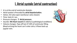
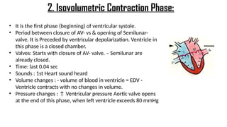
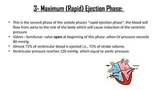
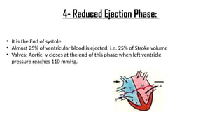
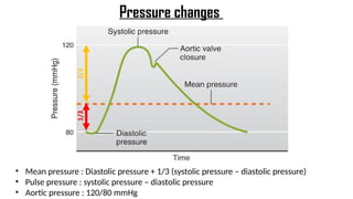
Moderator: Dr. Misbah (Senior Resident)
Presenter- Dr. Harshitha Gulur
CARDIAC CYCLE
Objectives :
• Main function of the heart.
• General principles of the cardiac cycle.
• Function of the atria, ventricles & valves.
• Different events that occur during the cardiac cycle.
The main function of the heart is to pump
blood throughout the body by pulmonary and
systemic circulations that work together .
Heart has two pump “double”:
• one of them called pulmonary- from the heart to the lung.
• The other one called Systemic - from the heart to the whole
body
• Both of them work together.
• Pulmonary carries deoxygenated blood.
• Systemic carries oxygenated blood.
• Any vessel goes out from the heart called artery, and any
one enter the heart called vein.
4 valves:
o aortic valve
o mitral valve
o pulmonary valve
o tricuspid valve
• Found at entry & exit of each ventricle
• Allow blood to flow in only ONE direction
• Opening & closure of vs occur as a result of pressure
gradient across the vs
• When AV-vs open, semilunar-vs close & vice versa
• AV cusps are held by chordae tendinea to muscular
projections called Papillary muscle
Valves of the heart
• Contraction of the heart generates pressure changes, & results in
orderly blood movement.
• Blood flows from an area of high pressure to an area of low
pressure. (Pressure gradient controls the opening and closure of the
cusps.)
• Events in the right & left sides of the heart are the same, but with
lower pressures in the right side
General Principles
CARDIAC CYCLE
Definition:
Sequences of the event
that takes place in the
heart in each beat
EVENTS:
I: Mechanical event
II: volume changes
III: Pressure changes
IV: Heart sounds
V: Electrical events
(ECG).
DURATION:
* Cardiac cycle duration=
0.8 sec, when HR is
72bpm
* the duration is
shortened when HR
becomes higher.
Mechanical events: mechanical means movement itself
which follow electrical signal “can’t happen by itself
without stimulus”.
Each heart beat consists of 2 major periods:
• Systole (contraction)
• Diastole (relaxation).
Systole and diastole happen on each chamber of the heart
so there is:
• Atrial systole & diastole
• Ventricular systole & diastole
I: Mechanical Events
Normally diastole is longer than systole.
Mechanical events periods
0.8 sec equals to the duration of cardiac cycle.
Importance of long ventricular diastole:
• Coronary blood flow.
• Ventricular filling
VENTRICLE ATRIUM
Ventricular systole= 0.3 sec Atrial systole= 0.1 sec
Ventricular diastole= 0.5 sec Atrial diastole= 0.7 sec
Systole “contraction” is shorter while Diastole “relaxation” is longer.
Because we need to wait for the heart to be filled with blood, another reason is because if you
keep the muscle in contraction for a long time this will cause cutting the blood supply for the
cardiac muscle”.
So in the ventricles the ratio is 0.5 (diastole) + 0.3 (systole) = 0.8 , however in the atrium the
ratio 0.7 (diastole) + 0.1 (systole)= 0.8
Intra-cardiac Pressure
Mechanical Phases
Mechanical Phases
PROTODIASTASIS :Very short Period between the end of ventricular systole and aortic valve closure. (As the ventricle
relax the pressure is reduced so the blood tend to flow back from aorta but prevented by closure of aortic valve)
• It is at the end of ventricular diastole .
• Atrial systole is Preceded by atrial depolarization.
• Valves: AV-valve open (semilunar valve closed.)
• Time: lasts 0.1 sec.
• Pressure changes: ↑ Atrial pressure.
• Sounds: 4th Heart sound (In elderly & pathological conditions)
• Volume changes: Tops off last 27-30% of ventricular filling.
Blood arriving the heart can’t enter atria, it flows back up
jugular vein.
I. Atrial systole (atrial contraction)
• It is the first phase (beginning) of ventricular systole.
• Period between closure of AV- vs & opening of Semilunar-
valve. It is Preceded by ventricular depolarization. Ventricle in
this phase is a closed chamber.
• Valves: Starts with closure of AV- valve. – Semilunar are
already closed.
• Time: last 0.04 sec
• Sounds : 1st Heart sound heard
• Volume changes : - volume of blood in ventricle = EDV -
Ventricle contracts with no changes in volume.
• Pressure changes : ↑ Ventricular pressure Aortic valve opens
at the end of this phase, when left ventricle exceeds 80 mmHg
2. Isovolumetric Contraction Phase:
• This is the second phase of the systole phases “rapid ejection phase”, the blood will
flow from aorta to the rest of the body which will cause reduction of the ventricle
pressure
• Valves : Semilunar- valve open at beginning of this phase ,when LV pressure exceeds
80 mmHg.
• Almost 75% of ventricular blood is ejected i.e., 75% of stroke volume.
• Ventricular pressure reaches 120 mmHg. which equal to aortic pressure.
3- Maximum (Rapid) Ejection Phase:
• It is the End of systole.
• Almost 25% of ventricular blood is ejected, i.e. 25% of Stroke volume
• Valves: Aortic- v closes at the end of this phase when left ventricle
pressure reaches 110 mmHg.
4- Reduced Ejection Phase:
• Period between closure of semilunar- vs & opening of AV- vs. Happens at the Beginning of
diastole.
• It is Preceded by ventricular repolarization.
• Time :Lasts 0.04 sec Sounds : 2nd Heart sound heard .
• Volume changes : -LV is a closed chamber, i.e. relax with no changes in volume .
• -Volume of blood in ventricle = ESV Valves : AV- vs open at the end of this
phase ; semilunar –vs close When ventricle pressure < arterial pressure → backflow of
blood → forces semilunar valves to close.
• Aortic pressure curve: INCISURA → when the aortic valve closes.
5- Isovolumetric Relaxation Phase:
• Pressure changes : Atrial pressure is higher than ventricular pressure.
• Valves: AV- vs open. -Semilunar valves are closed
• From the atrium 60-70% of blood passes passively move to the ventricles along
pressure gradient.
• Sounds :3rd Heart sound heard. (children only)
6- Rapid Filling Phase:
• Remaining atrial blood flows slowly into
ventricles.
• Valves : AV- vs still open - Semilunar valves
are closed
7- Reduced Filling Phase (Diastasis):
Definitions
Ventricular volume changes
Pressure changes
Pressure change
Descending/Catacrotid Limb
• Mean pressure : Diastolic pressure + 1/3 (systolic pressure – diastolic pressure)
• Pulse pressure : systolic pressure – diastolic pressure
• Aortic pressure : 120/80 mmHg
Pressure changes
Arterial pressure changes Pulmonary artery pressure
changes
• 110-130/70-85
• Similar to aortic pressure
waves, but sharper.
• (Its waves are Sharper than
aortic pressure waves.)
• Reflects a systolic peak
pressure of 110-130 mmHg &
a diastolic pressure 70-85
mmHg
• 25-30/4-12
• Similar to aortic pressure
changes, but with difference
in magnitude.
• Differs from aortic pressure
changes by magnitude.
• a wave is caused by atrial
contraction.
• c wave occurs when the ventricles
begin to contract; it is caused
partly by slight backflow of blood
into the atria at the onset of
ventricular contraction but mainly
by bulging of the A-V valves
backward toward the atria
because of increasing pressure in
the ventricles.
• v wave occurs toward the end of
ventricular contraction; when
blood flows into the atria from
the veins while the A-V valves are
closed during ventricular
contraction
Atrial pressure changes
Causes of Atrial pressure waves
• The 3 waves a, c & v are equal to ONE cardiac cycle = 0.8 sec.
• When ventricular contraction is over, the A-V valves open, allowing the stored atrial
blood to flow rapidly into the ventricles and causing the v wave to disappear.
Jugular venous pressure (JVP) changes
• 3 upward waves: a, c, & v 2
• downward waves: x & y
Heart Sounds Recorded During the Cardiac Cycle
These sound can be detected either by :
• Auscultation (using a Stethoscope)
• Phonocardiography (using a sound recording device)
•S1 – “lub” caused by the closing of the AV valves
•S2 – “dub” caused by the closing of semilunar valves
•S3 – linked with flow of blood into the ventricles
•S4 – linked with atrial contraction
•Generally, the heart makes two sounds – “lub” and “dub”.
•The third and fourth sounds are audible in individuals, however, they could show
abnormalities in the functioning of the heart.
•While the S1 and S2 are high-pitched, S3 and S4 are low-pitched sounds.
• 1st sound “lub”> Long, Low, Loud > best
heard at mitral & tricuspid
• 2 nd sound “dub”> Short, Soft, Sharp >
best heard at aortic & pulmonary.
• If we ask the patient to take deep
inspiration and hold it, then hear his
heart sound we’ll hear splitting “lub
tadub” in the 2nd sound, and this is
normal, but if it occurs in other than
deep inspiration then this is abnormal.
• When listening to the heart with a stethoscope, one does not hear the opening of the
valves because this is a relatively slow process that normally makes no noise.
• However, when the valves close, the vanes of the valves and the surrounding fluids
vibrate under the influence of sudden pressure changes, giving off sound that travels in
all directions through the chest.
• When the ventricles contract, one first hears a sound caused by closure of the A-V valves.
• The vibration pitch is low and relatively long-lasting and is known as the first heart sound.
• When the aortic and pulmonary valves close at the end of systole, one hears a rapid snap
because these valves close rapidly, and the surroundings vibrate for a short period.
• This is sound is called the second heart sound.
Relationship of the Heart Sounds to Heart Pumping
Cardiac Cycle Vs Heart Sounds
THANK YOU

cardiac cycle basics and anesthetic implication

  • 1.
    Moderator: Dr. Misbah(Senior Resident) Presenter- Dr. Harshitha Gulur CARDIAC CYCLE
  • 2.
    Objectives : • Mainfunction of the heart. • General principles of the cardiac cycle. • Function of the atria, ventricles & valves. • Different events that occur during the cardiac cycle.
  • 3.
    The main functionof the heart is to pump blood throughout the body by pulmonary and systemic circulations that work together . Heart has two pump “double”: • one of them called pulmonary- from the heart to the lung. • The other one called Systemic - from the heart to the whole body • Both of them work together. • Pulmonary carries deoxygenated blood. • Systemic carries oxygenated blood. • Any vessel goes out from the heart called artery, and any one enter the heart called vein.
  • 4.
    4 valves: o aorticvalve o mitral valve o pulmonary valve o tricuspid valve • Found at entry & exit of each ventricle • Allow blood to flow in only ONE direction • Opening & closure of vs occur as a result of pressure gradient across the vs • When AV-vs open, semilunar-vs close & vice versa • AV cusps are held by chordae tendinea to muscular projections called Papillary muscle Valves of the heart
  • 5.
    • Contraction ofthe heart generates pressure changes, & results in orderly blood movement. • Blood flows from an area of high pressure to an area of low pressure. (Pressure gradient controls the opening and closure of the cusps.) • Events in the right & left sides of the heart are the same, but with lower pressures in the right side General Principles
  • 6.
    CARDIAC CYCLE Definition: Sequences ofthe event that takes place in the heart in each beat EVENTS: I: Mechanical event II: volume changes III: Pressure changes IV: Heart sounds V: Electrical events (ECG). DURATION: * Cardiac cycle duration= 0.8 sec, when HR is 72bpm * the duration is shortened when HR becomes higher.
  • 7.
    Mechanical events: mechanicalmeans movement itself which follow electrical signal “can’t happen by itself without stimulus”. Each heart beat consists of 2 major periods: • Systole (contraction) • Diastole (relaxation). Systole and diastole happen on each chamber of the heart so there is: • Atrial systole & diastole • Ventricular systole & diastole I: Mechanical Events
  • 8.
    Normally diastole islonger than systole. Mechanical events periods 0.8 sec equals to the duration of cardiac cycle. Importance of long ventricular diastole: • Coronary blood flow. • Ventricular filling VENTRICLE ATRIUM Ventricular systole= 0.3 sec Atrial systole= 0.1 sec Ventricular diastole= 0.5 sec Atrial diastole= 0.7 sec
  • 9.
    Systole “contraction” isshorter while Diastole “relaxation” is longer. Because we need to wait for the heart to be filled with blood, another reason is because if you keep the muscle in contraction for a long time this will cause cutting the blood supply for the cardiac muscle”. So in the ventricles the ratio is 0.5 (diastole) + 0.3 (systole) = 0.8 , however in the atrium the ratio 0.7 (diastole) + 0.1 (systole)= 0.8
  • 10.
  • 11.
  • 12.
    Mechanical Phases PROTODIASTASIS :Veryshort Period between the end of ventricular systole and aortic valve closure. (As the ventricle relax the pressure is reduced so the blood tend to flow back from aorta but prevented by closure of aortic valve)
  • 13.
    • It isat the end of ventricular diastole . • Atrial systole is Preceded by atrial depolarization. • Valves: AV-valve open (semilunar valve closed.) • Time: lasts 0.1 sec. • Pressure changes: ↑ Atrial pressure. • Sounds: 4th Heart sound (In elderly & pathological conditions) • Volume changes: Tops off last 27-30% of ventricular filling. Blood arriving the heart can’t enter atria, it flows back up jugular vein. I. Atrial systole (atrial contraction)
  • 14.
    • It isthe first phase (beginning) of ventricular systole. • Period between closure of AV- vs & opening of Semilunar- valve. It is Preceded by ventricular depolarization. Ventricle in this phase is a closed chamber. • Valves: Starts with closure of AV- valve. – Semilunar are already closed. • Time: last 0.04 sec • Sounds : 1st Heart sound heard • Volume changes : - volume of blood in ventricle = EDV - Ventricle contracts with no changes in volume. • Pressure changes : ↑ Ventricular pressure Aortic valve opens at the end of this phase, when left ventricle exceeds 80 mmHg 2. Isovolumetric Contraction Phase:
  • 15.
    • This isthe second phase of the systole phases “rapid ejection phase”, the blood will flow from aorta to the rest of the body which will cause reduction of the ventricle pressure • Valves : Semilunar- valve open at beginning of this phase ,when LV pressure exceeds 80 mmHg. • Almost 75% of ventricular blood is ejected i.e., 75% of stroke volume. • Ventricular pressure reaches 120 mmHg. which equal to aortic pressure. 3- Maximum (Rapid) Ejection Phase:
  • 16.
    • It isthe End of systole. • Almost 25% of ventricular blood is ejected, i.e. 25% of Stroke volume • Valves: Aortic- v closes at the end of this phase when left ventricle pressure reaches 110 mmHg. 4- Reduced Ejection Phase:
  • 17.
    • Period betweenclosure of semilunar- vs & opening of AV- vs. Happens at the Beginning of diastole. • It is Preceded by ventricular repolarization. • Time :Lasts 0.04 sec Sounds : 2nd Heart sound heard . • Volume changes : -LV is a closed chamber, i.e. relax with no changes in volume . • -Volume of blood in ventricle = ESV Valves : AV- vs open at the end of this phase ; semilunar –vs close When ventricle pressure < arterial pressure → backflow of blood → forces semilunar valves to close. • Aortic pressure curve: INCISURA → when the aortic valve closes. 5- Isovolumetric Relaxation Phase:
  • 18.
    • Pressure changes: Atrial pressure is higher than ventricular pressure. • Valves: AV- vs open. -Semilunar valves are closed • From the atrium 60-70% of blood passes passively move to the ventricles along pressure gradient. • Sounds :3rd Heart sound heard. (children only) 6- Rapid Filling Phase:
  • 19.
    • Remaining atrialblood flows slowly into ventricles. • Valves : AV- vs still open - Semilunar valves are closed 7- Reduced Filling Phase (Diastasis):
  • 20.
  • 21.
  • 22.
  • 23.
  • 24.
  • 25.
    • Mean pressure: Diastolic pressure + 1/3 (systolic pressure – diastolic pressure) • Pulse pressure : systolic pressure – diastolic pressure • Aortic pressure : 120/80 mmHg Pressure changes
  • 26.
    Arterial pressure changesPulmonary artery pressure changes • 110-130/70-85 • Similar to aortic pressure waves, but sharper. • (Its waves are Sharper than aortic pressure waves.) • Reflects a systolic peak pressure of 110-130 mmHg & a diastolic pressure 70-85 mmHg • 25-30/4-12 • Similar to aortic pressure changes, but with difference in magnitude. • Differs from aortic pressure changes by magnitude.
  • 27.
    • a waveis caused by atrial contraction. • c wave occurs when the ventricles begin to contract; it is caused partly by slight backflow of blood into the atria at the onset of ventricular contraction but mainly by bulging of the A-V valves backward toward the atria because of increasing pressure in the ventricles. • v wave occurs toward the end of ventricular contraction; when blood flows into the atria from the veins while the A-V valves are closed during ventricular contraction Atrial pressure changes
  • 28.
    Causes of Atrialpressure waves • The 3 waves a, c & v are equal to ONE cardiac cycle = 0.8 sec. • When ventricular contraction is over, the A-V valves open, allowing the stored atrial blood to flow rapidly into the ventricles and causing the v wave to disappear.
  • 29.
    Jugular venous pressure(JVP) changes • 3 upward waves: a, c, & v 2 • downward waves: x & y
  • 30.
    Heart Sounds RecordedDuring the Cardiac Cycle These sound can be detected either by : • Auscultation (using a Stethoscope) • Phonocardiography (using a sound recording device) •S1 – “lub” caused by the closing of the AV valves •S2 – “dub” caused by the closing of semilunar valves •S3 – linked with flow of blood into the ventricles •S4 – linked with atrial contraction •Generally, the heart makes two sounds – “lub” and “dub”. •The third and fourth sounds are audible in individuals, however, they could show abnormalities in the functioning of the heart. •While the S1 and S2 are high-pitched, S3 and S4 are low-pitched sounds.
  • 33.
    • 1st sound“lub”> Long, Low, Loud > best heard at mitral & tricuspid • 2 nd sound “dub”> Short, Soft, Sharp > best heard at aortic & pulmonary. • If we ask the patient to take deep inspiration and hold it, then hear his heart sound we’ll hear splitting “lub tadub” in the 2nd sound, and this is normal, but if it occurs in other than deep inspiration then this is abnormal.
  • 34.
    • When listeningto the heart with a stethoscope, one does not hear the opening of the valves because this is a relatively slow process that normally makes no noise. • However, when the valves close, the vanes of the valves and the surrounding fluids vibrate under the influence of sudden pressure changes, giving off sound that travels in all directions through the chest. • When the ventricles contract, one first hears a sound caused by closure of the A-V valves. • The vibration pitch is low and relatively long-lasting and is known as the first heart sound. • When the aortic and pulmonary valves close at the end of systole, one hears a rapid snap because these valves close rapidly, and the surroundings vibrate for a short period. • This is sound is called the second heart sound. Relationship of the Heart Sounds to Heart Pumping
  • 35.
    Cardiac Cycle VsHeart Sounds
  • 36.

Editor's Notes

  • #6 The total duration of the cardiac cycle, including systole and diastole, is the reciprocal of the heart rate. e. When heart rate increases, the duration of each cardiac cycle decreases, including the contraction and relaxation phases. The duration of the action potential and the period of contraction (systole) also decrease, but not by as great a percentage as does the relaxation phase (diastole).
  • #14 isometric means ventricles are closed chambers, when the chambers are closed it means there’s no blood enter or get out of the heart ”this blood called enddiastolic volume”, this will increase the pressure inside the left ventricles until it’s reach the 80 so it will open the aortic valve
  • #15 Then the blood will rapidly be ejected out of the ventricles (eject 75% of the blood of the ventricles), and the pressure will reach 120 which equal to aorta pressure, this is the second phase of the systole phases “rapid ejection phase”, the blood will flow from aorta to the rest of the body which will cause reduced of the ventricle pressure
  • #16 - It will be reduced until it will reach 110, This reduction will cause the 3rd and last phase of systole phases “reduced ejection phase”, at this pressure the semilunar valves will close
  • #17 all the chambers are closed, the blood remaining in the ventricles in this case called end-systolic volume, ventricles keep relaxing and relaxing until the pressure decreased and become higher in atrium more than ventricle
  • #19 After rapid filling the blood will pass through AV valve passively and this cause the 3rd phase of diastole “diastasis (reduced filling phase)”, and this is the last phase in the circulation. All the sounds of the heart are occurring in the diastole phases.