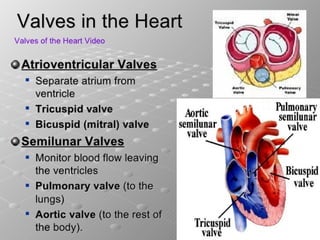
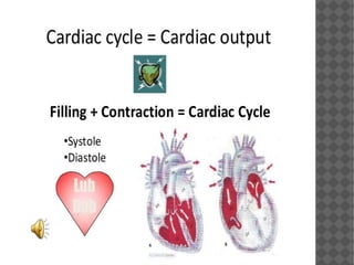
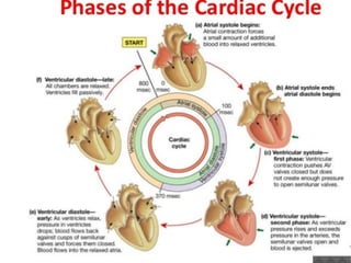
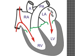
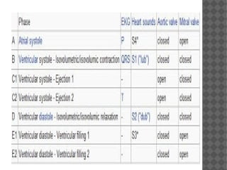
CARDIAC CYCLE
By,
Dr.Nithil Ann Varughese
 The cardiac cycle refers to a complete
heartbeat from its generation to the
beginning of the next beat, and so includes
the diastole, the systole and the intervening
pause. The frequency of the cardiac cycle is
described by the heart rate, which is
typically expressed as beats per minute
 A single cycle of cardiac activity can be divided
into two basic phases - diastole and systole. 
 Diastole represents the period of time when the
ventricles are relaxed .
 Throughout most of this period, blood is passively
flowing from the left atrium (LA) and right atrium
(RA) into the left ventricle (LV) and right ventricle
(RV), respectively .
The blood flows through atrio ventricular valves
(mitral and tricuspid) that separate the atria
from the ventricles. The RA receives venous blood
from the body through the superior vena cava
(SVC) and inferior vena cava (IVC). The LA
receives oxygenated blood from lungs through
four pulmonary veins that enter the LA.
At the end of diastole, both atria contract, which
propels an additional amount of blood into the
ventricles.
 An average adult has a heart rate of
about 70 beats per minute. At this rate, a
complete cardiac cycle takes roughly 0.8
seconds to complete. About 0.1s is atrial
systole, followed by about 0.3s of ventricular
systole, followed by about 0.4s of atrial and
ventricular diastole.
Cardiac Cycle - Atrial Contraction
(Phase 1)
§ As the atria contract, the pressure within the atrial
chambers increases, which forces more blood flow
across the open atrioventricular (AV) valves, leading
to a rapid flow of blood into the ventricles.
 Atrial contraction normally accounts for
about 10% of left ventricular filling when a
person is at rest because most of ventricular
filling occurs prior to atrial contraction as
blood passively flows from the pulmonary
veins, into the left atrium, then into the left
ventricle through the open mitral valve
 At high heart rates when there is less time
for passive ventricular filling, the atrial
contraction may account for up to 40% of
ventricular filling. This is sometimes referred
to as the "atrial kick." 
 After atrial contraction is complete, the atrial
pressure begins to fall causing a pressure gradient
reversal across the AV valves.  This causes the
valves to float upward (pre-position) before
closure.  At this time, the ventricular volumes are
maximal, which is termed the end-diastolic
volume (EDV). 
 The left ventricular EDV (LVEDV), which is typically
about 120 ml, represents the ventricular preload
 and is associated with end-diastolic pressures of 8-
12 mmHg and 3-6 mmHg in the left and right
ventricles, respectively.
.
A heart sound is sometimes noted during atrial
contraction (fourth heart sound, S4).  This sound is
caused by vibration of the ventricular wall during
atrial contraction.  Generally, it is noted when the
ventricle compliance is reduced ("stiff" ventricle) as
occurs in ventricular hypertrophy and in many older
individuals
Cardiac Cycle - Isovolumetric
Contraction (Phase 2)
 This phase of the cardiac cycle begins with
the appearance of the QRS complex of the
ECG, which represents ventricular
depolarization.
 The AV valves close when intraventricular pressure
exceeds atrial pressure. Ventricular contraction also
triggers contraction of the papillary muscles with
their chordae tendineae that are attached to the
valve leaflets. This tension on the the AV valve
leaflets prevent them from bulging back into the atria
and becoming incompetent (i.e., “leaky”). Closure of
the AV valves results in the first heart sound (S1).
This sound is normally split (~0.04 sec) because mitral
valve closure precedes tricuspid closure
 During the time period between the closure
of the AV valves and the opening of the
aortic and pulmonic valves, ventricular
pressure rises rapidly without a change in
ventricular volume (i.e., no ejection
occurs). Ventricular volume does not change
because all valves are closed during this
phase. Contraction, therefore, is said to be
"isovolumic" or "isovolumetric."  
Cardiac Cycle - Rapid Ejection
(Phase 3)
 This phase represents initial, rapid ejection of blood
into the aorta and pulmonary arteries from the left
and right ventricles, respectively. Ejection begins
when the intraventricular pressures exceed the
pressures within the aorta and pulmonary artery,
which causes the aortic and pulmonic valves to
open. During this phase, ventricular pressure
normally exceeds outflow tract pressure by a few
mmHg. This pressure gradient across the valve is
ordinarily low because of the relatively large valve
opening (i.e., low resistance). Maximal outflow
velocity is reached early in the ejection phase, and
maximal (systolic) aortic and pulmonary artery
pressures are achieved.
No heart sounds are ordinarily noted during ejection
because the opening of healthy valves is silent. The
presence of sounds during ejection (i.e., systolic
murmurs) indicate valve disease or intracardiac
shunts.
●
Left atrial pressure initially decreases as the atrial
base is pulled downward, expanding the atrial
chamber. Blood continues to flow into the atria
from their respective venous inflow tracts and the
atrial pressures begin to rise. This rise in pressure
continues until the AV valves open at the end of
phase 5.
Cardiac Cycle - Reduced
Ejection (Phase 4)
 Ventricular pressure falls slightly below
outflow tract pressure; however, outward
flow still occurs due to 
kinetic (or inertial) energy of the blood.
 Left atrial and right atrial pressures gradually
rise due to continued venous return from the
lungs and from the systemic circulation,
respectively.
Cardiac Cycle - Isovolumetric
Relaxation (Phase 5)
 When the intraventricular pressures fall
sufficiently at the end of phase 4, the aortic
and pulmonic valves abruptly close (aortic
precedes pulmonic) causing the second
heart sound (S2) and the beginning of
isovolumetric relaxation. Valve closure is
associated with a small backflow of blood
into the ventricles and a characteristic notch
(incisura or dicrotic notch) in the aortic and
pulmonary artery pressure tracings.
 After valve closure, the aortic and pulmonary
artery pressures rise slightly (dicrotic wave)
following by a slow decline in pressure.
 Although ventricular pressures decrease
during this phase, volumes do not change
because all valves are closed. The volume of
blood that remains in a ventricle is called
the end-systolic volume and is ~50 ml in the
left ventricle. The difference between the
end-diastolic volume and the end-systolic
volume is ~70 ml and represents the stroke
volume.
 Left atrial pressure (LAP) continues to rise
because of venous return from the lungs
Cardiac Cycle - Rapid Filling
(Phase 6)
 As the ventricles continue to relax at the end
of phase 5, the intraventricular pressures will
at some point fall below their respective
atrial pressures. When this occurs, the AV
valves rapidly open and passive ventricular
filling begins. Despite the inflow of blood
from the atria, intraventricular pressure
continues to briefly fall because the
ventricles are still undergoing relaxation.
Once the ventricles are completely relaxed,
their pressures will slowly rise as they fill
with blood from the atria.
 Ventricular filling is normally silent. When
a third heart sound (S3) is audible during
rapid ventricular filling, it may represent
tensing of chordae tendineae and AV ring
during ventricular relaxation and filling. This
heart sound is normal in children; but is
often pathological in adults and caused by
 ventricular dilation
Cardiac Cycle - Reduced Filling
(Phase 7)
 As the ventricles continue to fill with blood
and expand, they become less compliant and
the intraventricular pressures rise. The
increase in intraventricular pressure reduces
the pressure gradient across the AV valves so
that the rate of filling falls late in diastole.
 In normal, resting hearts, the ventricle is
about 90% filled by the end of this phase. In
other words, about 90% of ventricular filling
occurs before atrial contraction (phase 1)
and therefore is passive.
 Aortic and pulmonary arterial pressures
THANK YOU…

Cardiac cycle Dr. Nithil

  • 1.
  • 6.
     The cardiac cycle refersto a complete heartbeat from its generation to the beginning of the next beat, and so includes the diastole, the systole and the intervening pause. The frequency of the cardiac cycle is described by the heart rate, which is typically expressed as beats per minute
  • 7.
     A singlecycle of cardiac activity can be divided into two basic phases - diastole and systole.   Diastole represents the period of time when the ventricles are relaxed .  Throughout most of this period, blood is passively flowing from the left atrium (LA) and right atrium (RA) into the left ventricle (LV) and right ventricle (RV), respectively .
  • 9.
    The blood flowsthrough atrio ventricular valves (mitral and tricuspid) that separate the atria from the ventricles. The RA receives venous blood from the body through the superior vena cava (SVC) and inferior vena cava (IVC). The LA receives oxygenated blood from lungs through four pulmonary veins that enter the LA. At the end of diastole, both atria contract, which propels an additional amount of blood into the ventricles.
  • 11.
     An averageadult has a heart rate of about 70 beats per minute. At this rate, a complete cardiac cycle takes roughly 0.8 seconds to complete. About 0.1s is atrial systole, followed by about 0.3s of ventricular systole, followed by about 0.4s of atrial and ventricular diastole.
  • 13.
    Cardiac Cycle -Atrial Contraction (Phase 1) § As the atria contract, the pressure within the atrial chambers increases, which forces more blood flow across the open atrioventricular (AV) valves, leading to a rapid flow of blood into the ventricles.
  • 15.
     Atrial contractionnormally accounts for about 10% of left ventricular filling when a person is at rest because most of ventricular filling occurs prior to atrial contraction as blood passively flows from the pulmonary veins, into the left atrium, then into the left ventricle through the open mitral valve
  • 16.
     At highheart rates when there is less time for passive ventricular filling, the atrial contraction may account for up to 40% of ventricular filling. This is sometimes referred to as the "atrial kick." 
  • 17.
     After atrialcontraction is complete, the atrial pressure begins to fall causing a pressure gradient reversal across the AV valves.  This causes the valves to float upward (pre-position) before closure.  At this time, the ventricular volumes are maximal, which is termed the end-diastolic volume (EDV).   The left ventricular EDV (LVEDV), which is typically about 120 ml, represents the ventricular preload  and is associated with end-diastolic pressures of 8- 12 mmHg and 3-6 mmHg in the left and right ventricles, respectively. .
  • 18.
    A heart sound is sometimesnoted during atrial contraction (fourth heart sound, S4).  This sound is caused by vibration of the ventricular wall during atrial contraction.  Generally, it is noted when the ventricle compliance is reduced ("stiff" ventricle) as occurs in ventricular hypertrophy and in many older individuals
  • 19.
    Cardiac Cycle -Isovolumetric Contraction (Phase 2)  This phase of the cardiac cycle begins with the appearance of the QRS complex of the ECG, which represents ventricular depolarization.  The AV valves close when intraventricular pressure exceeds atrial pressure. Ventricular contraction also triggers contraction of the papillary muscles with their chordae tendineae that are attached to the valve leaflets. This tension on the the AV valve leaflets prevent them from bulging back into the atria and becoming incompetent (i.e., “leaky”). Closure of the AV valves results in the first heart sound (S1). This sound is normally split (~0.04 sec) because mitral valve closure precedes tricuspid closure
  • 20.
     During thetime period between the closure of the AV valves and the opening of the aortic and pulmonic valves, ventricular pressure rises rapidly without a change in ventricular volume (i.e., no ejection occurs). Ventricular volume does not change because all valves are closed during this phase. Contraction, therefore, is said to be "isovolumic" or "isovolumetric."  
  • 21.
    Cardiac Cycle -Rapid Ejection (Phase 3)  This phase represents initial, rapid ejection of blood into the aorta and pulmonary arteries from the left and right ventricles, respectively. Ejection begins when the intraventricular pressures exceed the pressures within the aorta and pulmonary artery, which causes the aortic and pulmonic valves to open. During this phase, ventricular pressure normally exceeds outflow tract pressure by a few mmHg. This pressure gradient across the valve is ordinarily low because of the relatively large valve opening (i.e., low resistance). Maximal outflow velocity is reached early in the ejection phase, and maximal (systolic) aortic and pulmonary artery pressures are achieved.
  • 22.
    No heart soundsare ordinarily noted during ejection because the opening of healthy valves is silent. The presence of sounds during ejection (i.e., systolic murmurs) indicate valve disease or intracardiac shunts. ● Left atrial pressure initially decreases as the atrial base is pulled downward, expanding the atrial chamber. Blood continues to flow into the atria from their respective venous inflow tracts and the atrial pressures begin to rise. This rise in pressure continues until the AV valves open at the end of phase 5.
  • 24.
    Cardiac Cycle -Reduced Ejection (Phase 4)  Ventricular pressure falls slightly below outflow tract pressure; however, outward flow still occurs due to  kinetic (or inertial) energy of the blood.  Left atrial and right atrial pressures gradually rise due to continued venous return from the lungs and from the systemic circulation, respectively.
  • 26.
    Cardiac Cycle -Isovolumetric Relaxation (Phase 5)  When the intraventricular pressures fall sufficiently at the end of phase 4, the aortic and pulmonic valves abruptly close (aortic precedes pulmonic) causing the second heart sound (S2) and the beginning of isovolumetric relaxation. Valve closure is associated with a small backflow of blood into the ventricles and a characteristic notch (incisura or dicrotic notch) in the aortic and pulmonary artery pressure tracings.  After valve closure, the aortic and pulmonary artery pressures rise slightly (dicrotic wave) following by a slow decline in pressure.
  • 27.
     Although ventricularpressures decrease during this phase, volumes do not change because all valves are closed. The volume of blood that remains in a ventricle is called the end-systolic volume and is ~50 ml in the left ventricle. The difference between the end-diastolic volume and the end-systolic volume is ~70 ml and represents the stroke volume.
  • 29.
     Left atrialpressure (LAP) continues to rise because of venous return from the lungs
  • 30.
    Cardiac Cycle -Rapid Filling (Phase 6)  As the ventricles continue to relax at the end of phase 5, the intraventricular pressures will at some point fall below their respective atrial pressures. When this occurs, the AV valves rapidly open and passive ventricular filling begins. Despite the inflow of blood from the atria, intraventricular pressure continues to briefly fall because the ventricles are still undergoing relaxation. Once the ventricles are completely relaxed, their pressures will slowly rise as they fill with blood from the atria.
  • 31.
     Ventricular fillingis normally silent. When a third heart sound (S3) is audible during rapid ventricular filling, it may represent tensing of chordae tendineae and AV ring during ventricular relaxation and filling. This heart sound is normal in children; but is often pathological in adults and caused by  ventricular dilation
  • 33.
    Cardiac Cycle -Reduced Filling (Phase 7)  As the ventricles continue to fill with blood and expand, they become less compliant and the intraventricular pressures rise. The increase in intraventricular pressure reduces the pressure gradient across the AV valves so that the rate of filling falls late in diastole.  In normal, resting hearts, the ventricle is about 90% filled by the end of this phase. In other words, about 90% of ventricular filling occurs before atrial contraction (phase 1) and therefore is passive.  Aortic and pulmonary arterial pressures
  • 37.