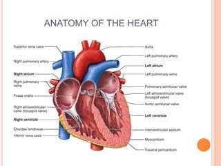
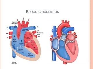
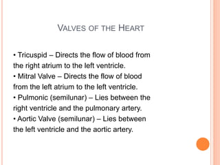
CARDIOVASCULAR
ASSESSMENT
Prepared by: Gianne T. Gregorio RN
INTRODUCTION
Cardiovascular disease is the every
State’s leading killer for both men and women among
all racial and ethnic groups.
•A thorough cardiovascular assessment
will help to identify significant factors that
can influence cardiovascular health such
as high blood cholesterol, cigarette use,
diabetes, or hypertension.
OBJECTIVES
1. Review anatomy and physiology of
cardiovascular system.
2. Describe physical assessment of
cardiovascular status.
3. Review diagnostic procedures.
ANATOMY OF THE HEART
FUNCTIONS OF THE HEART & CV SYSTEM
• Pumps blood to
tissues to
supply O2 &
nutrients
• Remove CO2 &
metabolic
wastes
BLOOD CIRCULATION
BLOOD CIRCULATION
1. Oxygen-poor blood (shown in blue) flows from the body
into the right atrium.
2. Blood flows through the right atrium into the right
ventricle.
3. The right ventricle pumps the blood to the lungs, where
the blood releases waste gases and picks up oxygen.
3. The newly oxygen-rich blood
(shown in red)returns to the heart and
enters the left atrium.
4. Blood flows through the left atrium
into the left ventricle.
5. The left ventricle pumps the oxygen-rich
blood to all parts of the body.
VALVES OF THE HEART
• Tricuspid – Directs the flow of blood from
the right atrium to the left ventricle.
• Mitral Valve – Directs the flow of blood
from the left atrium to the left ventricle.
• Pulmonic (semilunar) – Lies between the
right ventricle and the pulmonary artery.
• Aortic Valve (semilunar) – Lies between
the left ventricle and the aortic artery.
CARDIOVASCULAR ASSESSMENT
History
The purpose of the cardiovascular health
history is to provide information about
your patient’s cardiovascular symptoms
and how they developed. A complete
cardiovascular history will give you
indications to potential or underlying
cardiovascular illnesses or disease states.
Past Health History
It is important to ask questions about your
patient’s past health history. The past
health history should elicit information
about the following issues: hypertension,
elevated blood cholesterol or
triglycerides, heart murmurs, congenital
heart disease, rheumatic fever or
unexplained joint pains
Current Lifestyle and Psychosocial Status
• Nutrition
• Smoking
• Alcohol
• Exercise
• Drugs
• Family History
INSPECTION
Eyes
The presence of yellowish plaques
on the eyelids (xanthelasma)
Could indicate hyperlipoproteinemia,
a risk factor for hypertension as well
as arteriolosclerosis.
Chest
• Observe the chest for
overall torso contour.
• Do you see pectus
excavatum (caved-in chest)?
• Do you see pectus
carinatum (pigeon chest)?
Skin
 Clubbing
The presence of
clubbing (broadening of the
extremities of the digits,
accompanied by nails which
are abnormally curved and
shiny) indicates chronic poor
oxygen perfusion to the
distal tissues of the hand
and feet.
 Cyanosis
• The presence of
cyanosis (bluish
colour) also denotes
chronic poor oxygen
delivery to the
peripheral tissues of
the hands and feet.
 Xanthomas
• The presence of
yellowish plaques
under the skin (noneruptive)
Excoriated through the skin
(eruptive) could indicate
hyperlipoproteinemia,
a risk factor for hypertension
As well as arteriolosclerosis.
 Edema
• The presence of edema
(tissue swelling) can be
caused by several
factors, although most
commonly is associated
with decreased cardiac
function leading to
decreased capillary flow.
PALPATION
• Use the palm of your hand to feel the
chest wall for the "Point of Maximal
Impulse" (PMI), which is usually found at
the apex of the heart. This apical pulse
is generally located in the 5th intercostal
space, about 7-9 cm (the width of your
palm) to the left of the midline.
Palpate the peripheral arteries.
These include the brachial, radial,
femoral, popliteal, dorsalis pedis, and
posterior tibial. Note the contour and
amplitude of each pulsation. These
should feel similar bilaterally.
CHEST PERCUSSION:
• Normally only the left border of heart can
be detected by percussion. It extends
from the sternum to mid clavicular line in
the third to fifth inter costal space. The
right border lies under the right margin of
the sternum and is not detectable.
Enlargement of the heart too either the
left or right usually can be noted.
AUSCULTATION
the action of listening
to sounds from the
heart, lungs, or other
organs, typically with
a stethoscope, as a
part of medical
diagnosis.
TIPS FOR HEART AUSCULTATION
Remember the mnemonic:
“All Patients Take Medicine”
 Aortic: found right of the sternal border in the 2nd
intercostal space REPRESENTS S2 “dub”
 Pulmonic: found left of the sternal border in the
2nd intercostal space REPRESENTS S2 “dub”
 Erb’s Point: found left of the sternal border in the
3rd intercostal space
 Tricuspid: found right of the sternal border in the
4th intercostal space REPRESENTS S1 “lub”
 Mitral: found midclavicular in the 5th intercostal
space REPRESENTS S1 “lub” (also the site of
point of maximal impulse)
PATIENT POSITIONING FOR HEART AUSCULTATION
Supine or sitting-up: Use the diaphragm and
listen at all 5 auscultation sites (noting S1 and S2
and if there are any splits presents). In addition,
distinguish S1 from S2. Then repeat with the bell of
the stethoscope…noting any other extra sounds.
Left side: turn the patient onto their left side and
auscultate with the bell of the stethoscope at the
APEX area and listen for S3, S4, or mitral stenosis
murmurs.
Sit up, lean forward, and have patient exhale:
Listen with the diaphragm at the aortic and
pulmonic sites for murmurs.
BLOOD PRESSURE
REFERENCES
 http://www.registerednursern.com/auscultating-the-
heart/
 https://www.slideshare.net/manalihsolanki/cardiac-
assessment-ppt

Cardiovascular assessment

  • 1.
  • 2.
    INTRODUCTION Cardiovascular disease isthe every State’s leading killer for both men and women among all racial and ethnic groups. •A thorough cardiovascular assessment will help to identify significant factors that can influence cardiovascular health such as high blood cholesterol, cigarette use, diabetes, or hypertension.
  • 3.
    OBJECTIVES 1. Review anatomyand physiology of cardiovascular system. 2. Describe physical assessment of cardiovascular status. 3. Review diagnostic procedures.
  • 4.
  • 5.
    FUNCTIONS OF THEHEART & CV SYSTEM • Pumps blood to tissues to supply O2 & nutrients • Remove CO2 & metabolic wastes
  • 6.
  • 7.
    BLOOD CIRCULATION 1. Oxygen-poorblood (shown in blue) flows from the body into the right atrium. 2. Blood flows through the right atrium into the right ventricle. 3. The right ventricle pumps the blood to the lungs, where the blood releases waste gases and picks up oxygen. 3. The newly oxygen-rich blood (shown in red)returns to the heart and enters the left atrium. 4. Blood flows through the left atrium into the left ventricle. 5. The left ventricle pumps the oxygen-rich blood to all parts of the body.
  • 8.
    VALVES OF THEHEART • Tricuspid – Directs the flow of blood from the right atrium to the left ventricle. • Mitral Valve – Directs the flow of blood from the left atrium to the left ventricle. • Pulmonic (semilunar) – Lies between the right ventricle and the pulmonary artery. • Aortic Valve (semilunar) – Lies between the left ventricle and the aortic artery.
  • 9.
    CARDIOVASCULAR ASSESSMENT History The purposeof the cardiovascular health history is to provide information about your patient’s cardiovascular symptoms and how they developed. A complete cardiovascular history will give you indications to potential or underlying cardiovascular illnesses or disease states.
  • 10.
    Past Health History Itis important to ask questions about your patient’s past health history. The past health history should elicit information about the following issues: hypertension, elevated blood cholesterol or triglycerides, heart murmurs, congenital heart disease, rheumatic fever or unexplained joint pains
  • 11.
    Current Lifestyle andPsychosocial Status • Nutrition • Smoking • Alcohol • Exercise • Drugs • Family History
  • 12.
    INSPECTION Eyes The presence ofyellowish plaques on the eyelids (xanthelasma) Could indicate hyperlipoproteinemia, a risk factor for hypertension as well as arteriolosclerosis.
  • 13.
    Chest • Observe thechest for overall torso contour. • Do you see pectus excavatum (caved-in chest)? • Do you see pectus carinatum (pigeon chest)?
  • 14.
    Skin  Clubbing The presenceof clubbing (broadening of the extremities of the digits, accompanied by nails which are abnormally curved and shiny) indicates chronic poor oxygen perfusion to the distal tissues of the hand and feet.
  • 15.
     Cyanosis • Thepresence of cyanosis (bluish colour) also denotes chronic poor oxygen delivery to the peripheral tissues of the hands and feet.
  • 16.
     Xanthomas • Thepresence of yellowish plaques under the skin (noneruptive) Excoriated through the skin (eruptive) could indicate hyperlipoproteinemia, a risk factor for hypertension As well as arteriolosclerosis.
  • 17.
     Edema • Thepresence of edema (tissue swelling) can be caused by several factors, although most commonly is associated with decreased cardiac function leading to decreased capillary flow.
  • 18.
    PALPATION • Use thepalm of your hand to feel the chest wall for the "Point of Maximal Impulse" (PMI), which is usually found at the apex of the heart. This apical pulse is generally located in the 5th intercostal space, about 7-9 cm (the width of your palm) to the left of the midline.
  • 20.
    Palpate the peripheralarteries. These include the brachial, radial, femoral, popliteal, dorsalis pedis, and posterior tibial. Note the contour and amplitude of each pulsation. These should feel similar bilaterally.
  • 21.
    CHEST PERCUSSION: • Normallyonly the left border of heart can be detected by percussion. It extends from the sternum to mid clavicular line in the third to fifth inter costal space. The right border lies under the right margin of the sternum and is not detectable. Enlargement of the heart too either the left or right usually can be noted.
  • 22.
    AUSCULTATION the action oflistening to sounds from the heart, lungs, or other organs, typically with a stethoscope, as a part of medical diagnosis.
  • 23.
    TIPS FOR HEARTAUSCULTATION Remember the mnemonic: “All Patients Take Medicine”
  • 24.
     Aortic: foundright of the sternal border in the 2nd intercostal space REPRESENTS S2 “dub”  Pulmonic: found left of the sternal border in the 2nd intercostal space REPRESENTS S2 “dub”  Erb’s Point: found left of the sternal border in the 3rd intercostal space  Tricuspid: found right of the sternal border in the 4th intercostal space REPRESENTS S1 “lub”  Mitral: found midclavicular in the 5th intercostal space REPRESENTS S1 “lub” (also the site of point of maximal impulse)
  • 25.
    PATIENT POSITIONING FORHEART AUSCULTATION Supine or sitting-up: Use the diaphragm and listen at all 5 auscultation sites (noting S1 and S2 and if there are any splits presents). In addition, distinguish S1 from S2. Then repeat with the bell of the stethoscope…noting any other extra sounds. Left side: turn the patient onto their left side and auscultate with the bell of the stethoscope at the APEX area and listen for S3, S4, or mitral stenosis murmurs. Sit up, lean forward, and have patient exhale: Listen with the diaphragm at the aortic and pulmonic sites for murmurs.
  • 26.
  • 27.