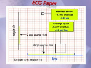
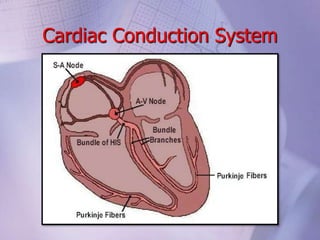
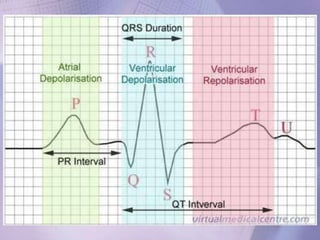
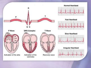
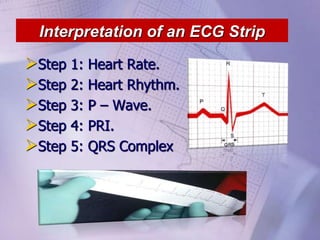
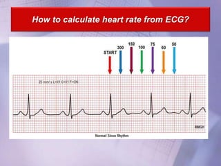
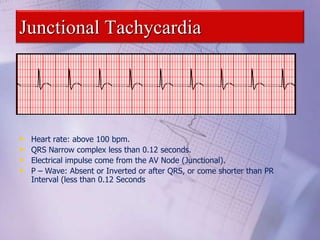
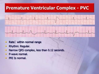
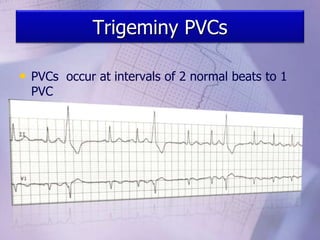
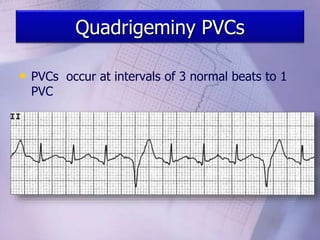
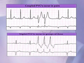
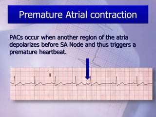
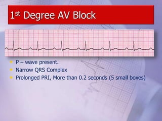
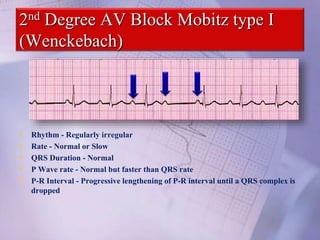
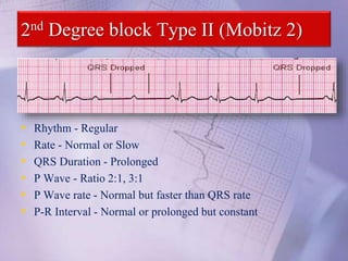
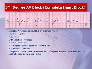
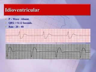
Cardiac Conduction System
Interpretation of an ECG Strip
Step 1: Heart Rate.
Step 2: Heart Rhythm.
Step 3: P – Wave.
Step 4: PRI.
Step 5: QRS Complex
How to calculate heart rate from ECG?
Calculating the heart rate when the rhythm is regular
• There are 300 large squares per minute
• If the rhythm is regular count the number of large squares between two
QRS complexes and divide it into 300.
6-Second Method: Have a six second strip, count the QRS
complexes and multiple by 10.
Heart Rhythm
Regular Irregular
To accurately determine the
regularity of the rhythm,
measure the R-R interval
across the entire strip .
 Is the distance between R-R
interval is constant?
P – Wave
5 Keys: Questions:
- Are P-wave presents?
- Are P-wave occurring Regularly ?
- Is there a P-wave for each QRS complex?
- Are p-waves upright or converted?
- Are they all similar in appearance?
PR Interval
• Normal length of the PR Interval is
(0.12 – 0.20 Seconds , 3-5 small
squares).
• Are PRI greater than 0.20 seconds?
• Are PRI smaller than 0.12 seconds?
• Are the PRI’s constant across the ECG
Strip?
QRS
Complex
Narrow
Supra
Ventricular
Wide
Ventricular
• Normal Duration: less than 0.12 seconds (less than 3 small
boxes).
• Are QRS same in shape?
• Are QRS Greater than 0.12 seconds?
• Are QRS Less than 0.12 seconds?
Dysrhthmias
Causes:
6 H’s & 5 T’s are mostly common:
6 H’s 5 T’s
Hypoxia Toxins (Drugs).
Hypothermia Tamponade
Hypoglycemia Tension Pneumothorax
Hypo/hyper kalemia Thrombosis (coronary, pulmonary)
Hypovolemia Trauma
Hydrogen Ion (Acidosis)
Normal Sinus Rhythm
Implies normal sequence of conduction, originating in the sinus node and
proceeding to the ventricles via the AV node and His-Purkinje system.
ECG Characteristics: Regular narrow-complex rhythm
Rate 60-100 bpm
Each QRS complex is proceeded by a P wave
Bradycardia
• HR< 60 bpm; every QRS narrow, preceded by p wave
• Can be normal in well-conditioned athletes
Bradycardia-Etiology
• Increased Vagal tone.
• Hypothermia.
• Hypothyroidism.
• Seizures.
• Increased intracranial
pressure.
• Aging
• Sleep.
• Intrinsic disease of SA Node (e.g. sick sinus
syndrome).
• Side effect of some drugs like digitalis, calcium
channel blockers
• Result of complication of some diseases like
rheumatic fever, viral myocaditis, diphtheria
• Bradycardia is Normal to Athletic people
ACLS – Bradycardia Drugs
• Atropine.
• Epinephrine.
• Dopamine.
Junctional Rhythm
• Heart rate: 40 – 60 bpm.
• QRS Narrow complex less than 0.12 seconds.
• Electrical impulse come from the AV Node (Junctional).
• P – Wave: Absent or Inverted or after QRS, or come shorter than PR
Interval (less than 0.12 Seconds
Accelerated Junctional Rhythm
• Heart rate: 60- 100 bpm.
• QRS Narrow complex less than 0.12 seconds.
• Electrical impulse come from the AV Node (Junctional).
• P – Wave: Absent or Inverted or after QRS, or come shorter than PR
Interval (less than 0.12 Seconds
Junctional Tachycardia
• Heart rate: above 100 bpm.
• QRS Narrow complex less than 0.12 seconds.
• Electrical impulse come from the AV Node (Junctional).
• P – Wave: Absent or Inverted or after QRS, or come shorter than PR
Interval (less than 0.12 Seconds
Premature Ventricular Complex - PVC
• Rate: within normal range
• Rhythm: Regular.
• Narrow QRS complex, less than 0.12 seconds.
• P-wave normal.
• PRI Is normal.
Bigeminy PVCs
• Premature Ventricular beats occurring after
every normal beat.
Trigeminy PVCs
• PVCs occur at intervals of 2 normal beats to 1
PVC
Quadrigeminy PVCs
• PVCs occur at intervals of 3 normal beats to 1
PVC
Premature Atrial contraction
PACs occur when another region of the atria
depolarizes before SA Node and thus triggers a
premature heartbeat.
Premature Junctional Contraction
1st Degree AV Block
• P – wave present.
• Narrow QRS Complex
• Prolonged PRI, More than 0.2 seconds (5 small boxes)
2nd Degree AV Block Mobitz type I
(Wenckebach)
• Rhythm - Regularly irregular
• Rate - Normal or Slow
• QRS Duration - Normal
• P Wave rate - Normal but faster than QRS rate
• P-R Interval - Progressive lengthening of P-R interval until a QRS complex is
dropped
2nd Degree block Type II (Mobitz 2)
• Rhythm - Regular
• Rate - Normal or Slow
• QRS Duration - Prolonged
• P Wave - Ratio 2:1, 3:1
• P Wave rate - Normal but faster than QRS rate
• P-R Interval - Normal or prolonged but constant
3rd Degree AV Block (Complete Heart Block)
• Complete AV disassociation, HR is a ventricular rate
• Rhythm - Regular
• Rate - Slow
• QRS Duration - Prolonged
• P Wave - Unrelated
• P Wave rate - Normal but faster than QRS rate
• P-R Interval - Variation
• Complete AV block. No atrial impulses pass through the atrioventricular node and the
ventricles generate their own rhythm
Idioventricular
• P – Wave : Absent.
• QRS: > 0.12 Seconds.
• Rate : 20 – 40
• HR > 100 bpm, regular
• Often difficult to distinguish p and t waves
Tachycardia-Etiology
• Fever
• Hyperthyroidism
• Anxiety
• Sepsis
• Anemia
• Hypotension and shock
• Pulmonary embolism
• Acute coronary ischemia
and myocardial infarction
• Heart failure
• Chronic pulmonary
disease
• Hypoxia
ACLS – Tachycardia Drugs
• Adenosine.
• Diltiazem.
• Beta Blockers.
• Amiodarone.
• Digoxin.
• Verapamil.
• Magnesium Sulphate
Atrial Fibrillation
• Irregularly irregular rhythm
• Absence of definite p waves
• Narrow QRS, less than 0.12 seconds
• Can be accompanied by rapid ventricular response
Atrial Flutter – Saw Teeth
Rate: 250 – 350 bpm.
Rhythm: Usually Regular.
No True P-waves are presents – saw teeth.
PRI – Usually constant
QRS Complex usually normal less than 0.12
seconds
Supraventricular Tachycardia
• Rate: Atrial Rate 150 – 250 bpm, Ventricular Rate 150- 250 bpm.
• Rhythm: Regular.
• Narrow QRS complex, less than 0.12 seconds.
• P-wave usually is discernable.
• PRI usually is discernable.
Ventricular Tachycardia
• Rhythm: regular.
• Rate: 100 – 220 bpm
• P - wave: non associated with QRS, Some time visible.
• PRI: None.
• QRS: Wide complex, greater than 0.12 seconds
Shock this rhythm if the patient is unconscious and without a
pulse
Non-sustained Ventricular tachycardia
• Defined as 3 or more consecutive ventricular beats
• Rate of >120 bpm, lasting less than 30 seconds
• May be discovered on Halter, or other exercise testing
Ventricular Fibrillation
Rhythm - Irregular
Rate - 300+, disorganized
QRS Duration - Not recognizable
P Wave - Not seen
This patient needs to be defibrillated!!
QUICKLY
ACLS – VF & VT Drugs
• Epinephrine.
• Vasopressin.
• Amiodarone.
• Lidocaine
Torsade's De Pointes
• QRS morphology of V Tach keeps
changing
A Systole
• Rhythm - Flat
• Rate - 0 Beats per minute
• QRS Duration - None
• P Wave - None
• Carry out CPR!!
PEA – Pulseless Electrical Activity
It is a rhythm appear on monitor but without
sensible pulse
All rhythms without pulse called PEA
EXCEPT:
1- VF
2- Pulseless VT.
3- A systole.
4- Torsade's De Pointes
Thank You

Cardiac arrhythmia

  • 3.
  • 6.
    Interpretation of anECG Strip Step 1: Heart Rate. Step 2: Heart Rhythm. Step 3: P – Wave. Step 4: PRI. Step 5: QRS Complex
  • 7.
    How to calculateheart rate from ECG?
  • 8.
    Calculating the heartrate when the rhythm is regular • There are 300 large squares per minute • If the rhythm is regular count the number of large squares between two QRS complexes and divide it into 300.
  • 9.
    6-Second Method: Havea six second strip, count the QRS complexes and multiple by 10.
  • 10.
    Heart Rhythm Regular Irregular Toaccurately determine the regularity of the rhythm, measure the R-R interval across the entire strip .  Is the distance between R-R interval is constant?
  • 11.
    P – Wave 5Keys: Questions: - Are P-wave presents? - Are P-wave occurring Regularly ? - Is there a P-wave for each QRS complex? - Are p-waves upright or converted? - Are they all similar in appearance?
  • 12.
    PR Interval • Normallength of the PR Interval is (0.12 – 0.20 Seconds , 3-5 small squares). • Are PRI greater than 0.20 seconds? • Are PRI smaller than 0.12 seconds? • Are the PRI’s constant across the ECG Strip?
  • 13.
    QRS Complex Narrow Supra Ventricular Wide Ventricular • Normal Duration:less than 0.12 seconds (less than 3 small boxes). • Are QRS same in shape? • Are QRS Greater than 0.12 seconds? • Are QRS Less than 0.12 seconds?
  • 14.
    Dysrhthmias Causes: 6 H’s &5 T’s are mostly common: 6 H’s 5 T’s Hypoxia Toxins (Drugs). Hypothermia Tamponade Hypoglycemia Tension Pneumothorax Hypo/hyper kalemia Thrombosis (coronary, pulmonary) Hypovolemia Trauma Hydrogen Ion (Acidosis)
  • 16.
    Normal Sinus Rhythm Impliesnormal sequence of conduction, originating in the sinus node and proceeding to the ventricles via the AV node and His-Purkinje system. ECG Characteristics: Regular narrow-complex rhythm Rate 60-100 bpm Each QRS complex is proceeded by a P wave
  • 17.
    Bradycardia • HR< 60bpm; every QRS narrow, preceded by p wave • Can be normal in well-conditioned athletes
  • 18.
    Bradycardia-Etiology • Increased Vagaltone. • Hypothermia. • Hypothyroidism. • Seizures. • Increased intracranial pressure. • Aging • Sleep. • Intrinsic disease of SA Node (e.g. sick sinus syndrome). • Side effect of some drugs like digitalis, calcium channel blockers • Result of complication of some diseases like rheumatic fever, viral myocaditis, diphtheria • Bradycardia is Normal to Athletic people
  • 19.
    ACLS – BradycardiaDrugs • Atropine. • Epinephrine. • Dopamine.
  • 20.
    Junctional Rhythm • Heartrate: 40 – 60 bpm. • QRS Narrow complex less than 0.12 seconds. • Electrical impulse come from the AV Node (Junctional). • P – Wave: Absent or Inverted or after QRS, or come shorter than PR Interval (less than 0.12 Seconds
  • 22.
    Accelerated Junctional Rhythm •Heart rate: 60- 100 bpm. • QRS Narrow complex less than 0.12 seconds. • Electrical impulse come from the AV Node (Junctional). • P – Wave: Absent or Inverted or after QRS, or come shorter than PR Interval (less than 0.12 Seconds
  • 23.
    Junctional Tachycardia • Heartrate: above 100 bpm. • QRS Narrow complex less than 0.12 seconds. • Electrical impulse come from the AV Node (Junctional). • P – Wave: Absent or Inverted or after QRS, or come shorter than PR Interval (less than 0.12 Seconds
  • 24.
    Premature Ventricular Complex- PVC • Rate: within normal range • Rhythm: Regular. • Narrow QRS complex, less than 0.12 seconds. • P-wave normal. • PRI Is normal.
  • 25.
    Bigeminy PVCs • PrematureVentricular beats occurring after every normal beat.
  • 26.
    Trigeminy PVCs • PVCsoccur at intervals of 2 normal beats to 1 PVC
  • 27.
    Quadrigeminy PVCs • PVCsoccur at intervals of 3 normal beats to 1 PVC
  • 29.
    Premature Atrial contraction PACsoccur when another region of the atria depolarizes before SA Node and thus triggers a premature heartbeat.
  • 31.
  • 32.
    1st Degree AVBlock • P – wave present. • Narrow QRS Complex • Prolonged PRI, More than 0.2 seconds (5 small boxes)
  • 33.
    2nd Degree AVBlock Mobitz type I (Wenckebach) • Rhythm - Regularly irregular • Rate - Normal or Slow • QRS Duration - Normal • P Wave rate - Normal but faster than QRS rate • P-R Interval - Progressive lengthening of P-R interval until a QRS complex is dropped
  • 34.
    2nd Degree blockType II (Mobitz 2) • Rhythm - Regular • Rate - Normal or Slow • QRS Duration - Prolonged • P Wave - Ratio 2:1, 3:1 • P Wave rate - Normal but faster than QRS rate • P-R Interval - Normal or prolonged but constant
  • 35.
    3rd Degree AVBlock (Complete Heart Block) • Complete AV disassociation, HR is a ventricular rate • Rhythm - Regular • Rate - Slow • QRS Duration - Prolonged • P Wave - Unrelated • P Wave rate - Normal but faster than QRS rate • P-R Interval - Variation • Complete AV block. No atrial impulses pass through the atrioventricular node and the ventricles generate their own rhythm
  • 36.
    Idioventricular • P –Wave : Absent. • QRS: > 0.12 Seconds. • Rate : 20 – 40
  • 38.
    • HR >100 bpm, regular • Often difficult to distinguish p and t waves
  • 39.
    Tachycardia-Etiology • Fever • Hyperthyroidism •Anxiety • Sepsis • Anemia • Hypotension and shock • Pulmonary embolism • Acute coronary ischemia and myocardial infarction • Heart failure • Chronic pulmonary disease • Hypoxia
  • 40.
    ACLS – TachycardiaDrugs • Adenosine. • Diltiazem. • Beta Blockers. • Amiodarone. • Digoxin. • Verapamil. • Magnesium Sulphate
  • 41.
    Atrial Fibrillation • Irregularlyirregular rhythm • Absence of definite p waves • Narrow QRS, less than 0.12 seconds • Can be accompanied by rapid ventricular response
  • 42.
    Atrial Flutter –Saw Teeth Rate: 250 – 350 bpm. Rhythm: Usually Regular. No True P-waves are presents – saw teeth. PRI – Usually constant QRS Complex usually normal less than 0.12 seconds
  • 43.
    Supraventricular Tachycardia • Rate:Atrial Rate 150 – 250 bpm, Ventricular Rate 150- 250 bpm. • Rhythm: Regular. • Narrow QRS complex, less than 0.12 seconds. • P-wave usually is discernable. • PRI usually is discernable.
  • 44.
    Ventricular Tachycardia • Rhythm:regular. • Rate: 100 – 220 bpm • P - wave: non associated with QRS, Some time visible. • PRI: None. • QRS: Wide complex, greater than 0.12 seconds Shock this rhythm if the patient is unconscious and without a pulse
  • 45.
    Non-sustained Ventricular tachycardia •Defined as 3 or more consecutive ventricular beats • Rate of >120 bpm, lasting less than 30 seconds • May be discovered on Halter, or other exercise testing
  • 46.
    Ventricular Fibrillation Rhythm -Irregular Rate - 300+, disorganized QRS Duration - Not recognizable P Wave - Not seen This patient needs to be defibrillated!! QUICKLY
  • 47.
    ACLS – VF& VT Drugs • Epinephrine. • Vasopressin. • Amiodarone. • Lidocaine
  • 48.
    Torsade's De Pointes •QRS morphology of V Tach keeps changing
  • 49.
    A Systole • Rhythm- Flat • Rate - 0 Beats per minute • QRS Duration - None • P Wave - None • Carry out CPR!!
  • 50.
    PEA – PulselessElectrical Activity It is a rhythm appear on monitor but without sensible pulse
  • 51.
    All rhythms withoutpulse called PEA EXCEPT: 1- VF 2- Pulseless VT. 3- A systole. 4- Torsade's De Pointes
  • 52.