Downloaded 62 times


![Incidence of lymph node involvement
• Depends on:
• Tumor grade: 30% G1 vs 40% G3
• Local stage : 60% in pT2 & 75% in pT3-4
• T1G2: 50% [Naumann BJU 2008]
• Type of local tumor: Basoloid vs Classic](https://image.slidesharecdn.com/capenisii-161229175859/85/Ca-penis-9-320.jpg)


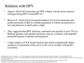
Circumcision early in life protects against carcinoma of the penis by reducing risk of HPV infection and preventing accumulation of smegma. Approximately half of penile carcinomas are HPV-related, with HPV-16 and HPV-18 being the most common types. Symptoms of penile carcinoma include masses, pain, bleeding, groin masses, and urinary symptoms. Diagnosis involves history, physical exam, biopsy, and imaging of lymph nodes. Staging uses the Jackson system or AJCC TNM system, with most cases being well-differentiated squamous cell carcinoma.


![Ca penis [edmond]](https://cdn.slidesharecdn.com/ss_thumbnails/capenisedmond-130318091738-phpapp01-thumbnail.jpg?width=640&height=640&fit=bounds)


