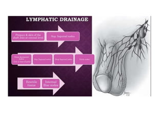
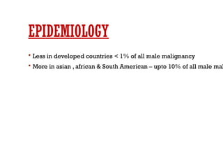
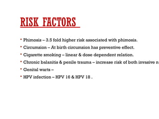
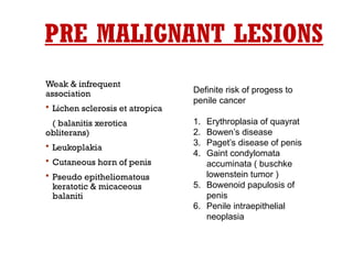
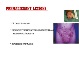
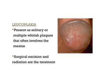
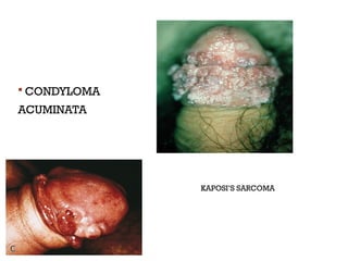
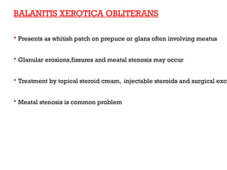
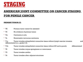
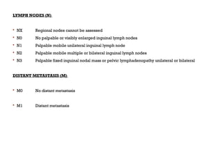
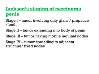
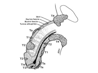
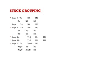
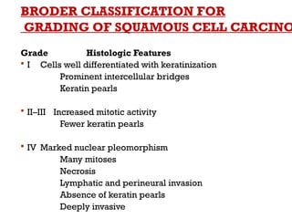
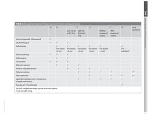
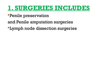
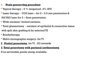
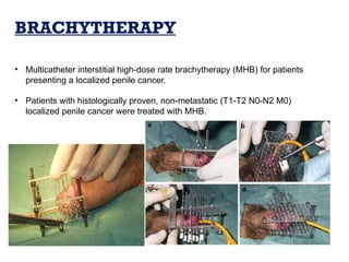
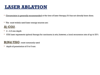
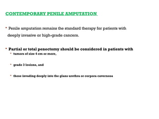
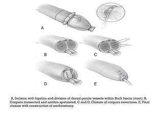
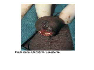
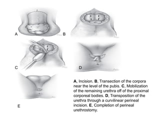
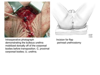
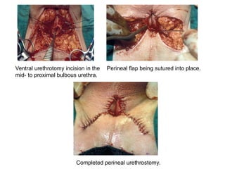
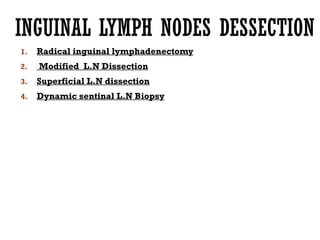
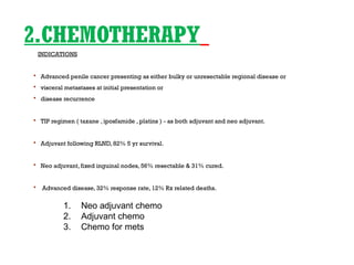
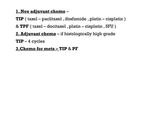
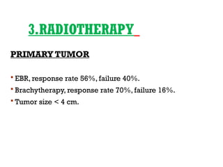
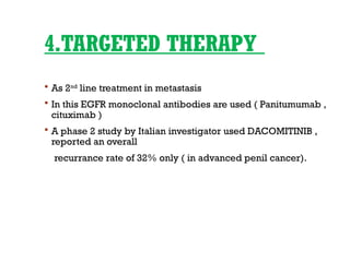
RECENT TRENDS IN MANAGEMENT OF
CARCINOMA PENIS
MODERATORS -
 Dr D. Borgohain
Associate professor
Dept. of general surgery
AMCH
 Dr B. Borah
Associate professor
Dept. of general surgery
AMCH
PRESENTED
BY -
Dr Niranjan kumar
PGT 1 Dept of General
Surgery AMCH
INTRODUCTION
 Rare malignancy in developed counties but more common in
India , Asia , South America, africa
 More than 95% SCC
 Peak age 5th
to 7th
decade of life
 Role of multidisciplinary approach
EPIDEMIOLOGY
 Less in developed countries < 1% of all male malignancy
 More in asian , african & South American – upto 10% of all male mal
RISK FACTORS
 Phimosis – 3.5 fold higher risk associated with phimosis.
 Circumsion – At birth circumsion has preventive effect.
 Cigarette smoking – linear & dose dependent relation.
 Chronic balanitis & penile trauma – increase risk of both invasive n
 Genital warts –
 HPV infection – HPV 16 & HPV 18 .
PRE MALIGNANT LESIONS
Weak & infrequent
association
 Lichen sclerosis et atropica
( balanitis xerotica
obliterans)
 Leukoplakia
 Cutaneous horn of penis
 Pseudo epitheliomatous
keratotic & micaceous
balaniti
Definite risk of progess to
penile cancer
1. Erythroplasia of quayrat
2. Bowen’s disease
3. Paget’s disease of penis
4. Gaint condylomata
accuminata ( buschke
lowenstein tumor )
5. Bowenoid papulosis of
penis
6. Penile intraepithelial
neoplasia
PREMALIGNANT LESIONS
 CUTANEOUS HORN
 PSEUDOEPITHELIOMATOUS MICACEOUS AND
KERATOTIC BALANITIS
 BOWENOID PAPULOSIS
LEUCOPLAKIA
 Present as solitary or
multiple whitish plaques
that often involves the
meatus
 Surgical excision and
radiation are the treatment
 CONDYLOMA
ACUMINATA
 KAPOSI’S SARCOMA
BALANITIS XEROTICA OBLITERANS
 Presents as whitish patch on prepuce or glans often involving meatus
 Glanular erosions,fissures and meatal stenosis may occur
 Treatment by topical steroid cream, injectable steroids and surgical exci
 Meatal stenosis is common problem
BUSCHKE-LOWENSTEIN TUMOUR
(VERRUCOUS CARCINOMA)
 True incidence is unknown.
 Invades locally.
 Compresses the adjacent tissues causing urethral erosion & fistulisation
 NEVER METASTASIZE
 SHOWS NO SIGN OF MALIGNANT CHANGE

 C.F. : bleeding, discharge & foul odour
 Treatment is excision.Total penectomy may be required.
 Laser therapy may be effective in some cases.
 Recurrence is common.
 Topical therapy with Podophyllin, 5FU, radiation and chemotherapy have all be
with no great success.
 Radiotherapy has rather been associated with malignant degeneration of the t
other sites
PENILE CANCER
 Squamous cell carcinoma. > 95%
 Mesenchymal tumors. < 3%
e.g Kaposi sarcoma, angiosarcoma etc
 Malignant Melanoma.
 Basal cell carcinoma.
 Metastasis.
CARCINOMA IN SITU
 Carcinoma in situ (Tis) of the penis is called erythroplasia of
Queyrat if it involves the glans penis and prepuce.
 Bowen’s disease if it involves the penile shaft or the remainder
of the genitalia or perineal region.
 Both are histologically similar.
 Clinically erythroplasia of Queyrat consists of a red, velvety, well-
marginated lesion of the glans penis or, less frequently, the prepuce of
the uncircumcised man.
 It may ulcerate and may be associated with discharge and pain
 Bowen’s disease is characterized by sharply defined plaques of scaly
erythema on the penile shaft. Crusted or ulcerated variants can occur.
 The appearance can be confused with bowenoid papulosis, nummular
eczema, psoriasis, and superficial basal cell carcinoma.
ERYTHROPLASIA OF QUEYRAT BOWEN’S DISEASE
NATURAL HISTORY
 Glans (48% ) > prepuce (21%) > glans + prepuce
( 9%) > coronal sulcus ( 6% ) > penile shaft ( <2%)
 Most common symptoms – growth , primary lesion is
usually painless
 A foul smelling discharge with/ without bleeding,
itching & burning may be present.
 Imperceptible induration  small growth pepules
 flat ulcerative lesion  exophytic lesion 
fungating ulceroproliferative growth in whole of penis
 Inguinal swelling ulceration & proliferative lesion -
 superimposed infection & seropurulent discharge.
 Spread of penile cancer
Glasns/shaft/ body  penetration of buck’s fascia & tunica albuginea
Corpus cavernosa  lymphtic system
 Lymphatic spread
Superficial lymph node  deep inguinal lymph node  pelvic nodes
Metasis ( lungs , liver , bone & brain)
 Most common landing site for mets –
superomedial group of superficial lymph nod
 Buck’s fascia acts as a temporary natural barrier to local extension
of the tumor, protecting the corporeal bodies from invasion.
 Penetration of Buck’s fascia and the tunica albuginea permits
invasion of the vascular corpora and establishes the potential for
vascular dissemination.
 Urethral and bladder involvement are rare
 Distant metastasis uncommon 1 – 10%
 Death within 2 years for most untreated cases.
CLINICAL FEATURES
 SYMPTOMS
 Pt. present with ulcer or a swelling on penis
 Pain does not develop in proportion to the extent of the local
destructive process and usually is not a presenting complaint.
 Weakness, weight loss, fatigue, and systemic malaise occur
secondary to chronic suppuration.
 On occasion, significant blood loss from the penile lesion, the
nodal lesion, or both may occur.
 Symptoms referable to metastases are rare.
SIGNS
 The presentation ranges from a relatively subtle induration or
small excrescence to a small papule, pustule, warty growth, or
exophytic lesion.
 It may appear as a shallow erosion or as a deeply excavated
ulcer with elevated or rolled-in edges.
 Erosion through the prepuce, foul preputial odor, and
discharge with or without bleeding
 Mass, ulceration, suppuration, or hemorrhage in the inguinal
area may be due to nodal metastases.
 Urinary retention or urethral fistula due to local corporeal
involvement is a rare presenting sign.
INVESTIGATIONS
1. Physical examination
2. Biopsy
3. Radiological investigation
4. Newer modalities
1.Physical examination
It includs –
 number of lesions
 Size
 Location
 Extent ( foreskin , glans , shaft)
 Appearance – flat, papillary , nodular , ulcerating , fungating
 Margins
 Involvement of adjacent structure ( CC , CS , urethera)
 edges
2) BIOPSY
 Confirmation of the diagnosis of carcinoma of the penis
 Asessment of the depth of invasion,
 Presence of vascular invasion, and
 Histologic grade of the lesion
Tissue for biopsy
 Excisional biopsy – for small lesion both diagnostic & therapeutic
 Incisional biopsy
 Brush biopsy
 Tissue core biopsy
 FNAC ( USG guided FNAC)
 Wedge biopsy – provides maximum information
3. Radiological investigations
A) CXR – for lung metastasis
B) PENILE ULTRASONOGRAPHY
 sensitivity 57% and specificity 91%
 can not delineate invasion into the subepithelial connective tissue of the glans penis from
corpus spongiosum involvement
C) CT SCAN
- sensitivity and specificity of CT are 36% and 100%
-Mainly for assessment of inguinal & pelvic LNs and abd. Sec.
- Role in examination of the inguinal region in obese patients in
those who have had prior inguinal surgery,
for whom the physical examination may be unreliable.
- CT-guided biopsy of enlarged pelvic nodes
D) MRI ( with artificial erection)
-sensitivity and specifity of 100% and 91% respectively
- assesses local staging of the tumor
- assessment of inguinal & pelvic LNs
-better with artificial erection
4. NEWER MODALITIES
Nanoparticle (Ferumoxtran-10 particles ) and
PET/CT of the inguinal region have shown promising
results in the detection of minimal inguinal metastases
when LNs are normal on CT/MRI but validation in larger
series is required
STAGING
AMERICAN JOINT COMMITTEE ON CANCER STAGING
FOR PENILE CANCER
PRIMARY TUMOR (T)
 TX Primary tumor cannot be assessed
 T0 No evidence of primary tumor
 Tis Carcinoma in situ
 Ta Noninvasive verrucous carcinoma
 T1a Tumor invades subepithelial connective tissue without lymph vascular invasion and
differentiated (i.e., grade 3-4)
 T1b Tumor invades subepithelial connective tissue without LVI and is poorly differentiated
 T2 Tumor invades corpus spongiosum or cavernosum
 T3 Tumor invades urethra
 T4 Tumor invades other adjacent structures
LYMPH NODES (N)
 NX Regional nodes cannot be assessed
 N0 No palpable or visibly enlarged inguinal lymph nodes
 N1 Palpable mobile unilateral inguinal lymph node
 N2 Palpable mobile multiple or bilateral inguinal lymph nodes
 N3 Palpable fixed inguinal nodal mass or pelvic lymphadenopathy unilateral or bilateral
DISTANT METASTASIS (M)
 M0 No distant metastasis
 M1 Distant metastasis
Jackson’s staging of carcinoma
penis
Stage I – tumor involving only glans / prepauce
/ both
Stage II – tumor extending into body of penis
Stage III – tumor having mobile inguinal nodes
Stage IV – tumor spreading to adjacent
structure/ fixed nodes
STAGE GROUPING
 Stage 0 Tis N0 M0
Ta N0 M0
 Stage I T1a N0 M0
 Stage II T1b N0 M0
T2 N0 M0
T3 N0 M0
 Stage IIIa T1-3 N1 M0
 Stage IIIb T1-3 N2 M0
 Stage IV T4 Any N M0
Any T N3 M0
Any T Any N M1
BRODER CLASSIFICATION FOR
GRADING OF SQUAMOUS CELL CARCINO
Grade Histologic Features
 I Cells well differentiated with keratinization
Prominent intercellular bridges
Keratin pearls
 II–III Increased mitotic activity
Fewer keratin pearls
 IV Marked nuclear pleomorphism
Many mitoses
Necrosis
Lymphatic and perineural invasion
Absence of keratin pearls
Deeply invasive
1. Surgeries
2. chemotherapy
3. Radiotherapy
4. Targeted therapy
5. Immunotherapy
6. Palliation for advance carcinoma
TREATMENT OF
PENILE LESION
1. SURGERIES INCLUDES
Penile preservation
and Penile amputation surgeries
Lymph node dissection surgeries
1. Penis preserving procedure
 Topical therapy – 5 % imiquinod , 5% 5FU
 Laser therapy - CO2 laser – for 2 – 2.5 mm penetration &
NdYAG laser for 3 – 5mm penetration.
 Wide excision/ limited excision.
 Total glansectomy - excision of epithelial & connective tissue
with split skin grafting & for selected T2
 Brachytherapy
 Moh’s micrographic surgery- for T1
2 . Pratial penectomy for T1 , G3 onwards
3. Total penectomy with perineal urethrostomy
If no serviceble penile stump available.
ORGAN PRESERVATION
GOAL
 to preserve glans sensation where possible
 to maximize penile shaft length
INDICATIONS
 primary tumors exhibiting favorable histologic features
 stages Tis,Ta,T1; grade 1 and grade 2 tumors
1) CIRCUMCISION AND LIMITED
EXCISION STRATEGIES
 2-cm surgical margin required for all patients undergoing partial penectomy has been
challenged
 The maximum proximal histologic extent of 5 mm for grade 1 and grade 2 tumors and 10
mm for grade 3 tumors is recommended.
Limitations of this approach include
 Proximal and distal deeply invasive tumors,
 High-grade tumors,
 Skip lesions and
 Patients with poor compliance who would not be candidates for salvage procedures
Recurrence rate is 4-6 % nowadays
Surgical glans defect covered
with outer preputial flap as
described by Ubrig and
colleagues. A, Superficial
glans tumor.
B, Outer preputial flap
outlined.
C, Tumor excised and
circumcision performed.
D, Glans defect filled with
outer preputial flap.
Finely meshed extragenital split-thickness skin graft quilted to glans
superficial tumor excision.
MOH’S MICROGRAPHIC SURGERY
 It involves layer-by-layer complete excision of the penile lesion in multiple
sessions (fixed tissue technique), with microscopic examination of the under
surface of each layer until the lesion is completely excised.
 Local recurrences rate of 8– 32% were highly associated with
 tumor size (3 cm),
 advanced stage, and
 failure of previous definitive therapy
• Mohs micrographic surgery best suited for patients with carcinoma in situ
or small superficially invasive tumors, with cure rates comparable to partial
penectomy.
• Complications include meatal stenosis and glans disfigurement.
BRACHYTHERAPY
• Multicatheter interstitial high-dose rate brachytherapy (MHB) for patients
presenting a localized penile cancer.
• Patients with histologically proven, non-metastatic (T1-T2 N0-N2 M0)
localized penile cancer were treated with MHB.
LASER ABLATION
 Circumcision is generally recommended at the time of laser therapy, if it has not already been done.
 The most widely used laser energy sources are:
A) CO2 :
 2 – 2.5 mm depth
 CO2 laser represents optimal therapy for carcinoma in situ; however, a local recurrence rate of up to 33%
B)Nd:YAG : most commonly used
 depth of penetration of 3 to 5 mm
CONTEMPORARY PENILE AMPUTATION
 Penile amputation remains the standard therapy for patients with
deeply invasive or high-grade cancers.
 Partial or total penectomy should be considered in patients with
 tumors of size 4 cm or more,
 grade 3 lesions, and
 those invading deeply into the glans urethra or corpora cavernosa
2. PARTIAL PENECTOMY
 Partial penectomy remains the most common surgical procedure for treatm
primary tumor in patients with invasive squamous cell carcinoma.
 Successful local control is accomplished in the majority of patients by ampu
penis at least 2 cm proximal to the tumor.
 Additional goals of the procedure are to preserve the ability to void in a sta
and possibly to allow sexual function.
A, Incision with ligation and division of dorsal penile vessels within Buck fascia (inset). B,
Corpora transected and urethra spatulated. C and D, Closure of corpora cavernosa. E, Final
closure with construction of urethrostomy.
A
B C
D E
Penile stump after partial penectomy.
3.TOTAL PENECTOMY
 Total penectomy is a bit of a misnomer, because the penis is
amputated at or near the level of the suspensory ligament of
the penis without removal of the corpora cavernosa more
proximally.
 It is indicated for penile tumors whose size or location would
not allow excision with an adequate surgical margin and
preservation of a remnant sufficient for upright voiding.
A, Incision. B, Transection of the corpora
near the level of the pubis. C, Mobilization
of the remaining urethra off of the proximal
corporeal bodies. D, Transposition of the
urethra through a curvilinear perineal
incision. E, Completion of perineal
urethrostomy.
A B
C D
E
Intraoperative photograph
demonstrating the bulbous urethra
mobilized dorsally off of the corporeal
bodies before transposition. C, proximal
corporeal bodies; U, urethra.
Incision for flap
perineal urethrostomy
Ventral urethrotomy incision in the
mid- to proximal bulbous urethra.
Perineal flap being sutured into place.
Completed perineal urethrostomy.
INGUINAL LYMPH NODES DESSECTION
1. Radical inguinal lymphadenectomy
2. Modified L.N Dissection
3. Superficial L.N dissection
4. Dynamic sentinal L.N Biopsy
2.CHEMOTHERAPY
INDICATIONS
 Advanced penile cancer presenting as either bulky or unresectable regional disease or
 visceral metastases at initial presentation or
 disease recurrence
 TIP regimen ( taxane , iposfamide , platins ) - as both adjuvant and neo adjuvant.
 Adjuvant following RLND, 82% 5 yr survival.
 Neo adjuvant, fixed inguinal nodes, 56% resectable & 31% cured.
 Advanced disease, 32% response rate, 12% Rx related deaths.
1. Neo adjuvant chemo
2. Adjuvant chemo
3. Chemo for mets
1. Neo adjuvant chemo –
TIP ( taxel – paclitaxel , ifosfamide , platin – cisplatin )
& TPF ( taxel – docitaxel , platin – cisplatin , 5FU )
2. Adjuvant chemo – if histologically high grade
TIP – 4 cycles
3.Chemo for mets – TIP & PF
3.RADIOTHERAPY
PRIMARY TUMOR
 EBR, response rate 56%, failure 40%.
 Brachytherapy, response rate 70%, failure 16%.
 Tumor size < 4 cm.
4.TARGETED THERAPY
 As 2nd
line treatment in metastasis
 In this EGFR monoclonal antibodies are used ( Panitumumab ,
cituximab )
 A phase 2 study by Italian investigator used DACOMITINIB ,
reported an overall
recurrance rate of 32% only ( in advanced penil cancer).
5.IMMUNOTHERAPY
 PDL1 inhibitor – NEVOLUMAB , IPILIMUMAB ,
CABOZATINIB
Has been seen to show partial response in 50%
of cases.
 Other on-going trails are underway with
NIVOLUMAB & PEMBROLIZUMAB in advanced
penil cancer.
6.PALLIATION IN ADVANCED
PENILE CANCER
AIM –
 To reduce pain
 To improve quality of life
 To optimize wound care
 To treat complications
Modalities employed in palliation
 multidrug chemo therapy
 Radiotherapy
 Hemipelvactomy ( In U/L gross disease / Bony involvement)
 Vascular stents for femoral vessel infiltration.
METASTASES
 bladder, prostate, and rectum
 The most frequent sign of penile metastasis is priapism; penile swelling,
nodularity, and ulceration have also been reported
 Prognosis is poor, and therapy should be directed toward the primary tumor’s histology or local palliatio
advised.
 Successful treatment may occasionally be possible in the case of solitary nodules or localized distal peni
involvement if complete excision by partial amputation succeeds in removing the entire area of malignan
infiltration
FOLLOW UP
 Crucial to pick up early recurrance or metastasis.
 Follow up protocol depends on
. stage & grade of primary lesion , status of inguinal lymph nodes ,
Treatment modality chosen.
CONCLUSION
 Lethal malignancy with significant high incidence in India.
 Huge inpact on longevity & quality of life of patient
 Recent data suggest that organ preserving approaches with much
Smaller surgical margins enable a better sexual function and quality
Of life and should be preferred in all feasible scenarios.
THANK YOU

Carcinoma penis power point presentation

  • 1.
    RECENT TRENDS INMANAGEMENT OF CARCINOMA PENIS MODERATORS -  Dr D. Borgohain Associate professor Dept. of general surgery AMCH  Dr B. Borah Associate professor Dept. of general surgery AMCH PRESENTED BY - Dr Niranjan kumar PGT 1 Dept of General Surgery AMCH
  • 2.
    INTRODUCTION  Rare malignancyin developed counties but more common in India , Asia , South America, africa  More than 95% SCC  Peak age 5th to 7th decade of life  Role of multidisciplinary approach
  • 7.
    EPIDEMIOLOGY  Less indeveloped countries < 1% of all male malignancy  More in asian , african & South American – upto 10% of all male mal
  • 8.
    RISK FACTORS  Phimosis– 3.5 fold higher risk associated with phimosis.  Circumsion – At birth circumsion has preventive effect.  Cigarette smoking – linear & dose dependent relation.  Chronic balanitis & penile trauma – increase risk of both invasive n  Genital warts –  HPV infection – HPV 16 & HPV 18 .
  • 9.
    PRE MALIGNANT LESIONS Weak& infrequent association  Lichen sclerosis et atropica ( balanitis xerotica obliterans)  Leukoplakia  Cutaneous horn of penis  Pseudo epitheliomatous keratotic & micaceous balaniti Definite risk of progess to penile cancer 1. Erythroplasia of quayrat 2. Bowen’s disease 3. Paget’s disease of penis 4. Gaint condylomata accuminata ( buschke lowenstein tumor ) 5. Bowenoid papulosis of penis 6. Penile intraepithelial neoplasia
  • 10.
    PREMALIGNANT LESIONS  CUTANEOUSHORN  PSEUDOEPITHELIOMATOUS MICACEOUS AND KERATOTIC BALANITIS  BOWENOID PAPULOSIS
  • 11.
    LEUCOPLAKIA  Present assolitary or multiple whitish plaques that often involves the meatus  Surgical excision and radiation are the treatment
  • 12.
  • 13.
    BALANITIS XEROTICA OBLITERANS Presents as whitish patch on prepuce or glans often involving meatus  Glanular erosions,fissures and meatal stenosis may occur  Treatment by topical steroid cream, injectable steroids and surgical exci  Meatal stenosis is common problem
  • 14.
    BUSCHKE-LOWENSTEIN TUMOUR (VERRUCOUS CARCINOMA) True incidence is unknown.  Invades locally.  Compresses the adjacent tissues causing urethral erosion & fistulisation  NEVER METASTASIZE  SHOWS NO SIGN OF MALIGNANT CHANGE   C.F. : bleeding, discharge & foul odour
  • 15.
     Treatment isexcision.Total penectomy may be required.  Laser therapy may be effective in some cases.  Recurrence is common.  Topical therapy with Podophyllin, 5FU, radiation and chemotherapy have all be with no great success.  Radiotherapy has rather been associated with malignant degeneration of the t other sites
  • 16.
    PENILE CANCER  Squamouscell carcinoma. > 95%  Mesenchymal tumors. < 3% e.g Kaposi sarcoma, angiosarcoma etc  Malignant Melanoma.  Basal cell carcinoma.  Metastasis.
  • 17.
    CARCINOMA IN SITU Carcinoma in situ (Tis) of the penis is called erythroplasia of Queyrat if it involves the glans penis and prepuce.  Bowen’s disease if it involves the penile shaft or the remainder of the genitalia or perineal region.  Both are histologically similar.
  • 18.
     Clinically erythroplasiaof Queyrat consists of a red, velvety, well- marginated lesion of the glans penis or, less frequently, the prepuce of the uncircumcised man.  It may ulcerate and may be associated with discharge and pain  Bowen’s disease is characterized by sharply defined plaques of scaly erythema on the penile shaft. Crusted or ulcerated variants can occur.  The appearance can be confused with bowenoid papulosis, nummular eczema, psoriasis, and superficial basal cell carcinoma.
  • 19.
    ERYTHROPLASIA OF QUEYRATBOWEN’S DISEASE
  • 20.
    NATURAL HISTORY  Glans(48% ) > prepuce (21%) > glans + prepuce ( 9%) > coronal sulcus ( 6% ) > penile shaft ( <2%)  Most common symptoms – growth , primary lesion is usually painless  A foul smelling discharge with/ without bleeding, itching & burning may be present.  Imperceptible induration  small growth pepules  flat ulcerative lesion  exophytic lesion  fungating ulceroproliferative growth in whole of penis  Inguinal swelling ulceration & proliferative lesion -  superimposed infection & seropurulent discharge.
  • 21.
     Spread ofpenile cancer Glasns/shaft/ body  penetration of buck’s fascia & tunica albuginea Corpus cavernosa  lymphtic system  Lymphatic spread Superficial lymph node  deep inguinal lymph node  pelvic nodes Metasis ( lungs , liver , bone & brain)  Most common landing site for mets – superomedial group of superficial lymph nod
  • 22.
     Buck’s fasciaacts as a temporary natural barrier to local extension of the tumor, protecting the corporeal bodies from invasion.  Penetration of Buck’s fascia and the tunica albuginea permits invasion of the vascular corpora and establishes the potential for vascular dissemination.  Urethral and bladder involvement are rare  Distant metastasis uncommon 1 – 10%  Death within 2 years for most untreated cases.
  • 23.
    CLINICAL FEATURES  SYMPTOMS Pt. present with ulcer or a swelling on penis  Pain does not develop in proportion to the extent of the local destructive process and usually is not a presenting complaint.  Weakness, weight loss, fatigue, and systemic malaise occur secondary to chronic suppuration.  On occasion, significant blood loss from the penile lesion, the nodal lesion, or both may occur.  Symptoms referable to metastases are rare.
  • 24.
    SIGNS  The presentationranges from a relatively subtle induration or small excrescence to a small papule, pustule, warty growth, or exophytic lesion.  It may appear as a shallow erosion or as a deeply excavated ulcer with elevated or rolled-in edges.  Erosion through the prepuce, foul preputial odor, and discharge with or without bleeding  Mass, ulceration, suppuration, or hemorrhage in the inguinal area may be due to nodal metastases.  Urinary retention or urethral fistula due to local corporeal involvement is a rare presenting sign.
  • 25.
    INVESTIGATIONS 1. Physical examination 2.Biopsy 3. Radiological investigation 4. Newer modalities
  • 26.
    1.Physical examination It includs–  number of lesions  Size  Location  Extent ( foreskin , glans , shaft)  Appearance – flat, papillary , nodular , ulcerating , fungating  Margins  Involvement of adjacent structure ( CC , CS , urethera)  edges
  • 27.
    2) BIOPSY  Confirmationof the diagnosis of carcinoma of the penis  Asessment of the depth of invasion,  Presence of vascular invasion, and  Histologic grade of the lesion
  • 28.
    Tissue for biopsy Excisional biopsy – for small lesion both diagnostic & therapeutic  Incisional biopsy  Brush biopsy  Tissue core biopsy  FNAC ( USG guided FNAC)  Wedge biopsy – provides maximum information
  • 29.
    3. Radiological investigations A)CXR – for lung metastasis B) PENILE ULTRASONOGRAPHY  sensitivity 57% and specificity 91%  can not delineate invasion into the subepithelial connective tissue of the glans penis from corpus spongiosum involvement C) CT SCAN - sensitivity and specificity of CT are 36% and 100% -Mainly for assessment of inguinal & pelvic LNs and abd. Sec. - Role in examination of the inguinal region in obese patients in those who have had prior inguinal surgery, for whom the physical examination may be unreliable. - CT-guided biopsy of enlarged pelvic nodes
  • 30.
    D) MRI (with artificial erection) -sensitivity and specifity of 100% and 91% respectively - assesses local staging of the tumor - assessment of inguinal & pelvic LNs -better with artificial erection
  • 31.
    4. NEWER MODALITIES Nanoparticle(Ferumoxtran-10 particles ) and PET/CT of the inguinal region have shown promising results in the detection of minimal inguinal metastases when LNs are normal on CT/MRI but validation in larger series is required
  • 32.
    STAGING AMERICAN JOINT COMMITTEEON CANCER STAGING FOR PENILE CANCER PRIMARY TUMOR (T)  TX Primary tumor cannot be assessed  T0 No evidence of primary tumor  Tis Carcinoma in situ  Ta Noninvasive verrucous carcinoma  T1a Tumor invades subepithelial connective tissue without lymph vascular invasion and differentiated (i.e., grade 3-4)  T1b Tumor invades subepithelial connective tissue without LVI and is poorly differentiated  T2 Tumor invades corpus spongiosum or cavernosum  T3 Tumor invades urethra  T4 Tumor invades other adjacent structures
  • 33.
    LYMPH NODES (N) NX Regional nodes cannot be assessed  N0 No palpable or visibly enlarged inguinal lymph nodes  N1 Palpable mobile unilateral inguinal lymph node  N2 Palpable mobile multiple or bilateral inguinal lymph nodes  N3 Palpable fixed inguinal nodal mass or pelvic lymphadenopathy unilateral or bilateral DISTANT METASTASIS (M)  M0 No distant metastasis  M1 Distant metastasis
  • 34.
    Jackson’s staging ofcarcinoma penis Stage I – tumor involving only glans / prepauce / both Stage II – tumor extending into body of penis Stage III – tumor having mobile inguinal nodes Stage IV – tumor spreading to adjacent structure/ fixed nodes
  • 36.
    STAGE GROUPING  Stage0 Tis N0 M0 Ta N0 M0  Stage I T1a N0 M0  Stage II T1b N0 M0 T2 N0 M0 T3 N0 M0  Stage IIIa T1-3 N1 M0  Stage IIIb T1-3 N2 M0  Stage IV T4 Any N M0 Any T N3 M0 Any T Any N M1
  • 37.
    BRODER CLASSIFICATION FOR GRADINGOF SQUAMOUS CELL CARCINO Grade Histologic Features  I Cells well differentiated with keratinization Prominent intercellular bridges Keratin pearls  II–III Increased mitotic activity Fewer keratin pearls  IV Marked nuclear pleomorphism Many mitoses Necrosis Lymphatic and perineural invasion Absence of keratin pearls Deeply invasive
  • 38.
    1. Surgeries 2. chemotherapy 3.Radiotherapy 4. Targeted therapy 5. Immunotherapy 6. Palliation for advance carcinoma TREATMENT OF PENILE LESION
  • 40.
    1. SURGERIES INCLUDES Penilepreservation and Penile amputation surgeries Lymph node dissection surgeries
  • 41.
    1. Penis preservingprocedure  Topical therapy – 5 % imiquinod , 5% 5FU  Laser therapy - CO2 laser – for 2 – 2.5 mm penetration & NdYAG laser for 3 – 5mm penetration.  Wide excision/ limited excision.  Total glansectomy - excision of epithelial & connective tissue with split skin grafting & for selected T2  Brachytherapy  Moh’s micrographic surgery- for T1 2 . Pratial penectomy for T1 , G3 onwards 3. Total penectomy with perineal urethrostomy If no serviceble penile stump available.
  • 42.
    ORGAN PRESERVATION GOAL  topreserve glans sensation where possible  to maximize penile shaft length INDICATIONS  primary tumors exhibiting favorable histologic features  stages Tis,Ta,T1; grade 1 and grade 2 tumors
  • 43.
    1) CIRCUMCISION ANDLIMITED EXCISION STRATEGIES  2-cm surgical margin required for all patients undergoing partial penectomy has been challenged  The maximum proximal histologic extent of 5 mm for grade 1 and grade 2 tumors and 10 mm for grade 3 tumors is recommended. Limitations of this approach include  Proximal and distal deeply invasive tumors,  High-grade tumors,  Skip lesions and  Patients with poor compliance who would not be candidates for salvage procedures Recurrence rate is 4-6 % nowadays
  • 44.
    Surgical glans defectcovered with outer preputial flap as described by Ubrig and colleagues. A, Superficial glans tumor. B, Outer preputial flap outlined. C, Tumor excised and circumcision performed. D, Glans defect filled with outer preputial flap.
  • 45.
    Finely meshed extragenitalsplit-thickness skin graft quilted to glans superficial tumor excision.
  • 46.
    MOH’S MICROGRAPHIC SURGERY It involves layer-by-layer complete excision of the penile lesion in multiple sessions (fixed tissue technique), with microscopic examination of the under surface of each layer until the lesion is completely excised.  Local recurrences rate of 8– 32% were highly associated with  tumor size (3 cm),  advanced stage, and  failure of previous definitive therapy • Mohs micrographic surgery best suited for patients with carcinoma in situ or small superficially invasive tumors, with cure rates comparable to partial penectomy. • Complications include meatal stenosis and glans disfigurement.
  • 47.
    BRACHYTHERAPY • Multicatheter interstitialhigh-dose rate brachytherapy (MHB) for patients presenting a localized penile cancer. • Patients with histologically proven, non-metastatic (T1-T2 N0-N2 M0) localized penile cancer were treated with MHB.
  • 48.
    LASER ABLATION  Circumcisionis generally recommended at the time of laser therapy, if it has not already been done.  The most widely used laser energy sources are: A) CO2 :  2 – 2.5 mm depth  CO2 laser represents optimal therapy for carcinoma in situ; however, a local recurrence rate of up to 33% B)Nd:YAG : most commonly used  depth of penetration of 3 to 5 mm
  • 49.
    CONTEMPORARY PENILE AMPUTATION Penile amputation remains the standard therapy for patients with deeply invasive or high-grade cancers.  Partial or total penectomy should be considered in patients with  tumors of size 4 cm or more,  grade 3 lesions, and  those invading deeply into the glans urethra or corpora cavernosa
  • 50.
    2. PARTIAL PENECTOMY Partial penectomy remains the most common surgical procedure for treatm primary tumor in patients with invasive squamous cell carcinoma.  Successful local control is accomplished in the majority of patients by ampu penis at least 2 cm proximal to the tumor.  Additional goals of the procedure are to preserve the ability to void in a sta and possibly to allow sexual function.
  • 51.
    A, Incision withligation and division of dorsal penile vessels within Buck fascia (inset). B, Corpora transected and urethra spatulated. C and D, Closure of corpora cavernosa. E, Final closure with construction of urethrostomy. A B C D E
  • 52.
    Penile stump afterpartial penectomy.
  • 53.
    3.TOTAL PENECTOMY  Totalpenectomy is a bit of a misnomer, because the penis is amputated at or near the level of the suspensory ligament of the penis without removal of the corpora cavernosa more proximally.  It is indicated for penile tumors whose size or location would not allow excision with an adequate surgical margin and preservation of a remnant sufficient for upright voiding.
  • 54.
    A, Incision. B,Transection of the corpora near the level of the pubis. C, Mobilization of the remaining urethra off of the proximal corporeal bodies. D, Transposition of the urethra through a curvilinear perineal incision. E, Completion of perineal urethrostomy. A B C D E
  • 55.
    Intraoperative photograph demonstrating thebulbous urethra mobilized dorsally off of the corporeal bodies before transposition. C, proximal corporeal bodies; U, urethra. Incision for flap perineal urethrostomy
  • 56.
    Ventral urethrotomy incisionin the mid- to proximal bulbous urethra. Perineal flap being sutured into place. Completed perineal urethrostomy.
  • 57.
    INGUINAL LYMPH NODESDESSECTION 1. Radical inguinal lymphadenectomy 2. Modified L.N Dissection 3. Superficial L.N dissection 4. Dynamic sentinal L.N Biopsy
  • 58.
    2.CHEMOTHERAPY INDICATIONS  Advanced penilecancer presenting as either bulky or unresectable regional disease or  visceral metastases at initial presentation or  disease recurrence  TIP regimen ( taxane , iposfamide , platins ) - as both adjuvant and neo adjuvant.  Adjuvant following RLND, 82% 5 yr survival.  Neo adjuvant, fixed inguinal nodes, 56% resectable & 31% cured.  Advanced disease, 32% response rate, 12% Rx related deaths. 1. Neo adjuvant chemo 2. Adjuvant chemo 3. Chemo for mets
  • 59.
    1. Neo adjuvantchemo – TIP ( taxel – paclitaxel , ifosfamide , platin – cisplatin ) & TPF ( taxel – docitaxel , platin – cisplatin , 5FU ) 2. Adjuvant chemo – if histologically high grade TIP – 4 cycles 3.Chemo for mets – TIP & PF
  • 60.
    3.RADIOTHERAPY PRIMARY TUMOR  EBR,response rate 56%, failure 40%.  Brachytherapy, response rate 70%, failure 16%.  Tumor size < 4 cm.
  • 61.
    4.TARGETED THERAPY  As2nd line treatment in metastasis  In this EGFR monoclonal antibodies are used ( Panitumumab , cituximab )  A phase 2 study by Italian investigator used DACOMITINIB , reported an overall recurrance rate of 32% only ( in advanced penil cancer).
  • 62.
    5.IMMUNOTHERAPY  PDL1 inhibitor– NEVOLUMAB , IPILIMUMAB , CABOZATINIB Has been seen to show partial response in 50% of cases.  Other on-going trails are underway with NIVOLUMAB & PEMBROLIZUMAB in advanced penil cancer.
  • 63.
    6.PALLIATION IN ADVANCED PENILECANCER AIM –  To reduce pain  To improve quality of life  To optimize wound care  To treat complications
  • 64.
    Modalities employed inpalliation  multidrug chemo therapy  Radiotherapy  Hemipelvactomy ( In U/L gross disease / Bony involvement)  Vascular stents for femoral vessel infiltration.
  • 65.
    METASTASES  bladder, prostate,and rectum  The most frequent sign of penile metastasis is priapism; penile swelling, nodularity, and ulceration have also been reported  Prognosis is poor, and therapy should be directed toward the primary tumor’s histology or local palliatio advised.  Successful treatment may occasionally be possible in the case of solitary nodules or localized distal peni involvement if complete excision by partial amputation succeeds in removing the entire area of malignan infiltration
  • 66.
    FOLLOW UP  Crucialto pick up early recurrance or metastasis.  Follow up protocol depends on . stage & grade of primary lesion , status of inguinal lymph nodes , Treatment modality chosen.
  • 68.
    CONCLUSION  Lethal malignancywith significant high incidence in India.  Huge inpact on longevity & quality of life of patient  Recent data suggest that organ preserving approaches with much Smaller surgical margins enable a better sexual function and quality Of life and should be preferred in all feasible scenarios.
  • 69.