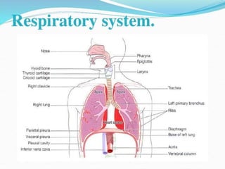
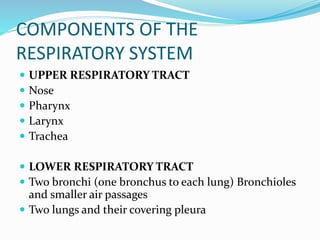
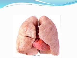
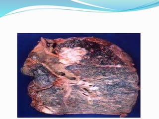
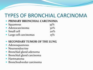
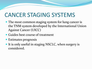
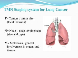
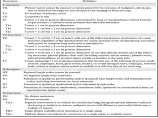
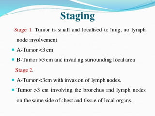
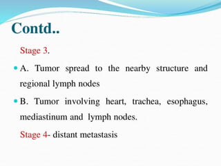
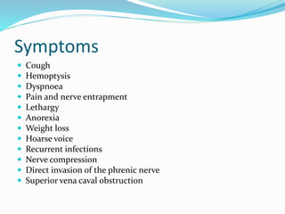
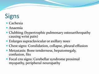
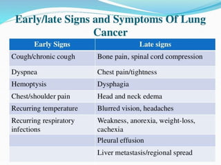
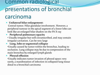
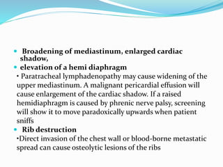
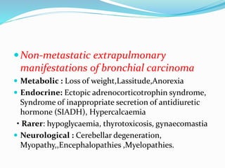
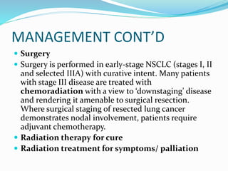
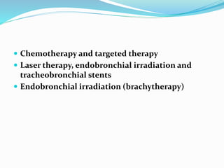
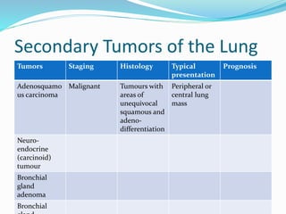
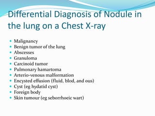
This document provides an outline and overview of lung cancer (bronchial carcinoma). It discusses the epidemiology, risk factors like smoking, pathogenesis, types, staging systems, clinical features, diagnosis, management including surgery and chemotherapy, complications, differential diagnosis, prognosis and conclusions. The respiratory system, normal physiology, and common radiological presentations are also outlined.
4. COMPONENTS OF THE
RESPIRATORY SYSTEM
UPPER RESPIRATORY TRACT
Nose
Pharynx
Larynx
Trachea
LOWER RESPIRATORY TRACT
Two bronchi (one bronchus to each lung) Bronchioles
and smaller air passages
Two lungs and their covering pleura
5. NORMAL PHYSIOLOGY OF
RESPIRATION
Normal respiration begins by inhaling air through the
mouth and nose.
This air flows down into trachea, which divides into
the left and right bronchi, and then to the alveoli.
The alveoli are responsible for oxygenating the blood
for circulation as well as removing carbon dioxide from
the blood.
9. Epidemiology
Lung cancer also called Bronchial carcinoma is the
most common malignant tumor worldwide, with
about
1.4million deaths annually.
It is the third most common cause of death in the UK
after ischemic heart disease and cerebrovascular
disease, and is now the most common cause of cancer-
related death in both men and women. Rates are
declining in men but still increasing overall, reflecting
the increasing incidence in women. The ratio in men
to women is now 1.2 : 1.
10. Aetiology
Cigarette smoking is by far the most important cause of lung
cancer. It is thought to be directly responsible for at least 90% of
lung carcinomas, the risk being proportional to the amount
smoked and to the tar content of cigarettes. The death rate from
the disease in heavy smokers is 40 times that in non-smokers.
The effect of ‘passive’ smoking is more difficult to quantify but
is currently thought to be a factor in 5% of all lung cancer deaths.
Exposure to naturally occurring radon is another risk. The
incidence of lung cancer is slightly higher in urban than in rural
dwellers, which may reflect differences in atmospheric pollution
(including tobacco smoke) or occupation, since a number of
industrial materials are associated with lung cancer.
OTHERS: Asbestos, chromium, arsenic, ironoxides
11. Pathogenesis
Bronchial carcinomas arise from the bronchial epithelium or mucous
gland.
When the tumor occurs in a large bronchus, symptoms arise early, but
tumors originating in a peripheral bronchus can grow very large
without producing symptoms, resulting in delayed diagnosis.
Peripheral squamous tumors may undergo central necrosis and
cavitation, and may resemble a lung abscess on X-ray. Bronchial
carcinoma may involve the pleura directly or by lymphatic spread, and
may extend into the chest wall, invading the intercostal nerves or the
brachial plexus and causing pain. Lymphatic spread to mediastinal and
supraclavicular lymph nodes often occurs before diagnosis. Blood-
borne metastases occur most commonly in liver, bone, brain, adrenals
and skin. Even a small primary tumor may cause widespread metastatic
deposits and this is a particular characteristic of small-cell lung
cancers.
12.
13. TYPES OF BRONCHIAL CARCINOMA
PRIMARY BRONCHIAL CARCINOMA
Squamous 35%
Adenocarcinoma 30%
Small cell 20%
Large cell carcinomas 15%
SECONDARY TUMOR OF THE LUNG
Adenosquamous
Neuroendocrine
Bronchial gland adenoma
Bronchial gland carcinoma
Hartmatoma
Bronchoalveolar carcinoma
14. CANCER STAGING SYSTEMS
The most common staging system for lung cancer is
the TNM system developed by the International Union
Against Cancer (UICC)
Guides best course of treatment
Estimates prognosis
It is only useful in staging NSCLC, when surgery is
considered.
15.
16.
17.
18.
19. Symptoms
Cough
Hemoptysis
Dyspnoea
Pain and nerve entrapment
Lethargy
Anorexia
Weight loss
Hoarse voice
Recurrent infections
Nerve compression
Direct invasion of the phrenic nerve
Superior vena caval obstruction
22. Common radiological
presentations of bronchial
carcinoma
Unilateral hilar enlargement:
Central tumor. Hilar glandular involvement. However, a
peripheral tumour in the apical segment of a lower lobe can
look like an enlarged hilar shadow on the PA X-ray
Peripheral pulmonary opacity
• Usually irregular but well circumscribed, and may contain
irregular cavitation. Can be very large
Lung, lobe or segmental collapse
•Usually caused by tumor within the bronchus, leading to
occlusion. Lung collapse may be due to compression of the
main bronchus by enlarged lymph glands
Pleural effusion
•Usually indicates tumor invasion of pleural space; very
rarely, a manifestation of infection in collapsed lung tissue
distal to a bronchial carcinoma
23. Broadening of mediastinum, enlarged cardiac
shadow,
elevation of a hemi diaphragm
• Paratracheal lymphadenopathy may cause widening of the
upper mediastinum. A malignant pericardial effusion will
cause enlargement of the cardiac shadow. If a raised
hemidiaphragm is caused by phrenic nerve palsy, screening
will show it to move paradoxically upwards when patient
sniffs
Rib destruction
•Direct invasion of the chest wall or blood-borne metastatic
spread can cause osteolytic lesions of the ribs
24. Non-metastatic extrapulmonary
manifestations of bronchial carcinoma
Metabolic : Loss of weight,Lassitude,Anorexia
Endocrine: Ectopic adrenocorticotrophin syndrome,
Syndrome of inappropriate secretion of antidiuretic
hormone (SIADH), Hypercalcaemia
• Rarer: hypoglycaemia, thyrotoxicosis, gynaecomastia
Neurological : Cerebellar degeneration,
Myopathy,,Encephalopathies ,Myelopathies.
28. MANAGEMENT
INVESTIGATIONS
The main aims of investigation are to confirm the diagnosis, establish
the histological cell type and define the extent of the disease.
Imaging:
CXR : from lobar collapse to mass lesions, effusion or malignant rib
destruction
CT : mediastinal or metastatic spread and to stage the tumor
Cytology : sputum and pleural fluid
Fine needle aspiration biopsy and histopathology :periphreal
leisons/lymph nodes
Bronchoscopy : to know histology and allows an assessment of
operability) +/- endobronchial ultrasound for assessment and biopsy
pleural aspiration and biopsy :in patients with pleural effusion
Lung function tests: help assess suitability for lobectomy
Radionuclide bone scan: if suspected metastasis
29.
30. MANAGEMENT CONT’D
Surgery
Surgery is performed in early-stage NSCLC (stages I, II
and selected IIIA) with curative intent. Many patients
with stage III disease are treated with
chemoradiation with a view to ‘downstaging’ disease
and rendering it amenable to surgical resection.
Where surgical staging of resected lung cancer
demonstrates nodal involvement, patients require
adjuvant chemotherapy.
Radiation therapy for cure
Radiation treatment for symptoms/ palliation
31. Chemotherapy and targeted therapy
Laser therapy, endobronchial irradiation and
tracheobronchial stents
Endobronchial irradiation (brachytherapy)
32. Secondary Tumors of the Lung
Tumors Staging Histology Typical
presentation
Prognosis
Adenosquamo
us carcinoma
Malignant Tumours with
areas of
unequivocal
squamous and
adeno-
differentiation
Peripheral or
central lung
mass
Neuro-
endocrine
(carcinoid)
tumour
Bronchial
gland
adenoma
Bronchial
33. Differential Diagnosis of Nodule in
the lung on a Chest X-ray
Malignancy
Benign tumor of the lung
Abscesses
Granuloma
Carcinoid tumor
Pulmonary hamartoma
Arterio-venous malformation
Encysted effusion (fluid, blod, and ous)
Cyst (eg hydatid cyst)
Foreign body
Skin tumour (eg seborrhoeic wart)
35. Palliative care
Patients dying of cancer of the lung need attention to
their overall wellbeing. Compared with patients who
have fatal cancers at other sites, patients with lung
cancer tend to remain relatively independent and
pain-free, but they die more rapidly once they reach
the terminal phase.
36. PROGNOSIS
The overall prognosis in bronchial carcinoma is very
poor, with around 70% of patients dying within a year
of diagnosis and only 6–8% of patients surviving 5
years after diagnosis. The best prognosis is with well
differentiated squamous cell tumors that have not
metastasized and are amenable to surgical resection.
37. CONCLUSION
A large trial carried out in the USA has demonstrated a
20% mortality benefit from low-dose helical CT
screening for lung cancer in high-risk populations of
smokers/ex-smokers between the ages of 55 and 74. A
similar trial is under way in Europe. It is likely that CT
screening will be employed in the future.