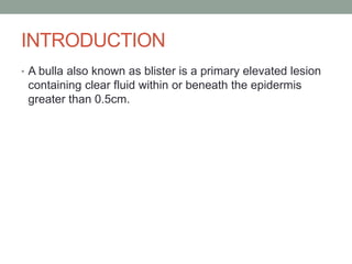
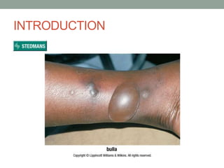
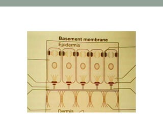
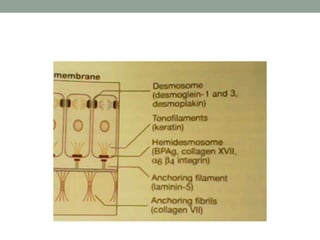
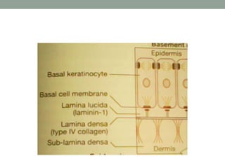
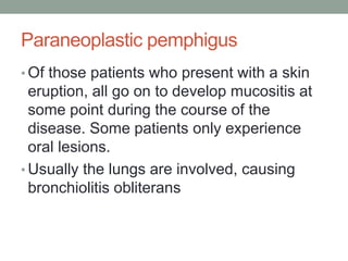
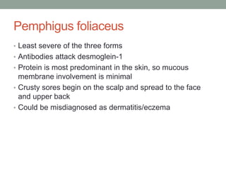
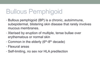
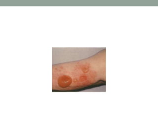
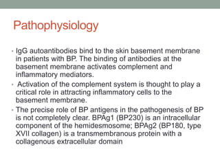
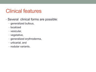
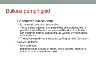
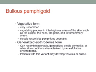
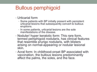
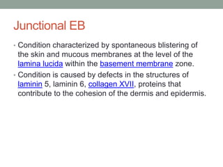
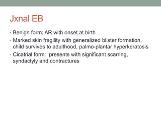
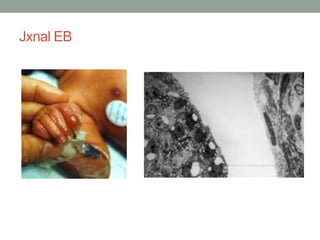
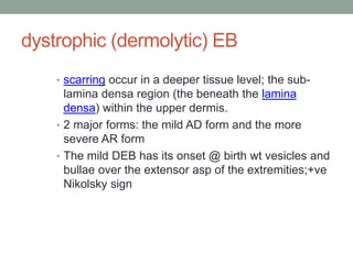
This document provides information on bullous skin disorders. It begins by defining a bulla or blister. Bullous skin disorders are characterized by the presence of blisters or erosions on the skin and mucous membranes. These disorders are usually due to autoimmune diseases or inherited abnormalities of structural proteins found in the epidermis and basement membrane. Bullous skin disorders can be divided into immunobullous or mechanobullous categories. Several specific immunobullous disorders are described in detail including their pathogenesis, clinical features, diagnosis, and treatment.
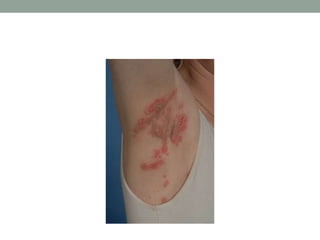
subcorneal
intraepidermal
subepidermal
pemphigus
nikolski's sign
pemphigus foliaceous
pemphigus vulgaris
pseudonikolski's sign
revision notes for dermatology based on lecture notes and high yield topic
based on previous year question
subcorneal
intraepidermal
subepidermal
pemphigus
nikolski's sign
pemphigus foliaceous
pemphigus vulgaris
pseudonikolski's sign
revision notes for dermatology based on lecture notes and high yield topic
based on previous year question
The skin is not only the largest organ of the body, but it also forms a living biological barrier with several functions.
Pyodermas are any pyogenic skin disease (has pus). Skin infections can be caused by bacteria (often Staphylococcal or Streptococcal) either invading normal skin, or affecting a compromised skin barrier
Some bacterial skin infections resolve without serious morbidity. However, skin infections can be severe and result in sepsis or death, particularly in vulnerable patient groups.
Molluscum contagiosum Made Extremely SimpleDrYusraShabbir
A brief description of a very common viral infection affecting children and adults. Molluscum Contagious is an infectious contagious disease. Useful information regarding the symptoms and treatment of the rash are available for medical students, doctors, dermatologists, ophthalmologists, gynaecologist, pediatricians and nurses. Helpful for studying for exams. Reference: Rooks, Textbook of Dermatology
The Indian Dental Academy is the Leader in continuing dental education , training dentists in all aspects of dentistry and
offering a wide range of dental certified courses in different formats.for more details please visit
www.indiandentalacademy.com
Current concepts of pemphigus with a deep insight into its molecular aspectishita1994
Pemphigus vulgaris is an autoimmune bullous disease involving both the skin and mucosal areas, which
is characterized by intraepithelial flaccid blisters and erosions. The pathogenesis of this disease is not
yet completely established, but novel intuitions into its pathogenesis have recently been published. An
unanswered question in its pathophysiology is the mechanism of acantholysis or loss of keratinocyte
cell adhesion. Acantholysis seems to result from a communal action of autoantibodies against numerous
keratinocyte self‑antigens, of which desmogleins 1 and 3, desmocollins and nondesmosome components,
such as the mitochondrion, might take part in the disease initiation. Lately, apoptosis was described as
a possible underlying mechanism of acantholysis. Likewise, apoptolysis is assumed to be the association
between suprabasal acantholytic and cell death pathways. Hence, the present review focuses on the current
concepts in the pathogenesis of the pemphigus in a nutshell.
Show drafts
volume_up
Empowering the Data Analytics Ecosystem: A Laser Focus on Value
The data analytics ecosystem thrives when every component functions at its peak, unlocking the true potential of data. Here's a laser focus on key areas for an empowered ecosystem:
1. Democratize Access, Not Data:
Granular Access Controls: Provide users with self-service tools tailored to their specific needs, preventing data overload and misuse.
Data Catalogs: Implement robust data catalogs for easy discovery and understanding of available data sources.
2. Foster Collaboration with Clear Roles:
Data Mesh Architecture: Break down data silos by creating a distributed data ownership model with clear ownership and responsibilities.
Collaborative Workspaces: Utilize interactive platforms where data scientists, analysts, and domain experts can work seamlessly together.
3. Leverage Advanced Analytics Strategically:
AI-powered Automation: Automate repetitive tasks like data cleaning and feature engineering, freeing up data talent for higher-level analysis.
Right-Tool Selection: Strategically choose the most effective advanced analytics techniques (e.g., AI, ML) based on specific business problems.
4. Prioritize Data Quality with Automation:
Automated Data Validation: Implement automated data quality checks to identify and rectify errors at the source, minimizing downstream issues.
Data Lineage Tracking: Track the flow of data throughout the ecosystem, ensuring transparency and facilitating root cause analysis for errors.
5. Cultivate a Data-Driven Mindset:
Metrics-Driven Performance Management: Align KPIs and performance metrics with data-driven insights to ensure actionable decision making.
Data Storytelling Workshops: Equip stakeholders with the skills to translate complex data findings into compelling narratives that drive action.
Benefits of a Precise Ecosystem:
Sharpened Focus: Precise access and clear roles ensure everyone works with the most relevant data, maximizing efficiency.
Actionable Insights: Strategic analytics and automated quality checks lead to more reliable and actionable data insights.
Continuous Improvement: Data-driven performance management fosters a culture of learning and continuous improvement.
Sustainable Growth: Empowered by data, organizations can make informed decisions to drive sustainable growth and innovation.
By focusing on these precise actions, organizations can create an empowered data analytics ecosystem that delivers real value by driving data-driven decisions and maximizing the return on their data investment.
Chatty Kathy - UNC Bootcamp Final Project Presentation - Final Version - 5.23...John Andrews
SlideShare Description for "Chatty Kathy - UNC Bootcamp Final Project Presentation"
Title: Chatty Kathy: Enhancing Physical Activity Among Older Adults
Description:
Discover how Chatty Kathy, an innovative project developed at the UNC Bootcamp, aims to tackle the challenge of low physical activity among older adults. Our AI-driven solution uses peer interaction to boost and sustain exercise levels, significantly improving health outcomes. This presentation covers our problem statement, the rationale behind Chatty Kathy, synthetic data and persona creation, model performance metrics, a visual demonstration of the project, and potential future developments. Join us for an insightful Q&A session to explore the potential of this groundbreaking project.
Project Team: Jay Requarth, Jana Avery, John Andrews, Dr. Dick Davis II, Nee Buntoum, Nam Yeongjin & Mat Nicholas
2. INTRODUCTION
• A bulla also known as blister is a primary elevated lesion
containing clear fluid within or beneath the epidermis
greater than 0.5cm.
4. • Bullous skin disorders are characterised by the
presence of blisters or erosions on the skin and
mucous membranes
• Bullous disorders are usually due to autoimmune
diseases or inherited abnormal structural proteins.
• These structural proteins are found within the
epidermis and the basement membrane.
5.
6.
7.
8. • The keratinocytes of the epidermis are tightly bound
together by desmosomes and intracellular substances to
form a barrier of high tensile strength and stability.
• Beneath the epidermis lies the basement membrane
zone, a specialized area of cell–extracellular matrix
adhesion.
9. • Specialized structures traversing this zone anchor the
epidermis to the dermis; these are Polysaccharides and
proteins (including collagens) linked to form
macromolecules (the adhesion complex).
10. • If any of these specialized attachments are malformed,
absent or damaged, cell–cell or cell–matrix adhesion is
impaired. Separation may then occur, leading to
accumulation of fluid in the extracellular space and blister
formation.
11. BULLOUS SKIN DISORDERS
They can be divided into two basic groups based on their
cause;
• Immunobullous and
• mechanobullous
12. BULLOUS SKIN DISORDERS
IMMUNOBULLOUS;
• Pemphigus
• Bullous pemphigoid
• Dermatitis herpitiformis
• Linear IgA disease(chronic bullous dermatosis of
childhood)
MECHANOBULLOUS;
• Epidermolysis bullosa
13. PEMPHIGUS
• Derived from the Greek word “pemphix”
• This is an autoimmune blistering disorder that
occurs due to loss of the integrity of the normal
intercellular attachments within epidermis and
mucosal epithelium.
• is mediated by auto-antibodies directed at the
keratinocyte cell surface.
• A relatively uncommon condition
14. Pemphigus
• The aetiology is related to the production of auto
antibodies directed at desmoglein, an adhesion
molecule found in the intercellular epidermis and
the dermo-epidermal basement membrane zone.
• Clinical and pathologic variants include;
• Pemphigus vulgaris
• Pemphigus foliaceus
• Paraneoplastic pemphigus
• Pemphigus Vegitans
• Endemic Pemphigus
• Pemphigus Erythematosus.
15. Pemphigus: pathophysiology
• An immunogenetic predisposition is well established.
• Blisters in PV are associated with the binding of IgG
autoantibodies to keratinocyte cell surface molecules.
• The binding of autoantibodies results in a loss of cell-cell
adhesion, a process termed acantholysis.
16. Pemphigus: pathophysiology
• PV antigen: Intercellular adhesion in the epidermis
involves several keratinocyte cell surface molecules.
• Pemphigus antibody binds to keratinocyte cell surface
molecules desmoglein 1 and desmoglein 3.
17. Pemphigus vulgaris
• Pemphigus vulgaris accounts for around 70% of all
cases of pemphigus.
• Arises due to the attack of desmoglein-3 by auto
antibodies, mostly IgG
• PV has been reported to occur worldwide, the
incidence varies from 0.5-3.2 cases per 100,000.
• PV incidence is increased in patients of Ashkenazi
Jewish descent and those of Mediterranean origin.
• PV is a potentially life-threatening autoimmune
mucocutaneous disease with a mortality rate of
approximately 5-15%.
• Prognosis is worse in patients with extensive
disease and in older patients.
18. Pemphigus vulgaris
• The condition is common in the middle aged,
particularly btw ages 40-60.
• It has predilection for mucosa and skin especially
on scalp, face, axilla, groin, trunk, and points of
pressure, sores commonly originate in the mouth.
• The oral ulcers persist for month before skin lesion
appears.
• Lesions rupture easily leaving a shallow erosion
covered with dried serum and crust.
19. • The large areas of denudation may become
infected and smelly, and severe oral ulcers
make eating painful.
23. Paraneoplastic pemphigus
• Most severe form and the least common
• Usually a complication of malignancy although it can
occur in association with benign neoplasms.
• The most common malignancy associated with
paraneoplastic pemphigus is non-Hodgkin lymphoma.
Other associated malignancies and conditions include
chronic lymphocytic leukemia, Castleman disease,
Waldenström macroglobulinemia, thymoma, sarcoma,
and lung carcinoma.
• The earliest and most constant clinical finding in
paraneoplastic pemphigus is painful oral erosions.
24. Paraneoplastic pemphigus
• Of those patients who present with a skin
eruption, all go on to develop mucositis at
some point during the course of the
disease. Some patients only experience
oral lesions.
• Usually the lungs are involved, causing
bronchiolitis obliterans
25. Pemphigus foliaceus
• Least severe of the three forms
• Antibodies attack desmoglein-1
• Protein is most predominant in the skin, so mucous
membrane involvement is minimal
• Crusty sores begin on the scalp and spread to the face
and upper back
• Could be misdiagnosed as dermatitis/eczema
26. Pemphigus vegetans
• Pemphigus vegetans is a variant of p.
vulgaris, Presents with a large moist
verrucous [wart-like] vegetating plaques
with pustules.
• by proliferative and verrucous lesions on
the erosions
• a rare form that do not present with blisters
• Distribution includes groin, axillae, and
flexural surfaces.
27.
28. PEMPHIGOID ERYTHEMATOSUS.
• This is a localised and less severe form of
pemphigus foliaceus.
• Areas of distributions include malar surface
of the face in a lupus erythematosus
fashion.
29. Pemphigus
• Pemphigus could also occur in pregnancy, although it is
uncommon
• The baby may be stillborn or may have clinical,
immunologic or histopathological features of pemphigus;
as trans-placental transmission of pemphigus is possible
30. pemphigus
• Could also be drug-induced
• Penicillamine is the most frequent cause, occurs in 3-10%
of users, pemphigus foliaceus more common in such
people.
• Piroxicam, captopril, phenytoin, phenobarbitone,
rifampicin also implicated
31. Pemphigus: diagnosis
• Classical morphologic xtics: flaccid bullae forming over a
normal or erythematous base; erosions; vegetations
• Nikolsky sign: firm sliding pressure on the blister reveals
normal looking epidermis, producing an erosion
• Bulla-spread sign:lateral pressure on the edge of a bulla
spreads it onto clinically unaffected skin
32. Pemphigus: diagnosis
• Histopathology:
• an intradermal blister.
• The earliest changes consist of intercellular edema with loss of
intercellular attachments in the basal layer.
• Basal cells are separated from one another and stand like a row of
tombstones on the floor of the blister, but they remain attached to
the basement membrane.
• Blister cells contain some acantholytic cells.
• Histopathology can help differentiate PV from pemphigus foliaceus,
which demonstrates a more superficial epidermal cleavage.
33. Pemphigus: diagnosis
• Immunofluorescence: direct and indirect
• DIF demonstrates in vivo deposits of antibodies and other
immunoreactants, such as complement.
• DIF usually shows IgG deposited on the surface of the
keratinocytes in and around lesions. IgG1 and IgG4 are
the most common subclasses.
• The best location for DIF is normal perilesional skin.
36. Pemphigus: treatment
• Corticosteroids
• High doses of oral prednisolone (80-120 mg daily).
Tests that assess a patients fitness for prolonged
steroid therapy should be carried out before
treatment.
• At remission, the dosage of the steroid is halved
and immunosuppresants are introduced-
methotrexate, azathioprine cyclophosphamide,
cyclosporine.
• Plasmapheresis
• Pulse therapy
37. Pemphigus:complications
• 10 percent may die due to complication.
• Secondary infection: immunosuppressants and the presence of
multiple erosions.
• Malignancies (resulting from immunosuppressants)
• Growth retardation: in children taking systemic corticosteroids and
immunosuppressants.
• Bone marrow suppression has been reported in patients receiving
immunosuppressants.
• Impaired immune responsiveness caused by corticosteroids and
other immunosuppressive drugs may result in the rapid spread of
infection.
• Osteoporosis may occur following the use of systemic corticosteroids.
• Adrenal insufficiency has been reported following prolonged use of
glucocorticoids.
38. complications
• Hyperparasitosis
• Pseudo tumor cerebri
• To prevent these, the side effect profile is monitored,
steroid sparing drugs like cyclophosphomide can be
administered.
39. Bullous Pemphigoid
• Bullous pemphigoid (BP) is a chronic, autoimmune,
subepidermal, blistering skin disease that rarely involves
mucous membranes.
• Xterised by eruption of multiple, tense bullae over
erythematous or normal skin
• Common in the elderly (6th-8th decade)
• Flexural areas
• Self-limiting, no sex nor HLA predilection
40.
41. Bullous pemphigoid
• Th aetiology is linked to the presence of immunoglobulin
G (IgG) autoantibodies specific for the hemidesmosomal
BP antigens BP230 (BPAg1) and BP180 (BPAg2).
• Other associated factors include age, Immunogenetics,
Epitope spreading, Complement activation & Chemokines
42. Bullous pemphigoid
• Immunogenetics: human leukocyte antigen (HLA)
haplotype, DQB1*0301, is increased in patients with BP.
• Age: Investigators have postulated that intrinsic changes
in the immune system with aging may be a factor in the
initiation of an autoimmune response against BP antigens.
43. Bullous pemphigoid
• Reports of BP arising in patients with inflammatory skin
diseases, such as psoriasis and lichen planus, or after
trauma, such as drug reactions, suggest that inflammation
may expose sequestered skin basement membrane
proteins and BP antigens and lead to the development of
an autoimmune response.
• Epitope spreading could occur in these conditions to
involve the BP autoantigens
44. Pathophysiology
• IgG autoantibodies bind to the skin basement membrane
in patients with BP. The binding of antibodies at the
basement membrane activates complement and
inflammatory mediators.
• Activation of the complement system is thought to play a
critical role in attracting inflammatory cells to the
basement membrane.
• The precise role of BP antigens in the pathogenesis of BP
is not completely clear. BPAg1 (BP230) is an intracellular
component of the hemidesmosome; BPAg2 (BP180, type
XVII collagen) is a transmembranous protein with a
collagenous extracellular domain
45. Clinical features
• Several clinical forms are possible:
• generalized bullous,
• localized
• vesicular,
• vegetative,
• generalized erythroderma,
• urticarial, and
• nodular variants.
46. Bullous pemphigoid
• Generalized bullous form
• is the most common presentation.
• Tense bullae arise on any part of the skin surface, with a
predilection on the flexural areas of the skin. The bullae
can occur on normal-appearing, as well as erythematous,
skin surfaces.
• The bullae usually heal without scarring or milia formation.
• Vesicular form
• less common.
• It manifests as groups of small, tense blisters, often on a
urticarial or erythematous base.
47. Bullous pemphigoid
• Vegetative form
• very uncommon
• vegetating plaques in intertriginous areas of the skin, such
as the axillae, the neck, the groin, and inframammary
areas.
• closely resembles pemphigus vegetans.
• Generalized erythroderma form
• Can resemble psoriasis, generalized atopic dermatitis, or
other skin conditions characterized by an exfoliative
erythroderma.
• Patients with this variant may develop vesicles or bullae.
48. Bullous pemphigoid
• Urticarial form
• Some patients with BP initially present with persistent
urticarial lesions that subsequently convert to bullous
eruptions.
• In some patients, urticarial lesions are the sole
manifestations of the disease.
• Nodular/ hyper keratotic form: This rare form,
termed pemphigoid nodularis, has clinical features
that resemble prurigo nodularis, with blisters
arising on normal-appearing or nodular lesional
skin.
• Acral form: In childhood-onset BP associated with
vaccination, the bullous lesions predominantly
affect the palms, the soles, and the face.
49. Diagnosis
• Histopathology:
• a subepidermal blister.
• The inflammatory infiltrate is typically polymorphous, with an
eosinophil predominance.
• Direct & indirect immunofluorescence studies
• Indirect immunoelectron microscopy, immunoblotting,
immunoprecipitation, and enzyme-linked
immunosorbent assay (ELISA).
• All these analyse IgG autoantibodies
50. • Direct immunofluorescence study performed on a perilesional
skin biopsy specimen from a patient with bullous pemphigoid
detects a linear band of immunoglobulin G deposit along the
dermoepidermal junction.
51.
52. Treatment
• As in other autoimmune bullous diseases, the goal of
therapy is to decrease blister formation, to promote
healing of blisters and erosions, and to determine the
minimal dose of medication necessary to control the
disease process.
• Anti-inflammatory:Prednisone;Tetracycline
• Immunosuppressants: azathioprine,rituzimab
53. Pemphigoid Complications
• Complications are similar to those of pemphigus
• Prognosis relates to the side-effects of therapy and the
presence/absence of other co-morbid conditions
55. Dermatitis herpetiformis
• Dermatitis herpetiformis (DH) is an autoimmune
blistering disorder associated with a gluten-
sensitive enteropathy (GSE).
• It’s a chronic, pruritic, papulovesicular eruption,
symmetric in distribution, wt a predilection for the
extensors of he extremities, shoulders, and
buttocks
• Mucosal involvement is infrequent
• DH occurs more frequently in individuals of
Northern European ancestry and is rare in Asians
and persons of African descent.
56. Dermatitis herpetiformis
• Male-to-female ratio is up to 2:1. In one
study of patients with GSE, 16% of the men
and 9% of the women had DH.
• Typically, the onset of DH is in the second
to fourth decade; however, persons of any
age may be affected.
• The aetiology is debatable; but it is believed
that it represents an allergic state in which
the antigen is presented to a genetically
predisposed individual via the GIT
57. Pathophysiology
• DH is a disease of the skin caused by the deposition of
IgA in the papillary dermis, which triggers an immunologic
cascade, resulting in neutrophil recruitment and
complement activation.
• It has been hypothesized that DH is the result of an
immunologic response to chronic stimulation of the gut
mucosa by dietary gluten with subsequent activation of
cutaneous endothelial cells and circulating inflammatory
cells, including neutrophils.
• An underlying genetic predisposition to the development
of DH has been demonstrated. Both DH and celiac
disease (CD) show an increased expression of HLA-A1,
HLA-B8, HLA-DR3, and HLA-DQ2 haplotypes
58. Pathophysiology
• Evidence is mounting that epidermal transglutaminase 3
(TGase3), a cytosolic enzyme involved in cell envelope
formation during keratinocyte differentiation, is the
autoantigen of DH.
• Hormonal factors may also play a role in the pathogenesis
of DH, and recent reports describe DH induced by
treatment with leuprolide acetate, a gonadotropin-
releasing hormone analog.
59. • Androgens have a suppressive effect
on immune activity, including
decreased autoimmunity, and
androgen deficient states may be a
potential trigger for DH exacerbation.
• Apoptosis may contribute to the
pathogenesis of epidermal changes in
DH
60. Clinical features
• Disease is heralded by intense itching
followed by eruption of urticarial wheals,
erythematous papules, papulo-vesicle or
groups of tiny vesicles
• Extensor asp of the limbs, knees, elbows,
buttocks and natal cleft
• mucosae are usually spared
• Ass wt autoimmune conditions,
malignancies, gastric surgery and certain
chemicals
62. Diagnosis
• skin biopsy
• Biopsy specimens of lesional skin reveal neutrophils in the
dermal papillae, with fibrin deposition, neutrophil fragments,
and edema. Eosinophils may be present.
• Vesicles form in the lamina lucida, the weakest portion of the
dermoepidermal junction, due to neutrophil lysosomal enzymes
• ELISA(IgA endomysial antibodies tissue transglutaminase)
• direct immunofluorescence studies taken from healthy-
appearing skin
63. Treatment
• Control of the skin disease can be achieved with
medications, dietary avoidance of gluten, or both.
• Dapsone (diaminodiphenyl sulfone) and sulfapyridine are
the primary medications used to treat DH.
• For patients unable to tolerate dapsone, particularly those
who develop hemolysis, sulfapyridine may be substituted.
• Other, less effective treatments for DH include colchicine,
cyclosporine, azathioprine, and prednisone. UV light may
provide some symptomatic relief.
64. Treatment
• Dietary intake of gluten causes the disease, and
elimination improves it.
• Dapsone 25-400 mg/d
• Sulfapyridine 1-1.5 g/d; 500 mg bid initially; increase by 1
g q1-2wk until disease is controlled; control may require
1-4 g/d
65. DH
Complications
• Complications are related to the GSE, the risk of
developing lymphomas, and the potential adverse effects
of medications (dapsone).
Prognosis
• DH is an ongoing disease process of variable severity.
• The prognosis is good for patients who can tolerate
dapsone and the few who can maintain a gluten-free diet
(which may decrease the risk of lymphoma).
66. Papillarymicroabscesses form and progress to subepidermalvacuolization and
vesicle formationin the lamina lucida,the weakestportionof the dermoepidermal
junction(hematoxylinand eosin stain).
67. LINEAR IgA DISEASE(CDC)
• It is an autoimmune subepidermal vesiculobullous
disease that may be idiopathic or drug-induced.
• Children and adults are affected .
• Linear IgA dermatosis is an autoimmune disease
histopathologically characterized by the linear
deposition of IgA at the basement membrane zone
(BMZ).
• Antibody deposition leads to complement activation
and neutrophil chemotaxis, which eventuates in
loss of adhesion at the dermal-epidermal junction
and in blister formation.
68. LINEAR IgA DISEASE(CDC)
• Remission occurs in most cases, the rate of
remission is higher in children compared to adults.
• Some case series have reported a slight female
preponderance; the female-to-male ratio is 1.6:1.
• The classic primary lesions of linear IgA dermatosis
are clear and/or hemorrhagic round or oval
vesicles or bullae on normal, erythematous, or
urticarial skin.
•
69. LINEAR IgA DISEASE(CDC)
• Cutaneous manifestations may also include
erythematous plaques, blanching macules and
papules, or targetoid erythema multiforme–like
lesions.
• Bullae may be discrete or arranged in a
herpetiform pattern,often described as the cluster
of jewels sign. Alternatively, vesicles and bullae
may be seen at the edge of annular or polycyclic
lesions, the appearance of which has been
described as the string of beads sign
70. LINEAR IgA DISEASE(CDC)
• The distribution of linear IgA dermatosis differs
between adults and children. Lesions in children
are typically localized to the lower abdomen and
anogenital areas with frequent involvement of the
perineum (see the image below). Other sites
include the feet, the hands, and the face,
particularly the perioral area.
• In adults, the trunk and the limbs are most
commonly affected. In adults, involvement of the
perineum and the perioral area is less frequent
than in children.
71. diagnosis
• Direct immunofluorescence study of both
perilesional skin and healthy skin typically shows
linear deposition of IgA at the BMZ. Linear
deposition of C3 may also be seen.
• Early urticarial papules or plaques reveal
neutrophils aligned along the BMZ accompanied by
vacuolar change. Neutrophilic microabscesses
may be seen in dermal papillae .
• Fully developed lesions reveal subepidermal
blistering with a predominantly polymorphonuclear
infiltrate, although mononuclear cells and
eosinophils may be present.
72. Treatment
• In cases of drug induced , the immidiate withdrawal
of the drug should cause ceasation of the symptom
e.g vancomycin.
• Most cases have been reported to respond to oral
dapsone or sulfapyridine.
• Other drugs that can be used are prednisone,
sulfamethoxypyridazine, colchicine, dicloxacillin,
mycophenolate mofetil .
73. Epidermolysis bullosa
• Is the name used to describe a group of disorders
characterised by skin fragility and blister formation
following seemingly insignificant trauma to the skin.
• These are due to inherited abnormalities in skin structural
proteins.
• These blisters appear at or shortly after birth.
• They are very rare conditions.
74. EPIDEMIOLOGY
• It is estimated that 2 to 4 out of every 100,000 people, or
up to 12,000 people in the United States, have some form
of EB.
• An estimated 50 in 1 million live births are diagnosed with
EB, and 9 in 1 million are in the population
• It occurs in all racial and ethnic groups and affects males
and females equally.
75. CLASSIFICATION
• 3 forms are identified:
• EB Simplex (EBS) -- above the basement membrane
• Junctional EB (JEB) -- through the basement membrane
• Dystrophic EB (DEB) -- under the basement membrane
76. Epidermolysis bullosa simplex
• Could occur either as the generalized (Koebner) or
localized (Weber-Cockayne) or hepertiform (EBS-DM)
or EBS-MP subtypes
• The generalized type has an AD mode of inheritance
• Vesicles and bullae form over the sites prone trauma:
hands, elbow, knees and feet; with minimal mucous
membrane involvement
• The localized form, inherited AD affects the palms and
soles 10, wt hot weather
77. Ruptured bulla and newly erupted bulla of the leg in a
newborn with epidermolysis bullosa simplex
78. Junctional EB
• Condition characterized by spontaneous blistering of
the skin and mucous membranes at the level of the
lamina lucida within the basement membrane zone.
• Condition is caused by defects in the structures of
laminin 5, laminin 6, collagen XVII, proteins that
contribute to the cohesion of the dermis and epidermis.
79. Junctional EB
• An Autosomal recessive disease
• 3 forms: lethalis, benign and cicatrial forms
• Lethalis form is characterised by severe generalised
blistering; present at birth
• Life threatening, long persistence, oral erosions,
dysplastic teeth, laryngeal & bronchial erosions,
growth retard, anemia
• Usually patient dies in childhood
80. Jxnal EB
• Benign form: AR with onset at birth
• Marked skin fragility with generalized blister formation,
child survives to adulthood, palmo-plantar hyperkeratosis
• Cicatrial form: presents with significant scarring,
syndactyly and contractures
82. dystrophic (dermolytic) EB
• scarring occur in a deeper tissue level; the sub-
lamina densa region (the beneath the lamina
densa) within the upper dermis.
• 2 major forms: the mild AD form and the more
severe AR form
• The mild DEB has its onset @ birth wt vesicles and
bullae over the extensor asp of the extremities;+ve
Nikolsky sign
83. DEB
• The AR form is the more severe type
• Onset at birth; with acral cutaneous and mucosal
blistering
• Growth retardation and anaemia
• Digital fusion with encasement of fingers and toes
85. DEB: diagnosis
• Prenatal diagnosis is possible
• skin biopsy (taking a small sample of skin
that is examined under a microscope).
• use of microscope and reflected light to
see if proteins needed for forming
connecting fibrils, filaments, or
hemidesmosomes are missing or reduced
in number.
• use of a high-power electron microscope,
which can greatly magnify tissue images, to
identify structural defects in the skin.
86. Treatment
• Preventing Blisters:
• avoiding overheating by keeping rooms at an even
temperature
• applying lubricants to the skin to reduce friction and keep
the skin moist
• using simple, soft clothing that requires minimal handling
when dressing a child
• using sheepskin on car seats and other hard surfaces
• wearing mittens at bedtime to help prevent scratching
87. Treatment
• Caring for Blistered Skin
• pat the blister with an alcohol pad before popping it at the sides
with a sterile needle or other sterile tool. The fluid can then drain
into a sterile gauze that is used to dab the blister. After opening and
draining, the doctor may suggest that an antibiotic ointment be
applied to the area of the blister before covering it with a sterile,
non sticking bandage.
• Treating Nutritional Problems
• Blisters that form in the mouth and oesophagus in some people
with EB are likely to cause difficulty in chewing and swallowing food
and drinks
• find recipes and prepare food that is nutritious and easy to
consume. identify high-caloric and protein-fortified foods and
beverages that help replace protein lost in the fluid from draining
blisters. They can suggest vitamin and mineral nutritional
supplements that may be needed,
88. Surgical Treatment
• Individuals with the severe forms of
autosomal recessive Dystrophic EB whose
oesophagus has been narrowed by scarring
may require dilation of their oesophagus for
food to travel from the mouth to the
stomach.
• Other individuals who are not getting proper
nutrition may need a feeding tube that
permits delivery of food directly to the
stomach.
• Also, patients whose fingers or toes are
fused together may require surgery to
release them.
89. Treatment
• Silver sulfadiazine – wound healing
• systemic antibiotics
• Phenyton inhibits collagenase and appears to be
beneficial in recessive dystrophic EB
• Steroids
90. Complications
• Secondary infections (eg, pseudomonal,
staphylococcal, and herpes organisms)
• Pseudosyndactyly of the fingers and toes
• Scarring of the skin
• Pigmentary changes
• Anonychia
• Dental anomalies
• Dysphagia