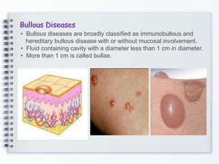
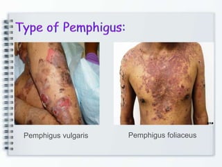
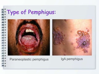
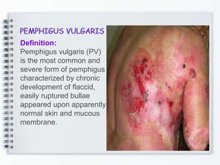
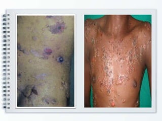
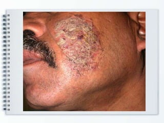
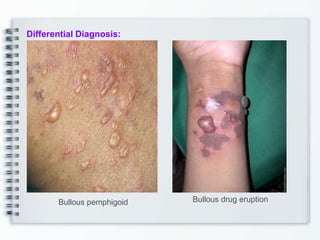
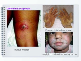
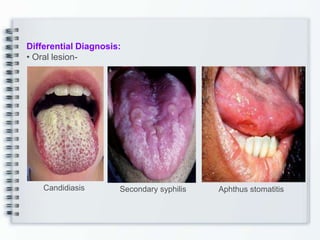
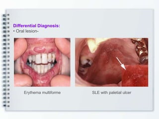
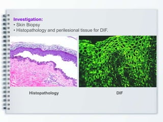
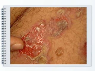
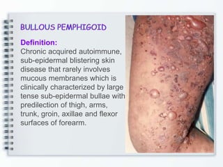
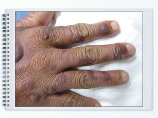
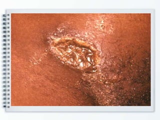
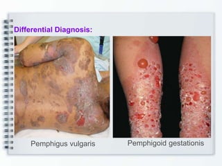
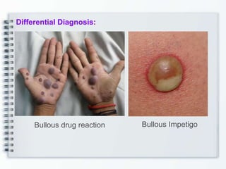
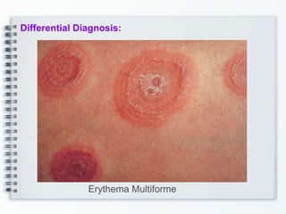
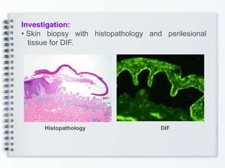
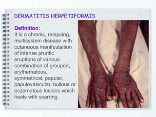
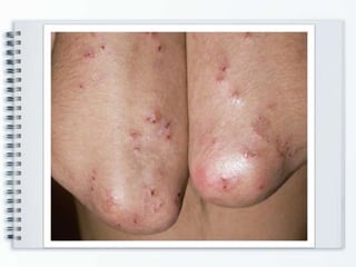
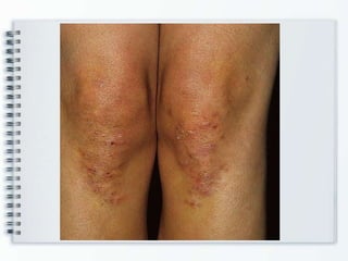
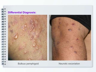
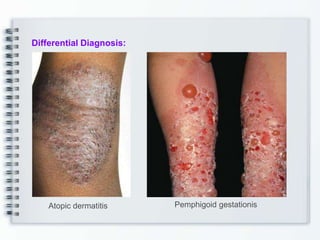
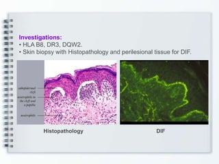
Top Rated Bangalore Call Girls Richmond Circle ⟟ 8250192130 ⟟ Call Me For Gen...
BULLOUS_DISEASES presentation and management
1. Speaker
Prof. M. U. Kabir Chowdhury FRCP
Visiting Professor of Dermatology
Dhaka Medical College
Holy Family Red Crescent Medical College Hospital
Adviser, Founder Principal and Visiting Professor
MH Samorita Hospital and Medical College
2. • Bullous diseases are broadly classified as immunobullous and
hereditary bullous disease with or without mucosal involvement.
• Fluid containing cavity with a diameter less than 1 cm in diameter.
• More than 1 cm is called bullae.
6. Definition:
Pemphigus vulgaris (PV)
is the most common and
severe form of pemphigus
characterized by chronic
development of flaccid,
easily ruptured bullae
appeared upon apparently
normal skin and mucous
membrane.
7. Epidemiology:
• Both sexes equally
• 40-60 years
• Rarely in children and younger persons
Aetiology:
• Unknown
• Autoimmune disease
• HLA-DR4, HLA-DR6 and HLA-DQ phenotypes
• Penicillamine, captopril, enalapril, penicillin, thiopronile,
nifedipine, piroxicam, interleukin-2, rifampicin
Pathogenesis:
• Auto immune
• Dsg 1 and Dsg 3
Mechanism of blister formation-
- Cellular and humoral autoimmunity
- Antibody can alone produce acantholysis
8.
9. Clinical features:
• Mucosal erosions and thin walled relatively flaccid, easily
ruptured bullae
• Fluid in the bulla is clear at first but in later may become
hemorrhagic or seropurulent even turbid.
• The bullae rupture to form erosions and large denuded areas.
• Rarely pruritic
• Often painful
• Hyperpigmentation and without scarring
• Usually appear first in the oral mucosa
• The conjunctiva, nasal mucosa, vaginal mucosa, penis and anus
may also be involved.
• Groin, scalp, face, neck, axillae and genitals are other common
sites.
• Nikolskys sign.
17. Management:
General:
• Severe patient to be hospitalized
• Adequate nutrition, maintenance of fluid electrolyte balance and
control of infection is important.
Topical treatment:
• Prolong daily bath is helpful in removing the crust.
• Antiseptic mouthwash.
• Topical corticosteroids and tacrolimas in localized disease.
18. Management:
Systemic therapy:
Corticosteroids:
• High dose (100-150mg)
• 1mg/kg/day
• Intravenous pulse therapy- 1gm/day over a period of 2 to 3 hours
daily for 5 days.
Immunosuppressive agents:
• Azathioprine
• Cyclophosphamide
• Azathioprin - It is relatively safer
• 2.5 to 5 mg /kg/day
• Mycophenolate mofetil
• Cyclophosphamide
Other modalities of treatment:
• Intravenous use of gamma globulin
• Infliximab and Rituximab
19.
20. Prognosis:
• Before the advent of glucocorticoid therapy,
pemphigus vulgaris was almost invariably fatal
• As soon as the diagnosis is established prognosis
is favorable.
21. Definition:
Chronic acquired autoimmune,
sub-epidermal blistering skin
disease that rarely involves
mucous membranes which is
clinically characterized by large
tense sub-epidermal bullae with
predilection of thigh, arms,
trunk, groin, axillae and flexor
surfaces of forearm.
22.
23. Epidemiology:
• Usally begins after age 60 years
Aetiology and Pathogenesis:
• This is an autoimmune disease and autoantibody is directed
against basement membrane zone (BMZ).
Clinical features:
• Bullous pemphigoid clinically manifested with large, tense, bullae
which arises on normal skin or on erythematous surface.
Eruption may be localized or generalized usually scattered
• Tendency to heal without scar.
30. Management:
Topical therapy
• Topical potent steroid like clobetasole propionate, halobetasole
propionate and topical tactolimus
Systemic therapy
• Prednisolone 50-100mg/day alone or combined with azathioprine
150mg daily till the lesion is cleared.
• Tetracycline and nicotinamide also successfully used.
• Dapsone and sulphapyridine also used with good control of
disease.
• Plasmapheresis, methotrexate, mycophenolate mofetil,
intravenous gama globulin, and chlorambucil is used if
unresponsive to above therapy.
Prognosis:
• Bullous pemphigoid is usually self limited disease
• Remission may occurred within 5-6 years
31. Definition:
It is a chronic, relapsing,
multisystem disease with
cutaneous manifestaition
of intense pruritic
eruptions of various
combination of grouped,
erythematous,
symmetrical, papular,
papulovasicular, bullous or
eczematous lesions which
heals with scarring.
32. Epidemiology:
• 3 to 4th decade but may occur in children.
• M:F - 2:1
Aetiology and pathogenesis:
• Unknown
• Gluten enteropathy
Clinical features:
• Manifested with recurrent pruritic lesion in symmetrical
distribution over extensor surface of body such as elbow,
knee, buttocks, shoulders and scalp.
• The eruptions on erythematous base which may be papular,
papulovesicular, vesicobullous, bullous, urticarial.
• The continuous appearance and disappearance of lesions
may results in hyper or hypopigmentation.
• A few patients may have coexistence gluten-sensitive
enteropathy manifested with diarrhoea.
39. Investigations:
• HLA B8, DR3, DQW2.
• Skin biopsy with Histopathology and perilesional tissue for DIF.
Histopathology DIF
40. Management:
• Dapsone is the drug of choice
• The dose varies between 50-300mg/day.
• Patients are strictly advised to avoid gluten in diet such as
wheat, barley, and rye which will help to suppress the disease
and to reduce dose of dapsone or sulfapyridine.
Prognosis:
• The disease is prolonged for many years with one third
patients may have spontaneous remission.