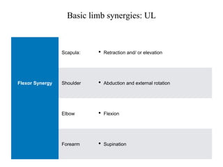
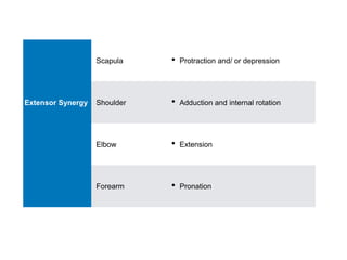
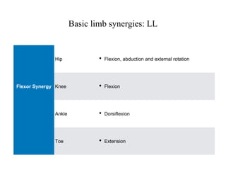
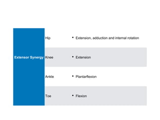
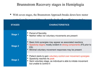
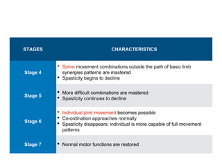
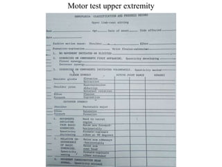
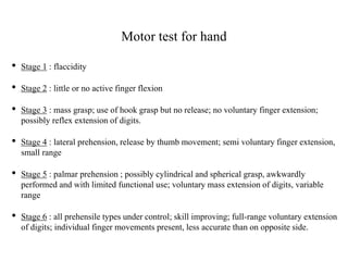
Brunnstrom Approach
Brunnstrom's Approach (SIGNE BRUNNSTROM)
Objectives: ➢ Discuss the concepts and principles underlying Brunnstrom’s approach ➢ Brunnstrom recovery stages ➢ Treatment principles & techniques
★ Brunnstrom’s approach was developed by the physical therapist from Sweden in the early 1950’s
★ Brunnstrom used motor control theory and observations of the patients'
★ Procedure: In a “trial & error” fashion ★ Later modified: in light of neurophysiological knowledge
Introduction: Reflex Theory Movement is controlled by stimulus-response. Reflexes are the basis for movement: reflexes are combined into actions that create behavior. Hierarchical Theory Characterized by a top-down structure, in which higher centers are always in charge of lower centers.
● When the CNS is injured, as, in a cerebrovascular accident, an individual goes through an “evolution in reverse”. Movement becomes primitive, reflexive, and automatic.
● Changes in tone and the presence of reflexes are considered a normal process of recovery.
● Movement recovery tends to be stereotypic.
● Patients exhibit only a few stereotypic movement patterns: Basic Limb Synergies.
● Based on observations of recovery following a stroke, this approach makes use of associated reactions, tonic reflexes, and the development of basic limb synergies to facilitate movements.
● The use of such a procedure is temporary.
Basic Limb Synergies:
● Normal synergistic movements are purposeful movements with maximum precision and minimum waste of energy.
● Basic limb synergy (BLS) does not permit the different combinations of muscles.
● BLS is considered primitive, automatic, and reflexive due to loss of inhibitory control from higher centers.
● Mass movement patterns in response to a stimulus or voluntary effort both Gross flexor movement (Flexor Synergy) Gross extensor movement (Extensor Synergy) Combination of the strongest component of the synergies (Mixed Synergy)
● Appear during the early spastic period of recovery
Upper Limb Flexor Synergy: Scapula: Retraction / Elevation Shoulder: Abduction and External rotation Elbow: Flexion Forearm: Supination Wrist and Finger: Flexion Lower Limb Extensor Synergy: Pelvis: posterior tilt Hip: Extension, Adduction & Internal rotation Knee: Extension Ankle: Plantarflexion Toes: Flexion Upper Limb Extensor Synergy: Scapula: Protraction / Depression Shoulder: Adduction and Internal rotation Elbow: Extension Forearm: Pronation Wrist: Extension Finger: Flexion Lower Limb Flexor Synergy: Pelvis: anterior tilt Hip: Flexion, Abduction & External rotation Knee: Flexion Ankle: Dorsiflexion Toes: Extension
Upper Limb Mixed Synergy: Scapula retraction Shoulder add.+IR Elbow flexion Forearm pronation Wrist & fingers flexion Lower Limb Mixed Synergy: Pelvis post tilt hip add.+IR Knee extension Ankle & toes plantarflexion
Rubrospinal tract Vestibulospinal tract
Associated Reactions
Primitive Reflexes
Hierachical theory- says that higher centers control on lower center; but when higher center damage then this inhibitory control from the higher center is loss which leads to exageration of the movt.
In normal individual, these occur a smooth, rhythmic movt. Because there is a presence of control from higher center on lower center.
Neurodevelopmental Therapy
Neurodevelopmental treatment (NDT) is a hands-on treatment approach used by physical therapists, occupational therapists, and speech-language pathologists
Without NDT interventions, the patient likely will develop a limited set of movement patterns that he or she will apply to nearly all tasks.
This presentation is detail about Volta therapy which is commonly used in paediatric neurological conditions and also for adults. this presentation explains what are the various techniques, methods of application of Volta therapy, indications, contraindications, etc.
constraint induced movement therapy.pptxibtesaam huma
Constraint induced movement therapy
Dr. Quazi Ibtesaam Huma (MPT)
Dr. Suvarna Ganvir (Phd, Prof & HOD)
Dept. of Neurophysiotherapy
DVVPF’s College of Physiotherapy
Content
Introduction
History of CIMT
Components of CIMT
Population for CIMT
Advantages of CIMT
Recent advances
Introduction
History of CIMT
CIMT is based on research by Edward Taub ,his hypothesize that the non use was a learning mechanism and calls this behavior “Learned non-use”.
It was observed that patients with hemiparesis did not use their affected extremity .
Overcoming learned non use
Mechanisms of CIMT
Population for CIMT
Stroke
Traumatic Brain Injury
Spinal Cord Injury
Multiple Sclerosis
Cerebral Palsy
Brachial Plexus Injury
Advantages of CIMT
Overall greater improvement in function than traditional treatment.
Highly researched and credible treatment approach.
There are brain activity and observed gray matter reorganization in primary motor, cortices and hippocampus.
Increase social participation
Components Of CIMT
Types of CIMT
Restraining Tools for CIMT
Minimal Requirement of hand function for CIMT
Recent Advances
The EXCITE Trial: Retention of Improved Upper Extremity Function Among Stroke Survivors Receiving CI Movement Therapy.(2008)
The Extremity Constraint Induced Movement Therapy Evaluation (EXCITE) demonstrated that CIMT administered 3-9 months post-stroke, resulted in statistically significant and clinically relevant improvement in upper extremity function during the first year compared to those achieved by participants undergoing usual and customary care.
This study was the first randomized clinical trial to examine retention and improvements for the 24 month period following CIMT therapy in a subacute sample.
Study design - single masked cross-over design, with participants undergoing adaptive randomization to balance ,gender, prestroke dominant side, side of stroke, and level of paretic arm function across sites.
CIMT was delivered up to 6 hours per day, 5 days per week for 2 weeks.
Subsequent evaluations were made after the two week period, and at 4, 8, and 12 months.
Because the control group was crossed over to receive CIMT after one year.
Primary outcome measures – Wolf Motor Function Test
Motor Activity Log
Secondary outcome measure - Stroke Impact Scale (SIS)
were assessed at each of these time intervals, was administered only at baseline, 4, 12, 16 and 24 month evaluations.
Result :There was no observed regression from the treatment effects observed at 12 months after treatment during the next 12 months for the primary outcome measures of WMFT and MAL.
In fact, the additional changes were in the direction of increased therapeutic effect. For the strength components of the WMFT the changes were significant (P < .05) Secondary outcome variables, including the SIS, exhibited a similar pattern.
Conclusion: Mild to moderately impaired patients who are 3-9 months post-stroke demonstrate
It is a technique developed by Janet H Carr and Roberta B Shepherd which provides physiotherapists and occupational therapists with an approach to stroke rehabilitation that is clear, relevant, and effective, building on the research-based model created by the authors
Basic Introduction about Joint Mobilisation and Manipulation, This article gives clear notes for the students to understand the Mobilisation techniques.
Concept given by Shacklock (modern concept) and Butler (old concept), a method of assessment as well as treatment of peripheral neurological system by physiotherapists.
Part-I: The current slideshow: theoretical aspect of neurodynamics.
Part-II: Assessment of peripheral nervous system on the basis of neurodynamic concepts: Date: 01/04/2020
Part-III: treatment part: Date: 03/04/2020
Part-IV: Self neurodynamics: 05/04/2020
“The ability of neurons to change their function, chemical profile or structure is referred to as neuroplasticity.”
Neuroplasticity includes :
- Habituation
- Learning & memory
- Cellular recovery after injury
At the end of the lecture, the students should be able to:
Discuss the theoretical basis of the neurodevelopmental approaches
Discuss the concepts and principles underlying the Bobath approach
Discuss the concepts and principles underlying the Brunnstrom approach
Vojta technique is neuromuscular approach deals with all the conditions of CNS and Musculoskeletal system.
Contents :
Introduction
Definition
What is REFLEX LOCOMOTION
Indication
Stimulating Points
Reflex locomotion
Reflex Rolling phase 1
Reflex Rolling phase 2
Reflex creeping
Effects of Vojta technique
Principles and application of various Neurological Approaches. Comprises of PNF, ROODS, NDT, BOBATH, SENSORY INTEGRATION, BRUNNSTORM, VOJTA, Motor Re-learning Approach , Neural Tissue Mobilization
Dynamic Neuro-Cognitive Imagery Improves Mental Imagery Ability, Disease Severity, and Motor and Cognitive Functions in People with Parkinson’s Disease
Couples presenting to the infertility clinic- Do they really have infertility...Sujoy Dasgupta
Dr Sujoy Dasgupta presented the study on "Couples presenting to the infertility clinic- Do they really have infertility? – The unexplored stories of non-consummation" in the 13th Congress of the Asia Pacific Initiative on Reproduction (ASPIRE 2024) at Manila on 24 May, 2024.
These lecture slides, by Dr Sidra Arshad, offer a quick overview of physiological basis of a normal electrocardiogram.
Learning objectives:
1. Define an electrocardiogram (ECG) and electrocardiography
2. Describe how dipoles generated by the heart produce the waveforms of the ECG
3. Describe the components of a normal electrocardiogram of a typical bipolar leads (limb II)
4. Differentiate between intervals and segments
5. Enlist some common indications for obtaining an ECG
Study Resources:
1. Chapter 11, Guyton and Hall Textbook of Medical Physiology, 14th edition
2. Chapter 9, Human Physiology - From Cells to Systems, Lauralee Sherwood, 9th edition
3. Chapter 29, Ganong’s Review of Medical Physiology, 26th edition
4. Electrocardiogram, StatPearls - https://www.ncbi.nlm.nih.gov/books/NBK549803/
5. ECG in Medical Practice by ABM Abdullah, 4th edition
6. ECG Basics, http://www.nataliescasebook.com/tag/e-c-g-basics
Ozempic: Preoperative Management of Patients on GLP-1 Receptor Agonists Saeid Safari
Preoperative Management of Patients on GLP-1 Receptor Agonists like Ozempic and Semiglutide
ASA GUIDELINE
NYSORA Guideline
2 Case Reports of Gastric Ultrasound
These simplified slides by Dr. Sidra Arshad present an overview of the non-respiratory functions of the respiratory tract.
Learning objectives:
1. Enlist the non-respiratory functions of the respiratory tract
2. Briefly explain how these functions are carried out
3. Discuss the significance of dead space
4. Differentiate between minute ventilation and alveolar ventilation
5. Describe the cough and sneeze reflexes
Study Resources:
1. Chapter 39, Guyton and Hall Textbook of Medical Physiology, 14th edition
2. Chapter 34, Ganong’s Review of Medical Physiology, 26th edition
3. Chapter 17, Human Physiology by Lauralee Sherwood, 9th edition
4. Non-respiratory functions of the lungs https://academic.oup.com/bjaed/article/13/3/98/278874
- Video recording of this lecture in English language: https://youtu.be/lK81BzxMqdo
- Video recording of this lecture in Arabic language: https://youtu.be/Ve4P0COk9OI
- Link to download the book free: https://nephrotube.blogspot.com/p/nephrotube-nephrology-books.html
- Link to NephroTube website: www.NephroTube.com
- Link to NephroTube social media accounts: https://nephrotube.blogspot.com/p/join-nephrotube-on-social-media.html
Flu Vaccine Alert in Bangalore Karnatakaaddon Scans
As flu season approaches, health officials in Bangalore, Karnataka, are urging residents to get their flu vaccinations. The seasonal flu, while common, can lead to severe health complications, particularly for vulnerable populations such as young children, the elderly, and those with underlying health conditions.
Dr. Vidisha Kumari, a leading epidemiologist in Bangalore, emphasizes the importance of getting vaccinated. "The flu vaccine is our best defense against the influenza virus. It not only protects individuals but also helps prevent the spread of the virus in our communities," he says.
This year, the flu season is expected to coincide with a potential increase in other respiratory illnesses. The Karnataka Health Department has launched an awareness campaign highlighting the significance of flu vaccinations. They have set up multiple vaccination centers across Bangalore, making it convenient for residents to receive their shots.
To encourage widespread vaccination, the government is also collaborating with local schools, workplaces, and community centers to facilitate vaccination drives. Special attention is being given to ensuring that the vaccine is accessible to all, including marginalized communities who may have limited access to healthcare.
Residents are reminded that the flu vaccine is safe and effective. Common side effects are mild and may include soreness at the injection site, mild fever, or muscle aches. These side effects are generally short-lived and far less severe than the flu itself.
Healthcare providers are also stressing the importance of continuing COVID-19 precautions. Wearing masks, practicing good hand hygiene, and maintaining social distancing are still crucial, especially in crowded places.
Protect yourself and your loved ones by getting vaccinated. Together, we can help keep Bangalore healthy and safe this flu season. For more information on vaccination centers and schedules, residents can visit the Karnataka Health Department’s official website or follow their social media pages.
Stay informed, stay safe, and get your flu shot today!
MANAGEMENT OF ATRIOVENTRICULAR CONDUCTION BLOCK.pdfJim Jacob Roy
Cardiac conduction defects can occur due to various causes.
Atrioventricular conduction blocks ( AV blocks ) are classified into 3 types.
This document describes the acute management of AV block.
Title: Sense of Smell
Presenter: Dr. Faiza, Assistant Professor of Physiology
Qualifications:
MBBS (Best Graduate, AIMC Lahore)
FCPS Physiology
ICMT, CHPE, DHPE (STMU)
MPH (GC University, Faisalabad)
MBA (Virtual University of Pakistan)
Learning Objectives:
Describe the primary categories of smells and the concept of odor blindness.
Explain the structure and location of the olfactory membrane and mucosa, including the types and roles of cells involved in olfaction.
Describe the pathway and mechanisms of olfactory signal transmission from the olfactory receptors to the brain.
Illustrate the biochemical cascade triggered by odorant binding to olfactory receptors, including the role of G-proteins and second messengers in generating an action potential.
Identify different types of olfactory disorders such as anosmia, hyposmia, hyperosmia, and dysosmia, including their potential causes.
Key Topics:
Olfactory Genes:
3% of the human genome accounts for olfactory genes.
400 genes for odorant receptors.
Olfactory Membrane:
Located in the superior part of the nasal cavity.
Medially: Folds downward along the superior septum.
Laterally: Folds over the superior turbinate and upper surface of the middle turbinate.
Total surface area: 5-10 square centimeters.
Olfactory Mucosa:
Olfactory Cells: Bipolar nerve cells derived from the CNS (100 million), with 4-25 olfactory cilia per cell.
Sustentacular Cells: Produce mucus and maintain ionic and molecular environment.
Basal Cells: Replace worn-out olfactory cells with an average lifespan of 1-2 months.
Bowman’s Gland: Secretes mucus.
Stimulation of Olfactory Cells:
Odorant dissolves in mucus and attaches to receptors on olfactory cilia.
Involves a cascade effect through G-proteins and second messengers, leading to depolarization and action potential generation in the olfactory nerve.
Quality of a Good Odorant:
Small (3-20 Carbon atoms), volatile, water-soluble, and lipid-soluble.
Facilitated by odorant-binding proteins in mucus.
Membrane Potential and Action Potential:
Resting membrane potential: -55mV.
Action potential frequency in the olfactory nerve increases with odorant strength.
Adaptation Towards the Sense of Smell:
Rapid adaptation within the first second, with further slow adaptation.
Psychological adaptation greater than receptor adaptation, involving feedback inhibition from the central nervous system.
Primary Sensations of Smell:
Camphoraceous, Musky, Floral, Pepperminty, Ethereal, Pungent, Putrid.
Odor Detection Threshold:
Examples: Hydrogen sulfide (0.0005 ppm), Methyl-mercaptan (0.002 ppm).
Some toxic substances are odorless at lethal concentrations.
Characteristics of Smell:
Odor blindness for single substances due to lack of appropriate receptor protein.
Behavioral and emotional influences of smell.
Transmission of Olfactory Signals:
From olfactory cells to glomeruli in the olfactory bulb, involving lateral inhibition.
Primitive, less old, and new olfactory systems with different path
Ethanol (CH3CH2OH), or beverage alcohol, is a two-carbon alcohol
that is rapidly distributed in the body and brain. Ethanol alters many
neurochemical systems and has rewarding and addictive properties. It
is the oldest recreational drug and likely contributes to more morbidity,
mortality, and public health costs than all illicit drugs combined. The
5th edition of the Diagnostic and Statistical Manual of Mental Disorders
(DSM-5) integrates alcohol abuse and alcohol dependence into a single
disorder called alcohol use disorder (AUD), with mild, moderate,
and severe subclassifications (American Psychiatric Association, 2013).
In the DSM-5, all types of substance abuse and dependence have been
combined into a single substance use disorder (SUD) on a continuum
from mild to severe. A diagnosis of AUD requires that at least two of
the 11 DSM-5 behaviors be present within a 12-month period (mild
AUD: 2–3 criteria; moderate AUD: 4–5 criteria; severe AUD: 6–11 criteria).
The four main behavioral effects of AUD are impaired control over
drinking, negative social consequences, risky use, and altered physiological
effects (tolerance, withdrawal). This chapter presents an overview
of the prevalence and harmful consequences of AUD in the U.S.,
the systemic nature of the disease, neurocircuitry and stages of AUD,
comorbidities, fetal alcohol spectrum disorders, genetic risk factors, and
pharmacotherapies for AUD.
New Directions in Targeted Therapeutic Approaches for Older Adults With Mantl...i3 Health
i3 Health is pleased to make the speaker slides from this activity available for use as a non-accredited self-study or teaching resource.
This slide deck presented by Dr. Kami Maddocks, Professor-Clinical in the Division of Hematology and
Associate Division Director for Ambulatory Operations
The Ohio State University Comprehensive Cancer Center, will provide insight into new directions in targeted therapeutic approaches for older adults with mantle cell lymphoma.
STATEMENT OF NEED
Mantle cell lymphoma (MCL) is a rare, aggressive B-cell non-Hodgkin lymphoma (NHL) accounting for 5% to 7% of all lymphomas. Its prognosis ranges from indolent disease that does not require treatment for years to very aggressive disease, which is associated with poor survival (Silkenstedt et al, 2021). Typically, MCL is diagnosed at advanced stage and in older patients who cannot tolerate intensive therapy (NCCN, 2022). Although recent advances have slightly increased remission rates, recurrence and relapse remain very common, leading to a median overall survival between 3 and 6 years (LLS, 2021). Though there are several effective options, progress is still needed towards establishing an accepted frontline approach for MCL (Castellino et al, 2022). Treatment selection and management of MCL are complicated by the heterogeneity of prognosis, advanced age and comorbidities of patients, and lack of an established standard approach for treatment, making it vital that clinicians be familiar with the latest research and advances in this area. In this activity chaired by Michael Wang, MD, Professor in the Department of Lymphoma & Myeloma at MD Anderson Cancer Center, expert faculty will discuss prognostic factors informing treatment, the promising results of recent trials in new therapeutic approaches, and the implications of treatment resistance in therapeutic selection for MCL.
Target Audience
Hematology/oncology fellows, attending faculty, and other health care professionals involved in the treatment of patients with mantle cell lymphoma (MCL).
Learning Objectives
1.) Identify clinical and biological prognostic factors that can guide treatment decision making for older adults with MCL
2.) Evaluate emerging data on targeted therapeutic approaches for treatment-naive and relapsed/refractory MCL and their applicability to older adults
3.) Assess mechanisms of resistance to targeted therapies for MCL and their implications for treatment selection
2. • The Brunnstrom Approach was developed in the 1970’s by Signe
Brunnstrom, physical and occupational therapist from Sweden, on her
book (movement therapy in Hemiplegia).
3. • Theoretical foundation
• Sherrington 1800s, (afferent - efferent mechanisms are retained)
• Jackson, late 1884, explained three different centres for evolution
of movement (lowest, middle (rolandic region), frontal lobes).
4. Basic Limb Synergies
• Normal movement — synergistic motor behaviour — the coupling together of
muscles in an orderly fashion as a means by which purposeful movement is achieved
with maximal precision and minimal waste of energy.
• In most patient the flaccidity that follows the acute episode is sooner or later replaced
by spasticity.
• Beevor stated “Muscles may be paralysed for one movement but not for another.”
5. Different synergies
• Mass movement patterns in response to stimulus or voluntary effort or both
• Flexor Synergy (Gross flexor movement)
• Extensor Synergy (Gross extensor movement)
• Mixed Synergy (Combination of the strongest components of the
synergies)
• Appear during the early spastic period of recovery
6. Basic limb synergies: UL
Scapula: • Retraction and/ or elevation
Flexor Synergy Shoulder • Abduction and external rotation
Elbow • Flexion
Forearm • Supination
15. The Typical Hemiplegic Posture
Head • Laterally flexed toward the affected side
UL
• Scapula- depressed, retracted
• Shoulder- adducted, IR
• Elbow- flexed
• Forearm- pronated
• Wrist- flexed, ulnarly deviated
• Fingers- flexed
Trunk • Laterally flexed toward the affected side
LL
• Pelvis- posteriorly elevated, retracted
• Hip- IR, adducted, extended
• Knee- extended
• Ankle- plantarflexed, inverted, supinated
• Toes- flexed
16. Principle of Treatment
• Treatment must progress developmentally (reflex, voluntary, functional)
• Movement is facilitated using
1. Reflexes
2. Associated reactions
3. Proprioceptive and exteroceptive stimuli
4. Resistance
20. • Tonic Labyrinthine Reflex
• Supine : extensor tone is maximal
• Prone : extensor tone is minimal
21. • Tonic Lumbar Reflex
• Trunk rotation to right :- Inc flexor tone rt UE and lft LE, inc extensor
tone in L UE and R LE
• Trunk rotation to left - opposite.
22. 2. Associated Reactions
• Riddoch and Buzzard defines associated reactions as “automatic activities
which fix or alter the posture of a part or parts when some other part of the body
is brought into action by either voluntary effort or reflex stimulation”
• In most patients voluntary forceful movements in other part of the body readily
elicit such reactions in the affected limb.
• More commonly elicited when spasticity is present than when condition is
flaccid.
• It may also be present years after the onset of hemiplegia.
23. • Brunnstrom observed (1951-1952)
• UE- movements employed, elicited the same reactions in the affected limb. Eg,
flexion tended to evoke flexion and extension tended to evoke extension.
• LE- movements employed, elicited opposite reactions in the affected limb
24. Associated reactions evoked by yawning, sneezing and coughing
• Yawn in a patient with hemiplegia — involuntary muscle contraction in the
affected upper limb.
• The stimulus must have an automatic character because if the patient
voluntarily initiates a yawn or a morning stretch the reactions do not
develop.
• Cough and sneezing evoke sudden muscular contractions of short duration.
25. • Homolateral Limb Synkinesis
• The response of one extremity to stimulus will elicit the same
response in its epsilateral extremity, i.e, efforts at flexion of an
upper extremity causes flexion of the lower extremity.
26. • Raimiste’s Phenomenon
• Resisted abduction or adduction of the sound limb evokes a similar response in
the affected limb
• In Raimiste’s phenomenon, in contrast, the stimulus and the response are of the
same type, i.e, adduction evokes adduction.
27. Hand reactions
• Proprioceptive Traction Response
• Stretch of any of the flexors muscles in the UL facilitates contraction of flexor
muscles in all other joints.
• Instinctive Grasp Reaction
• Closure of hand in response to contact of stationary object with palm, and
unable to release the object.
• Without object in the hand person has no difficulty in opening and closing of
the hand.
28. • Instinctive Avoiding Reaction
• Stroking over palmar surface in distal direction cause hyperextension of the fingers
• Pt reaches out to grasp the object, as the affected hand approach the object, the
fingers hyperextend.
29. • Souque’s Phenomenon
• Elevation of the affected arm 90degree (shoulder) causes the
paralysed fingers to extend automatically
• Used to facilitate release of fingers
30. • Tonic thumb reflex
• When affected UE is elevated above horizontal with forearm
supination, thumb extension is facilitated.
• Imitation Synkinesis
• Mirroring of movements occur in the affected side when movements
are attempted or performed on the unaffected side.
• Flexion of the unaffected side will evoke flexion of the affected side.
31. Principles for evaluating progress in hemiplegia
• It should be based on typical recovery stages of these patients, as an indication of
the approximate extent of the recovery of the central nervous system.
• It should be brief and easy to administer so as not to overly fatigue the patient.
• It should avoid complicated equipment, yet function with a considered amount of
objectivity.
• It should be standardised and administered by personnel familiar with the motor
behaviour of patients with hemiplegia.
32. Brunnstrom Recovery stages in Hemiplegia
• With seven stages, the Brunnstrom Approach breaks down how motor
control can be restored throughout the body after suffering a stroke.
STAGES CHARACTERISTICS
Stage 1
• Period of flaccidity
• Neither reflex nor voluntary movements are present
Stage 2
• Basic limb synergies may appear as associated reactions
• Spasticity begins mostly evident in strong components (FS prior to
ES)
• Minimal voluntary movement responses may be present
Stage 3
• Patient starts to gain voluntary control over movement synergies
• Spasticity reaches its peak
• Semi voluntary stage, as individual is able to initiate movement
but unable to control it
33. STAGES CHARACTERISTICS
Stage 4
• Some movement combinations outside the path of basic limb
synergies patterns are mastered
• Spasticity begins to decline
Stage 5
• More difficult combinations are mastered
• Spasticity continues to decline
Stage 6
• Individual joint movement becomes possible
• Co-ordination approaches normally
• Spasticity disappears: individual is more capable of full movement
patterns
Stage 7 • Normal motor functions are restored
38. Gross testing for Sensory loss
1. Joint sense :
• Patient is seated and blindfolded after explanation of the procedure.
• Affected UE is supported by examiner and moved to different positions.
• Therapists ask patient to perform identical position with unaffected UE.
39. 2. Touch sensation
• The palmar aspect of the finger - tips are touched with the rubber end of a
pencil.
• Patient is asked to determine without looking which fingertip is touched
on affected UE.
3. Sole sensation
• Without looking, the patient is asked to determine if an object is touching
and pressing against sole of foot or not and where it is being touched.
40. Speed test :
• Used to assess spasticity during any recovery stage as long as patient has sufficient
AROM.
• Patient seated on a chair w/o armrest, and keeping head erect.
• Two movements studied :
• Hand moved from lap to chin requiring complete range of elbow flexion.
• Hand is moved from lap to opposite knee requiring full range of elbow
extension.
• Number of full strokes completed in 5 seconds is recorded, first on unaffected and
then on affected.
41.
42. Motor test for hand
• Stage 1 : flaccidity
• Stage 2 : little or no active finger flexion
• Stage 3 : mass grasp; use of hook grasp but no release; no voluntary finger extension;
possibly reflex extension of digits.
• Stage 4 : lateral prehension, release by thumb movement; semi voluntary finger extension,
small range
• Stage 5 : palmar prehension ; possibly cylindrical and spherical grasp, awkwardly
performed and with limited functional use; voluntary mass extension of digits, variable
range
• Stage 6 : all prehensile types under control; skill improving; full-range voluntary extension
of digits; individual finger movements present, less accurate than on opposite side.
43. • Treatment
1. Bed positioning
• Starts when the pt is in flaccid state.
• Should be placed opposite the pattern of greater amount of
muscle tone.
2. Bed Exercises
• Passive and active assisted movements
• Turning supine to side lying (affected side, unaffected side)
44.
45. 3. Trunk movement and balance
• Early goal in treatment is to gain sitting balance
• Most of hemiplegic patient demonstrate listing to the affected side which may result in a fall, if
appropriate equilibrium responses do not occur.
• To train, disturb the patient’s erect sitting posture (forward-backward and side-to-side direction.
47. 4. Upper limb training
• Stage 1 and 2 : aim is to elicit muscle tone and synergic pattern in reflex
basis using variety of facilitation procedure like associated reactions, tonic
reflex.
• Flexor synergy : proximal traction response, tapping over the biceps.
(usually appears first so begin with elicit flexor synergy)
• Extensor synergy : Tapping over the triceps.
• Quick stretch and surface stroking of the skin over muscles— activate
muscles.
48. • Stage 2 and 3
• Aim : to achieve voluntary control of the synergic patterns.
• Accomplished by repetition of alternating performance of the synergy
patterns, first with assistance and facilitation of therapist.
• Facilitation is provided through resistance to voluntary motion,
verbal commands, tapping and cutaneous stimulation
• Followed by without facilitation.
• Bilateral Rowing movements with the therapist holding the patient’s
hand is useful activity for reciprocal motion of the synergies that should
be started during this time.
49. • Stage 4 and 5
• Aim : to break the synergy by combining antagonistic synergy.
• Stage 5 and 6
• Aim : to achieve ease in performance of movement combinations and
isolated motions and to increase speed of movement.
• Complex movement combinations and isolated movements.
50. 5. Lower limb training
• Aim : to modify the gross movement synergies and facilitate movement
combination
• It includes trunk balance and activation of specific muscle groups followed by
gait training.
51. 6. Hand training
• Hand training separately as recovery of hand function does not always
coincide with arm recovery.
• First goal : Achieve mass grasp
• Proximal traction response,
• Maintain wrist in extension, arm and elbow supported by the therapist.
• Command patient to squeeze.
52. • Second goal : Achieve wrist fixation for grasp
• Percussion of wrist extensors and ask patient to squeeze simultanously.
• Alternate “squeeze” and “stop squeeze”
• Repeat until active response from wrist extensors is achieved.
• Support is removed, and patient holds the contraction (facilitated by tapping)
• If successful, ask patient to perform eccentric contraction followed by
concentric contraction.
53. Stage 3 and 4
• (1st series of manipulation)
• Position : therapist seats in front of patient
• Pull thumb out of palm by grasping thenar eminence
• Passively supinate the forearm
• Alternate pronation and supination (emphasizing supination)
• Decrease pressure on thumb (pronation)
• Facilitate cutaneous stimulation over dorsum of hand (supination)
54. • (2nd series of manipulation)
• Same position as 1st series
• Rapid stroking over phalanges distally (PIP and DIP)
• (3rd series of manipulation)
• Facilitates forearm pronation and finger extension
• Pull thumb out of palm
• Perform souque’s position
• Gradually discontinue support as active response is achieved.
55. Stage 4
• Patient pulls thumb away from index finger
• Percussion at abductor pollicis longus extensor pollicis brevis.
• Functional use of lateral prehension is encouraged. eg., holding cards, using
a key
56. • Stage 5
• Encourage advances prehension pattern through activities
• In order of increasing difficulty: palmar prehension, cylindrical grasp,
spherical grasp.
• Stage 6
• Individual finger movement
• Provide home program of activities to encourage individual finger use,
speed and accuracy