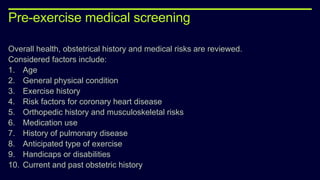
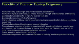
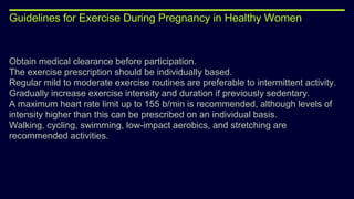
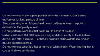
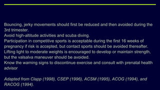
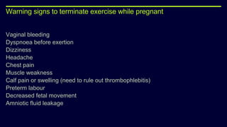
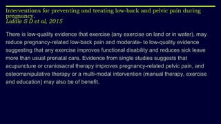
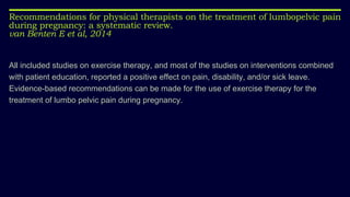
2. OVERVIEW
Physiology of Pregnancy
Physical and physiological changes during each trimester
Guidelines for Exercise in Pregnancy
Evidences for antenatal exercises
Postnatal
Evidences for postnatal exercises
4. STRUCTURAL CHANGES
UTERUS :
Volume- increases gradually. Reaches to about 5-7liters.
Size- Increase in size is due to the hyperplasia and hypertrophy of the myometrium
and due to the growth of the foetus.
Weight- non-pregnant uterus-> 30-50g
end of 3rd trimester- 1,000- 1,200g.
Shape- non-pregnant uterus- pyriform shape.
12th week- globular
3rd trimester- pyriform
5. VAGINA :
increased size
increased blood supply.
Deposition of glycogen increases in the epithelial calls due to the
increased secretion of estrogen.
The pH in vagina becomes less than 3.5
6. CERVIX :
Increase in number of cervical glands.
The blood supply of cervix is increased.
Mucus secretion increased.
The tough cervix becomes soft.
7. FALLOPIAN TUBE :
Hyperplasia of epithelial cells.
Blood supply is increased.
Because of the increase size of uterus, the fallopian tubes are pushed
upwards.
8. OVARIES :
Follicular changes are not seen in ovary and ovulation does not occur.
Corpus luteum enlarges and secretes a large quantity of progesterone
and little estrogen which is essential for maintaining pregnancy.
This continues for 3 months and then corpus luteum degenerates. By this
time placenta develops fully and takes over the function of secreting
estrogen and progesterone.
This continues for the entire term
9. MAMMARY GLANDS :
Changes occur due to the activity of estrogen and progesterone.
Development of new and more ducts.
Formation of more number of alveoli.
Deposition of fat
Increased in size
Increase in vascularization
10. Physiological changes
CARDIOVASCULAR SYSTEM
The blood volume increases by 40%, from 4.0 to 5.5 litres.
greater increase in plasma than in red cells, haemoglobin level falls to
about 80%.
effect is called a dilution anaemia
physiological anaemia of pregnancy
Supine hypotension
Diastolic and systolic blood pressure
Alteration in the venous blood pressure
20% decrease in peripheral resistance
11.
12. RESPIRATORY SYSTEM
Shortness of breath
Increased basal oxygen consumption and minute ventilation
Increase their ventilation by breathing more deeply
Subcoastal angle – 68 to 103 degree
13.
14. IMMUNE SYSTEM
slightly depressed
More prone for pneumococcal pneumonia, influenza or poliomyelitis
Reactivation of latent virus CMV or herpes
Passive antibodies from 6th week of pregnancy to 9 months of age
15. DIGESTIVE SYSTEM
Hyperemesis gravidarum
Esophagus: decreased competence of lower oesophageal sphincter
which leads to heart burn.
Stomach: decreased gastric secretions. Decreased motility leading to
slower emptying times. Progesterone causes decreased tone leading to
nausea.
16.
17.
18. URINARY SYSTEM
Progesterone dilation of renal pelves and ureters
Later, uterus compresses the ureters at the pelvic brim, causing a
slowing of the urine flow.
The musculature of the ureters is slightly hypotonic so they dialate and
also seem to elongate to circumvent the enlarging uterus. This leads to
pooling and stagnation of urine causing a predisposition to urinary tract
infections.
19. MUSCULOSKELETAL SYSTEM
Relaxin, progesterone, oestrogen and cortisols increase in joint laxity
and range.
Postural adaptations that occur at pregnancy, such as a forward shift in
the centre of gravity anterior pelvic tilt increase in lumbar lordosis and
thoracic kyphosis
In fact, 75% of women demonstrate a more posterior posture, one in
which the weight of the uterus is carried posterior to the normal centre of
gravity.
20. Despite major anatomical changes, the kinematics of gait during
pregnancy were found to be remarkably unchanged.
There is a significant increased use of hip extensor, hip abductor and
ankle plantar flexor.
21.
22. NERVOUS SYSTEM
Mood liability, anxiety, insomnia, nightmares, food fads and aversions,
slight reductions in cognitive ability and amnesia
Water retention causes unusual pressure on nerves, particularly those
passing through canals formed of inelastic materials like bone and fibrous
tissue for eg the carpal tunnel resulting in neuropraxia
Occasionally pregnant women complain of traction on nerves which can
be due to increased weight
23. Common discomforts of pregnancy
Urinary frequency
Constipation
Haemorrhoids
Nausea/ vomiting
Heartburn
Fainting
Varicose vein
Vulval varicosities
26. ANTENATAL
The antenatal period is defined as before birth, during or relating to
pregnancy according to the Oxford Dictionary (2014).
Hence, it can be said that the antenatal period would consist of the 40
weeks of gestation endured during a full term pregnancy.
27. ANTENATAL CARE
is a holistic package provided for an expectant mother by healthcare
professionals during the pregnancy.
A series of services will be provided from various healthcare practitioners
such as general practitioners (GP), gynecologists, obstetricians, midwives
and also physiotherapists specializing in women’s health (NHS Choices,
2013).
28. importance of antenatal care
is that it constitutes of health and socioeconomic condition screenings of the
expecting family. It is vital for the following reasons:
To increase the possibility of specific adverse pregnancy outcomes.
To provide effective therapeutic interventions.
To ensure maternal education of planning and going through a safe birth.
To prepare parents of pregnancy emergencies and how to deal with them.
29. ROUTINE
Routine antenatal check up
Antenatal screening
Plan an antenatal care
During the first visit, pregnant woman should be given information regarding:
Folic acid supplements
Food hygiene, including how to reduce the risk of a food-acquired infection
Lifestyle, including smoking cessation and the risks of recreational drug use and
alcohol consumption
Antenatal screening tests
30. During the next visit
pregnant woman should be given information regarding:
How the baby develops during pregnancy
Nutrition and diet, including vitamin D supplements
Exercise, including pelvic floor exercises
Antenatal screening tests
Pregnancy care
Breastfeeding and workshops
Antenatal classes
Maternity benefits.
31. Plan the care that patient will get throughout pregnancy
Identify any occupation related potential risks
Measure height and weight and calculate patient's body mass index (BMI)
Measure blood pressure and test urine for protein
Find out whether patient is at increased risk of gestational diabetes or pre-eclampsia
Ask of any known mental illness and signs of depression
Offer an ultrasound scan at 18 to 20 weeks to check the physical development of the baby.
Antenatal classes can be started as soon as possible, as it will be ease and prepare the
woman for delivery
32. At 16th week
The doctor should give information about the ultrasound scan which will be offered at
18 to 20 weeks and help with any concerns or questions that the patient's have.
Review, discuss and record the results of any screening tests
Measure blood pressure and test patient's urine for protein
Consider an iron supplement if patient is anaemic.
33. 16th – 20th weeks
Ultrasound scan will be done to check the physical development of the baby.
25th weeks
Check abdominal size
Measure blood pressure and test urine for protein.
34. 28th weeks
Check the size of the abdomen
Measure blood pressure and test urine for protein
Offer more blood screening tests
Offer first anti-D treatment if patient is rhesus D-negative.
35. 31st weeks
Review, discuss and record the results of any screening tests from the last
appointment
Check the size of abdomen
Measure blood pressure and test urine for protein.
36. 34th week
Doctor should give patient information about preparing for labour and birth, including
how to recognise active labour, ways of coping with pain in labour and patient's birth
plan.
Review, discuss and record the results of any screening tests from the last
appointment
Check the size of abdomen
Measure blood pressure and test urine for protein
Offer second anti-D treatment if patient is rhesus D-negative.
37. 36th week
Information regarding:
Breastfeeding, including hints and tips for success
Caring for the newborn baby
Vitamin K and screening tests for newborn baby
Patient's own health after the baby is born
Being aware of the ‘baby blues’ and postnatal depression.
Check the size of abdomen
Check the position of the baby and discuss options to turn the baby if he or she is
bottom first (breech position)
Measure blood pressure and test urine for protein.
38. 38th week
Doctor should give patient information about what happens if pregnancy lasts longer
than 41 weeks.
Check the size of abdomen
Measure blood pressure and test urine for protein.
40 weeks
Doctor should give patient information about what happens if pregnancy lasts longer
than 41 weeks.
Check the size of abdomen
Measure blood pressure and test urine for protein.
39. 41st weeks
Check the size of abdomen
Measure blood pressure and test urine for protein
Offer induction of labour
40. Pre-exercise medical screening
Overall health, obstetrical history and medical risks are reviewed.
Considered factors include:
1. Age
2. General physical condition
3. Exercise history
4. Risk factors for coronary heart disease
5. Orthopedic history and musculoskeletal risks
6. Medication use
7. History of pulmonary disease
8. Anticipated type of exercise
9. Handicaps or disabilities
10. Current and past obstetric history
41. Benefits of Exercise During Pregnancy
Maintain healthy body weight and avoid excess fat accumulation
Maintain or improve cardiovascular fitness, muscular strength and endurance, and flexibility
Decreased musculoskeletal complaints such as back pain
Decreased minor discomforts of pregnancy
Improved posture and body mechanics, which may improve coordination, balance, and body
awareness
Reinforced principles of breath awareness and relaxation
Prevention and treatment of problems associated with gestational diabetes, hypertension,
and preeclampsia (hypertension due to (cause being placenta) 20 weeks later, protein in
urine , eclampsia - with seizures (magnesium sulphate)
Stress reduction and enhanced self-image
Possible easing of labor with fewer complications of delivery and faster postnatal recovery
42. Guidelines for Exercise During Pregnancy in Healthy Women
Obtain medical clearance before participation.
The exercise prescription should be individually based.
Regular mild to moderate exercise routines are preferable to intermittent activity.
Gradually increase exercise intensity and duration if previously sedentary.
A maximum heart rate limit up to 155 b/min is recommended, although levels of
intensity higher than this can be prescribed on an individual basis.
Walking, cycling, swimming, low-impact aerobics, and stretching are
recommended activities.
43. Do not exercise in the supine position after the 4th month. Don't stand
motionless for long periods of time.
Stop exercising when fatigued and do not deliberately reach a point of
exhaustion. Get plenty of rest.
Do not perform exercises that could cause a loss of balance.
Eat an additional 150–300 calories a day and drink plenty of fluids before,
during, and after exercise. Emphasize complex carbohydrates to replace
muscle glycogen stores.
Do not exercise when it is hot or humid or when febrile. Wear clothing that is
cool and allows ventilation.
44. Bouncing, jerky movements should first be reduced and then avoided during the
3rd trimester.
Avoid high-altitude activities and scuba diving.
Participation in competitive sports is acceptable during the first 16 weeks of
pregnancy if risk is accepted, but contact sports should be avoided thereafter.
Lifting light to moderate weights is encouraged to develop or maintain strength,
but the valsalva maneuver should be avoided.
Know the warning signs to discontinue exercise and consult with prenatal health
advisor
Adapted from Clapp (1998), CSEP (1996), ACSM (1995), ACOG (1994), and
RACOG (1994).
45. Warning signs to terminate exercise while pregnant
Vaginal bleeding
Dyspnoea before exertion
Dizziness
Headache
Chest pain
Muscle weakness
Calf pain or swelling (need to rule out thrombophlebitis)
Preterm labour
Decreased fetal movement
Amniotic fluid leakage
46. Absolute Contraindications
Severe cardiovascular, respiratory or systemic disease
Uncontrolled hypertension, diabetes or thyroid disease
Ruptured membranes
Incompetent cervix
Preeclampsia or toxaemia
Multiple pregnancy (triplets, etc.)??
Poor fetal growth
Previous uterine rupture
47. Relative Contraindications
History of repeated (3 or more) miscarriage or premature labor
Diabetes
History of rapid labor or poor fetal growth
Early pregnancy bleeding
Sedentary lifestyle with very poor fitness
Breech presentation after 28 weeks
Palpitations or arrhythmias
Anemia or iron deficiency
Extreme under or over weight
48. 1st Trimester - 0-12 weeks
AIMS AND OBJECTIVES:
1. Antenatal Education
2. Maintenance of Anaerobic and Aerobic capacity – antenatal exercises.
3. Nutritional Education
4. Ergonomic/Body mechanic
53. 2nd Trimester - 13- 24 week
Continuation of first trimester exercises
Ergonomic advises
Core stabilization exercises
Aerobic exercises
54.
55.
56.
57. 3rd Trimester - 25- 38weeks
Progression of exercises.
Aiming to treat Neuromusculoskeletal discomforts.
Ergonomic advise
Education on Stages of Delivery
Teach and train Positions for Delivery
Teach relaxation techniques to use during
pregnancy.
59. Absolute contraindications to aerobic exercise during pregnancy
Haemodynamically significant heart disease
Restrictive lung disease
Incompetent cervix/cerclage
Multiple gestation at risk for premature labour
Persistent second or third trimester bleeding
Placenta praevia after 26 weeks gestation
Premature labour during the current pregnancy
Ruptured membranes
Pregnancy induced hypertension
60. Relative contraindications to aerobic exercise during pregnancy
Unevaluated maternal cardiac arrhythmia
Chronic bronchitis
Poorly controlled type I diabetes
Extreme morbid obesity
Extreme underweight (body mass index <12)
History of extremely sedentary lifestyle
Intrauterine growth restriction in current pregnancy
Poorly controlled hypertension/pre-eclampsia
Orthopaedic limitations
Poorly controlled seizure disorder
Poorly controlled thyroid disease
Heavy smoker
Severe anaemia
61. Pregnant women should be encouraged to perform combined ( aerobic + resistance )
exercise to improve an important health outcome as in cardiorespiratory fitness and
urinary incontinence.
Benefits of aerobic or resistance training during pregnancy on maternal health
and perinatal outcomes: A systematic review.
Maria Perales et al, 2016
62. Effects of aerobic exercise training on maternal and neonatal outcome: a randomized controlled trial on
pregnant women in Iran
Zahra ghodsi et al , 2014
Exercising on a bicycle ergometer during pregnancy seems to be safe for the mother
and the neonate
63. Aerobic exercise for women during pregnancy
Kramer M S et al, 2006
Regular aerobic exercise during pregnancy appears
to improve (or maintain) physical fitness.
This review of 14 trials involving 1014 pregnant
women, found that pregnant women who engage in
vigorous exercise at least two or three times per
65. Treatments for pregnancy-related lumbopelvic pain: a systematic review of physiotherapy modalities
Gutke A et al, 2015
For lumbopelvic pain during pregnancy, the evidence was strong for positive
effects of acupuncture and pelvic belts.
The evidence was low for exercise in general and for specific stabilizing
exercises.
The evidence was very limited for efficacy of water gymnastics, progressive
muscle relaxation, a specific pelvic tilt exercise, osteopathic manual therapy,
craniosacral therapy, electrotherapy and yoga.
For postpartum lumbopelvic pain, the evidence was very limited for clinic-
based treatment concepts, including specific stabilizing exercises, and for
self-management interventions for women with severe disabilities.
67. Interventions for preventing and treating low-back and pelvic pain during
pregnancy.
Liddle S D et al, 2015
There is low-quality evidence that exercise (any exercise on land or in water), may
reduce pregnancy-related low-back pain and moderate- to low-quality evidence
suggesting that any exercise improves functional disability and reduces sick leave
more than usual prenatal care. Evidence from single studies suggests that
acupuncture or craniosacral therapy improves pregnancy-related pelvic pain, and
osteomanipulative therapy or a multi-modal intervention (manual therapy, exercise
and education) may also be of benefit.
68. Recommendations for physical therapists on the treatment of lumbopelvic pain
during pregnancy: a systematic review.
van Benten E et al, 2014
All included studies on exercise therapy, and most of the studies on interventions combined
with patient education, reported a positive effect on pain, disability, and/or sick leave.
Evidence-based recommendations can be made for the use of exercise therapy for the
treatment of lumbo pelvic pain during pregnancy.
70. Effect of pelvic floor muscle training during pregnancy and after childbirth on prevention
and treatment of urinary incontinence: a systematic review.
Morkved S et al,2014
22 randomised or quasiexperimental trials
PFMT during pregnancy and after delivery can prevent and treat UI.
A supervised training protocol following strength-training principles, emphasising close to
maximum contractions and lasting at least 8 weeks is recommended.
PFMT is effective when supervised training is conducted.
Given the prevalence of female UI and its impact on exercise participation, PFMT should be
incorporated as a routine part of women's exercise programmes in general.
71. Effect of Kegel exercise to prevent urinary and fecal incontinence in antenatal
and postnatal women: systematic review
Park S H et al, 2013
Kegel exercise can prevent urinary and fecal
incontinence.
Therefore, a priority task is to develop standardized
Kegel exercise programs for Korean pregnant and
postpartum women and make efficient use of these
programs.
72. Pelvicfloor muscle training for prevention and treatment of urinary and fecal incontinence in antenatal
and postnatal women: a short version Cochrane review.
Boyle R et al, 2014
For women who are continent during pregnancy, PFMT may
prevent urinary incontinence up to 6 months after delivery.
The extent to which
mixed prevention and treatment approaches to PFMT in the
postnatal period are effective is less clear that is, offering
advice on PFMT to all pregnant or postpartum women
whether they have incontinence symptoms or not.
74. Diet and exercise interventions for preventing gestational diabetes mellitus.
Bain E et al, 2015
Results from 13 RCTs (of moderate quality)
suggest no clear difference in the risk of
developing GDM for women receiving a
combined diet and exercise intervention compared
with women receiving no intervention.
75. Exercise for pregnant women for preventing gestational diabetes mellitus
Han S et al, 2015
77. 3 phases of postnatal rehabilitation
Phase 1: First 8weeks
Phase 2: 8-12 weeks
Phase 3: After 12 weeks
Postnatal Status:
Normal Delivery
Forceps Delivery
CS
78. Stage 1 : 6 to 8 weeks
NORMAL DELIVERY
Breathing exercises
Relaxation exercises
Epidural pain relief
Isometrics
Pelvic floor exercises
• Chest care
• Limb movements
• DVT
• Post surgical pain
• Relaxation techniques
• Pelvic floor ex
79. Stage 2 and 3
Warm up exercises
Mild aerobic training
FITT principle
Moderate aerobic exercise
80. AMERICAN COLLEGE OF OBSTETRICIANS AND
GYNECOLOGIST
Type Mode Intensity Frequency Duration Progression
Aerobic
walking, aerobic
dance, swimming,
cycling.
Moderate 3-4 METS.
50-60% VO2 max.
RPE: 12-16
30 minutes per day
most days of the
week.
20-60 minutes as
per patient
tolerance.
Increase exercise
duration slowly.
Flexibility
Aquatic exercise
ROM ex’s full
mobility
Strength
Light
weights/increased
reps.
12 reps, individually
tailored with
monitoring.
81. REFERENCES
Vairajothi K, Chitra TV, Baranitharan R, Mahalakshmi V. A comparative study of the
therapeutic effect of pelvic floor exercises and perineometer among women with
urinary stress Incontinence. IJOPT, 2005, volume 5; number 1, pg 33-36.
Amar TA. Stabilization exercises in postnatal low back pain. Indian Journal of
Physiotherapy and Occupational Therapy. 2011, Vol. 5, No.1.
Mantle J et al. Physiotherapy in Obstetrics & Gynaecology, 2nd edition.
Ferreira CWS, Alburquerque-Sendn F. Effectiveness of physical therapy for
pregnancy-related low back and/or pelvic pain after delivery: A systematic Review.
Physiotherapy Theory and Practice, 2012; 1-3. (published online).
82. Stuge B, Lærum E , Kirkesola G, Vøllestad P. The Efficacy of a Treatment Program
Focusing on Specific Stabilizing Exercises for Pelvic Girdle Pain After Pregnancy A
Randomized Controlled Trial. SPINE Volume 29, Number 4, pp 351–359, 2004