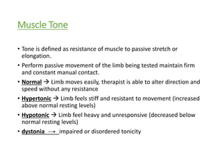
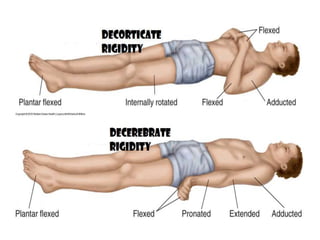
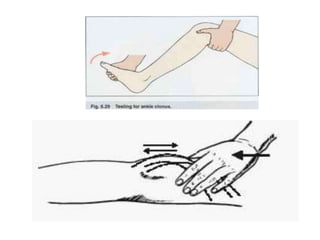
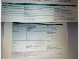
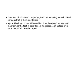
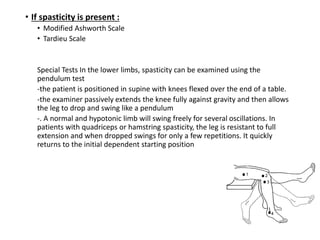
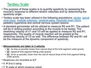
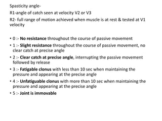
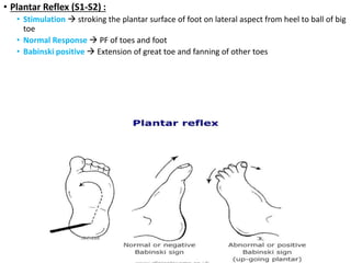
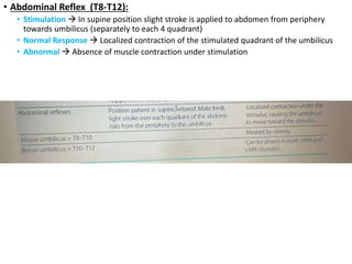
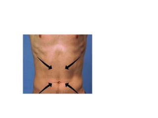
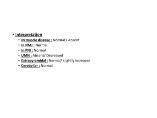
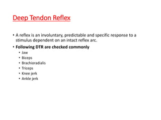
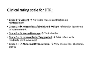
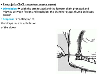
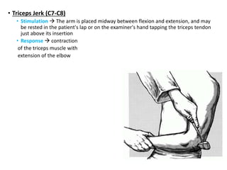
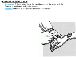
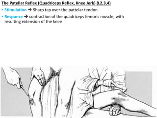
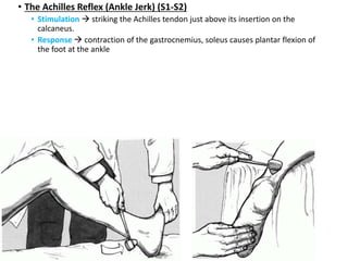
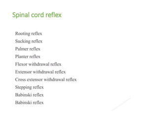
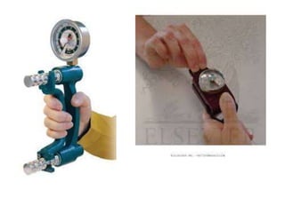
3. Muscle Tone
• Tone is defined as resistance of muscle to passive stretch or
elongation.
• Perform passive movement of the limb being tested maintain firm
and constant manual contact.
• Normal Limb moves easily, therapist is able to alter direction and
speed without any resistance
• Hypertonic Limb feels stiff and resistant to movement (increased
above normal resting levels)
• Hypotonic Limb feel heavy and unresponsive (decreased below
normal resting levels)
• dystonia impaired or disordered tonicity
5. Spasticity Rigidity
Increased muscle tone (hypertonia) Increased muscle tone (hypertonia)
Motor disorder characterized by resistance catch and release,
mid range or end range.
Hypertonic state characterized by resistance throughout
ROM.
Velocity dependent
As speed of passive movement is increased increase in
resistance to passive movement
Velocity independent
Resistance to passive movement is constant
Lesion of Pyramidal tracts (corticospinal tract) Lesion of extrapyramial system
Only one group of muscles are affected
(In chronic stages both agonist and antagonist are affected
but one group is always stronger)
Both agonist and antagonist muscle have increased tone
(equal variation)
6. Rigidity types :
1. Lead pipe rigidity – constant resistant throughout range
2. Cog wheel rigidity – Alternate letting go & increasing
resistance to movement
3. Decorticate rigidity – trunk and LL in extension & UL in
flexion (corticospinal tract at level of diencephalon
above superior coliculus)
4. Decerebrate rigidity – trunk and limb in full extension
(corticospinal lesion in brainstem between superior
colliculus & vestibular nucleus)
Additional signs include :
Clonus : Rhythmical contraction of muscle or group of
muscle in response to sudden, passive and sustained
stretching of spastic muscle
Ankle clonus-
Jaw clonus
Wrist clonus
Additional signs include :
Tremor
Bradykinesia
Postural instability
7.
8.
9.
10. • Dystonia :
• Prolonged involuntary movement disorder characterized by twisting repetitive
movements and increased muscle tone
• Hypotonia/flaccidity :
• Decreased or absent muscle tone
• Resistance to passive movement is diminished
• Stretch reflex diminished or absent
• Hyperextensibility of joints is common. & limbs are easily moved(floppy)
• Seen in LMN lesion
• Acute UMN lesion produces temporary hypotonia : known as spinal shock or cerebral
shock
• Which is commonly followed by Typical UMN signs
11. • Comparison should made between upper and lower extremity and
right and left extremities.
• Symmetrical/asymmetrical
12. EXAMINATION OF TONE
• An examination of tone consists of- (1) initial observation of resting posture and
palpation
(2) passive motion testing, and
(3) active motion testing.
• Initial observation- of the patient can reveal abnormal posturing of the limbs or
body. Careful inspection should be made regarding the position of the limbs,
trunk, and head.
• Palpation- of the muscle belly may yield additional information about the resting
state of muscle. Consistency, firmness, and turgor should all be examined.
Hypotonic muscles will feel soft and flabby, whereas hypertonic muscles will feel
taut and harder than normal.
• Passive motion testing reveals information about the responsiveness of muscles
to stretch. Because these responses should be examined in the absence of
voluntary control, the patient is instructed to relax, letting the therapist support
& move the limb
13.
14. • Clonus- a phasic stretch response, is examined using a quick stretch
stimulus that is then maintained
• eg- ankle clonus is tested by sudden dorsiflexion of the foot and
maintaining the foot in dorsiflexion. he presence of a clasp-knife
response should also be noted
15. Clinical rating scale for tone
• 0 No response (Flaccidity)
• 1+ Decreased response (Hypotonia)
• 2+ Normal response
• 3 + Exaggerated response (mild o moderate hypertonia)
• 4+ Sustained response (Severe hypertonia)
16. • If spasticity is present :
• Modified Ashworth Scale
• Tardieu Scale
Special Tests In the lower limbs, spasticity can be examined using the
pendulum test
-the patient is positioned in supine with knees flexed over the end of a table.
-the examiner passively extends the knee fully against gravity and then allows
the leg to drop and swing like a pendulum
-. A normal and hypotonic limb will swing freely for several oscillations. In
patients with quadriceps or hamstring spasticity, the leg is resistant to full
extension and when dropped swings for only a few repetitions. It quickly
returns to the initial dependent starting position
17. Modified Ashworth Scale
0 No increase in muscle tone
1 Slight increase in muscle tone, manifested by a catch and release or
by minimal resistance at the end of the range of motion when the
affected part(s) is moved in flexion or extension
1+ Slight increase in muscle tone, manifested by a catch, followed by
minimal resistance throughout the remainder (less than half) of the
ROM
2 More marked increase in muscle tone through most of the ROM,
but affected part(s) easily moved
3 Considerable increase in muscle tone, passive movement difficult
4 Affected part(s) rigid in flexion or extension
18. Tardieu Scale
• The purpose of these scales is to quantify spasticity by assessing the
muscle's response to different stretch velocities and by determining the
spasticity angle .
• Tardieu scale has been utilized in the following populations: stroke, spinal
cord injury, multiple sclerosis, cerebral palsy, traumatic brain injury,
pediatric hypertonia and central nervous system lesions
• A standard goniometer will be utilized to measure R2 and R1. The patient
will be in testing position according to the muscle to be tested. The
stretching velocity of V1 and V3 will be applied to measure R2 and R1,
respectively. The quality of muscle reaction will be graded at the
stretching velocity of V3 as well. The difference between R2 and R1 will
be the measure of the dynamic component of spasticity.
• Measurements are taken in 3 velocity
• V1 : As slow as possible slower than natural drop of the limb segment under gravity
• V2 : Speed of limb segment falling under gravity
• V3 : As far as possible, faster than the rate of natural drop of the limb segment falling
under gravity
• Responses are recorded as X/Y
• X 0 to 5 rating
• Y angle at which response noted
19. Spasticity angle-
R1-angle of catch seen at velocity V2 or V3
R2- full range of motion achieved when muscle is at rest & tested at V1
velocity
• 0 :- No resistance throughout the course of passive movement
• 1 :- Slight resistance throughout the course of passive movement, no
clear catch at precise angle
• 2 :- Clear catch at precise angle, interrupting the passive movement
followed by release
• 3 :- Fatigable clonus with less than 10 sec when maintaining the
pressure and appearing at the precise angle
• 4 :- Unfatiguable clonus with more than 10 sec when maintaining the
pressure and appearing at the precise angle
• 5 :- Joint is immovable
22. • Plantar Reflex (S1-S2) :
• Stimulation stroking the plantar surface of foot on lateral aspect from heel to ball of big
toe
• Normal Response PF of toes and foot
• Babinski positive Extension of great toe and fanning of other toes
23. • Abdominal Reflex (T8-T12):
• Stimulation In supine position slight stroke is applied to abdomen from periphery
towards umbilicus (separately to each 4 quadrant)
• Normal Response Localized contraction of the stimulated quadrant of the umbilicus
• Abnormal Absence of muscle contraction under stimulation
24.
25. • Interpretation
• IN muscle disease : Normal / Absent
• In NMJ : Normal
• In PNI : Normal
• UMN : Absent/ Decreased
• Extrapyramidal : Normal/ slightly increased
• Cerebellar : Normal
26. Deep Tendon Reflex
• A reflex is an involuntary, predictable and specific response to a
stimulus dependent on an intact reflex arc.
• Following DTR are checked commonly
• Jaw
• Biceps
• Brachioradialis
• Triceps
• Knee jerk
• Ankle jerk
27. Clinical rating scale for DTR :
• Grade 0 Absent No visible muscle contraction on
reinforcement
• Grade 1+ Hyporeflexia/diminished Slight reflex with little or no
joint movement.
• Grade 2+ Normal/average Typical reflex
• Grade 3+ Hyperreflexia/Exaggerated Brisk reflex with
moderate joint movement
• Grade 4+ Abnormal (hyperreflexia) Very brisk reflex, abnormal,
clonus
28. • Biceps jerk (C5-C6 musculocutaneous nerve)
• Stimulation With the arm relaxed and the forearm slight pronated and
midway between flexion and extension, the examiner places thumb on biceps
tendon
• Response contraction of
the biceps muscle with flexion
of the elbow
29. • Triceps Jerk (C7-C8)
• Stimulation The arm is placed midway between flexion and extension, and may
be rested in the patient's lap or on the examiner's hand tapping the triceps tendon
just above its insertion
• Response contraction
of the triceps muscle with
extension of the elbow
30. • Brachiradialis reflex (C5-C6)
• Stimulation Tapping just above the styloid process of the radius with the
forearm in semiflexion and semipronation
• Response flexion of the elbow, with variable supination
31. The Patellar Reflex (Quadriceps Reflex, Knee Jerk) (L2,3,4)
• Stimulation Sharp tap over the pattelar tendon
• Response contraction of the quadriceps femoris muscle, with
resulting extension of the knee
32. • The Achilles Reflex (Ankle Jerk) (S1-S2)
• Stimulation striking the Achilles tendon just above its insertion on the
calcaneus.
• Response contraction of the gastrocnemius, soleus causes plantar flexion of
the foot at the ankle
33. Interpretation :
• Muscle pathology : Decreased parallel to reduced muscle strength
• NMJ : Normal to decreased parallel to reduced muscle strength
• Peripheral nerve : Diminished or absent
• Spinal cord : hypoactive at level of lesion and hyperactive below the
level of lesion
• Cortico spinal tract : Hyperactive on contralateral side
• Extrapyramidal : Usually Normal
• Cerebellar : pendular
34. Primitive & tonic reflex
• Primitive and tonic reflexes are present during infancy as a stage in normal
development and become integrated by the CNS at an early age
• Patients who exhibit these reflexes typically present with extensive brain
damage (e.g., stroke, TBI) and other UMN signs
• CLASSIFICATION-
Automatic reflexes
Spinal reflexes
Brain stem reflexes
Midbrain reflexes
Cortical reflexes
42. Sucking reflex
TESTING POSITION: supine lying
STIMULUS: initiates when roof of an infant's mouth is touche the nipple or
finger
RESPONSE: infant will automatically start sucking the nipple or finger>
ONSET: birth(32 weeks of pregnancy)
INTEGRATED: 4 months
43.
44.
45. • Flexor withdrawal
Stimulus- Noxious stimulus (pinprick) to sole of foot. Tested in supine
or sitting position
Response- Toes extend, foot dorsiflexes, entire LE flexes uncontrollably.
Onset: 28 weeks of gestation.
Integrated: 1–2 months
60. Cortical reflex
They are highly integrated & complex mechanism which are automatic response to
changes in posture & movement aimed to restore balance
61.
62. Muscle bulk
• Atrophy the loss of muscle bulk (wasting), occurs as a result of the loss of
functional mobility (disuse atrophy), LMN disease (neurogenic atrophy), or
protein-calorie malnutrition
• Disuse atrophy is evident after periods of inactivity, developing in weeks or
months. It is generally widespread and affects antigravity muscles to a
greater extent.
• Neurogenic atrophy accompanies LMN injury (e.g., peripheral nerve injury,
spinal root injury) and occurs rapidly, generally within 2 to 3 weeks. Atrophy
is also accompanied by other signs of LMN injury (e.g., decreased or absent
tone or decreased or absent DTRs, fasciculations, weak or absent voluntary
movement
Examination of Muscle Bulk- the therapist should visually inspect the muscle
symmetry and shapes, comparing and contrasting their size and contour
• Muscles that look flat or concave are indicative of atrophy. Comparisons
should be made between and within limbs. Is the atrophy unilateral or
bilateral?
• Palpation at rest and during muscle contraction is used to determine muscle
tension. Girth measurements or volumetric displacement measures (e.g.,
hands or feet) can be used to confirm visual inspection findings.
63. Muscle Strength Examination
• MMT of b/l UL, LL and trunk
• Performed if VC grading is > or equal to 4
• Grip strength and pinch strength assessment by dynamometer
64.
65. • In patients with peripheral sensorimotor neuropathy (e.g., chronic diabetic
neuropathy) or acute motor neuropathy (e.g., Guillain-Barré), strength losses
are typically greater in distal segments (i.e., foot and ankle) than proximal
with involvement of more proximal segments as the disease progresses
• Documentation of strength and power changes should include a
determination of the specific muscles and body segments tested and tests
used; the type and degree of changes present (e.g., paresis, paralysis)
66. Voluntary Control Assessment (brunnstrom
approach)
• Criteria for voluntary control assessment
• Presence of spasticity
• Positive babinski’s sign
• Presence of synergy patterns
• Quality of performance in any position is the criteria of scoring i.e. the degree of
isolation
• Mid range of motion is usually selected as it is the most functional range &
easiest for a muscle group to work
• Chosen position must be in antigravity position to assess the muscle power of
function
67. Voluntary control grades
• 0 –no contraction
• 1- flicker of contraction
• 2- half range of motion in pattern
• 3- full range of motion in pattern
• 4- initial half in isolation and latter half in pattern
• 5-full ROM in isolation but goes in pattern on resistance
• 6-full ROM in isolation with resistance
Editor's Notes
Tone is influenced by no of factors includes – 1) physical inertia 2) intrinsic mechanical elastic stiffness of muscle & connective tissue 3) spinal reflex muscle contraction
Spasticity velocity dependent -the larger & quicker the stretch the stronger the resistance of the spastic muscle during rapid movement , initial high resistance (spastic catch) maybe followed by a sudden inhibition or letting go of the limb relaxation in response to a stretched stimulus – clasp knife response
CHRONIC SPASTICITY- associated with contracture ,abnormal posturing & deformity , functional limitation & disability
LESION- arises from injury to descending motor pathways from cortex (pyramidal) or brainstem (medial & lateral vestibulospinal tracts, reticulospinal tract) producing disinhibition of spinal reflex with hhyperactive tonic stretch reflex or a failure of reciprocal inhibition the result is HYPEREXCITABILITY OF ALPHA MOTOR NEURON POOL, occurs as part of UMN syndrome
RIGIDITY- lesion- basal ganglia (extrapyramida;) usually it is seen in parkinsons disease
decorticate
Severe brain injury can result in coma with decorticate or decerebrate rigidity
Decorticate rigidity refers to sustained contraction and posturing of the upper limbs in flexion and the lower limbs in extension. he elbows, wrists, and fingers are held in flexion with shoulders adducted tightly to the sides while the legs are held in extension, internal rotation, and plantarflexion
. Decerebrate rigidity (abnormal extensor response) refers to sustained contraction and posturing of the trunk and limbs in a position of full extension. he elbows are extended with shoulders adducted, forearms pronated, and wrist and fingers flexed. he legs are held in stiff extension with plantarflexion.
Dystonic posturing refers to sustained abnormal postures caused by co-contraction of muscles that may last for several mins for hours, or permanently
Hypotonia -results from lesions that affect the anterior horn cell and peripheral nerve (e.g., peripheral neuropathy, cauda equina lesion, radiculopathy).
-It produces symptoms of decreased or absent tone, decreased or absent reflexes, paresis, muscle fasciculations and fibrillations with denervation, and neurogenic atrophy. Mild decreases in tone along with asthenia (weakness) can also be seen in cerebellar lesions
-Acute UMN lesions (e.g., hemiplegia, tetraplegia, paraplegia) can produce temporary hypotonia, termed spinal shock or cerebral shock depending on the location of the lesion.
the duration of CNS depression and hypotonia that occurs with shock is highly variable, lasting days or weeks. It is typically followed by the development of spasticity and classic UMN signs
-
For example, patients with spasticity can vary in their presentation from morning to afternoon, day to day, or even hour to hour depending on a number of factors, including (1) volitional effort and movement, (2) anxiety and pain, (3) position and interaction of tonic reflexes, (4) medications, (5) general health, (6) ambient temperature, and (7) state of CNS arousal or alertness. In addition, urinary bladder status (full or empty), fever and infection, and metabolic and/or electrolyte imbalance can also influence tone he therapist should therefore consider the impact of each of these factors in arriving at a determination of tone.
Passive- During a passive motion test, the therapist should maintain firm and constant manual contact, moving the limb in all motions. When tone is normal, the limb moves easily and the therapist is able to alter direction and speed without feeling abnormal resistance. he limb is responsive and feels light. Hypertonic limbs generally feel stiff and resistant to movement, whereas flaccid limbs feel heavy and unresponsive
All limbs and body segments are examined, All limbs and body segments are examined,
. he pendulum test can be quantified using an isokinetic dynamometer, an electrogoniometer, or computerized video equipment with high test–retest reliability.27,28
A normal response consists of flexion of the big toe; sometimes the other toes will demonstrate a downgoing (flexion) response, or no response at all. An abnormal response (positive Babinski sign) consists of extension dorsiflexion (upgoing) of the big toe, with fanning of the lateral four toes. It is indicative of a corticospinal (UMN) lesion.
Nueromuscular junction
peripheralnerveinjury
DTRs are increased in UMN syndrome (e.g., stroke) and decreased in LMN syndrome (e.g., peripheral neuropathy, nerve root compression), cerebellar syndrome, and muscle disease
-If DTRs are difficult to elicit, responses can be enhanced by specific reinforcement maneuvers. In the Jendrassik maneuver, the patient hooks together the fingers of the hands and strongly pulls them apart. While this pressure is maintained, LE reflexes are tested.
-Maneuvers that can be used to reinforce responses in the upper extremities (UEs) include squeezing the knees together, clenching the teeth, or making a fist with the contralateral extremity
If persistent- problem with head control & sitting
May cause delay of symmetrical trunk stability & difficulty with walking standing & sitting
If persistent- interfere with normal tounge movement which later cause speech problems
If persistent- difficulty in reaching for and grasping object once grasp will have difficulty in releasing & weight bearing on open hand
If perisitent- toe clawing inability to stand flat decreased balance decreased gait ability
Delay in ability to stand
If persistent – motor difficulty in rolling & sitting
Patients with stroke demonstrate significant changes in muscle performance, including altered recruitment patterns, abnormal times to achieve force, and decreased motor unit firing rates.34-36 hey also demonstrate up to a 50% decrease in motor units of affected extremities within 2 months after insult with greater losses of Type II (fast twitch) fibers.37,38 Impairments in grip strength impairments are observed, including an exaggeration of grip force, altered times to achieve grip, and difficulty maintaining grip.39 Muscle performance in patients with stroke is influenced by the presence of other UMN impairments including spasticity, disordered synergistic activity/mass patterns of movements, abnormal muscle co-contraction, and/or profound sensory deficit.40-42
Strength testing measures (MMT) were originally developed to examine motor function in patients with polio (an LMN disease). There are validity issues when used in the clinical examination of patients with UMN lesions
isokinetic protocols, may not be possible in the presence of UMN lesion where stereotypic abnormal movement patterns (obligatory synergies) are present.
2-(e.g., myopathies) typically experience proximal weakness whereas patients with myasthenia gravis experience decremental strength losses. hus, the first contraction of a muscle may start out strong and then each succeeding contraction gets weaker and weaker
4-whether the changes are symmetrical or asymmetrical, distal or proximal; presence of associated signs (e.g., UMN or LMN); presence of atrophy; and factors that modify muscle performance
The principles of Movement therapy are as follows :-
Treatment progresses in a developmental sequence from reflexes to voluntary to functional movements.
When no motion exists, movement can be facilitated using reflexes, associated reactions, proprioceptive/exteroceptive stimuli to develop muscle tension.
Reflex and associated reactions are combined with voluntary effort which produces semi-voluntary movement thus providing sensory feedback and satisfaction.
Various stimuli given assist in eliciting movement. Resistance (proprioceptive stimuli) promotes a spread of impulses to other muscles to produce an associated reaction whereas a tactile stimuli facilitates muscles only to stimulated area.
The patient is asked to hold (isometric) a response if voluntary effort is done. If possible, he is asked for an eccentric (controlled lengthening) followed by concentric (controlled shortening) contraction.
Even if a partial movement is possible, reversal of movement is stressed within each session.
Facilitation techniques are dropped a soon as the patient shows voluntary control. Responses to exteroceptive stimuli are least stereotyped, so tactile stimuli are last to be eliminated. No primitive reflexes are used beyond stage 3.
Emphasis is more on voluntary movement and similar ADL's are encouraged to perform.
Correct movement, once elicited, should be repeated and practiced.