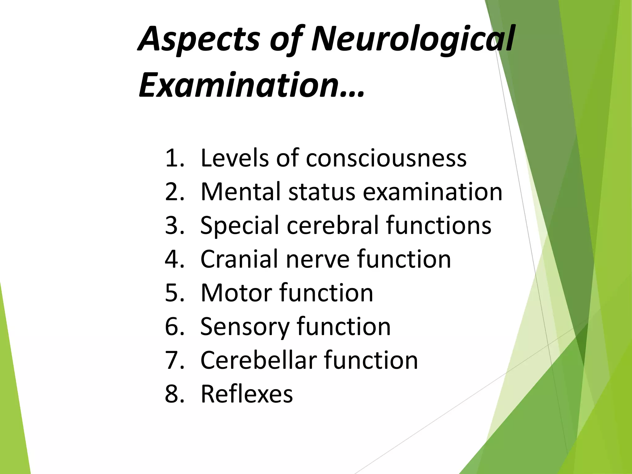
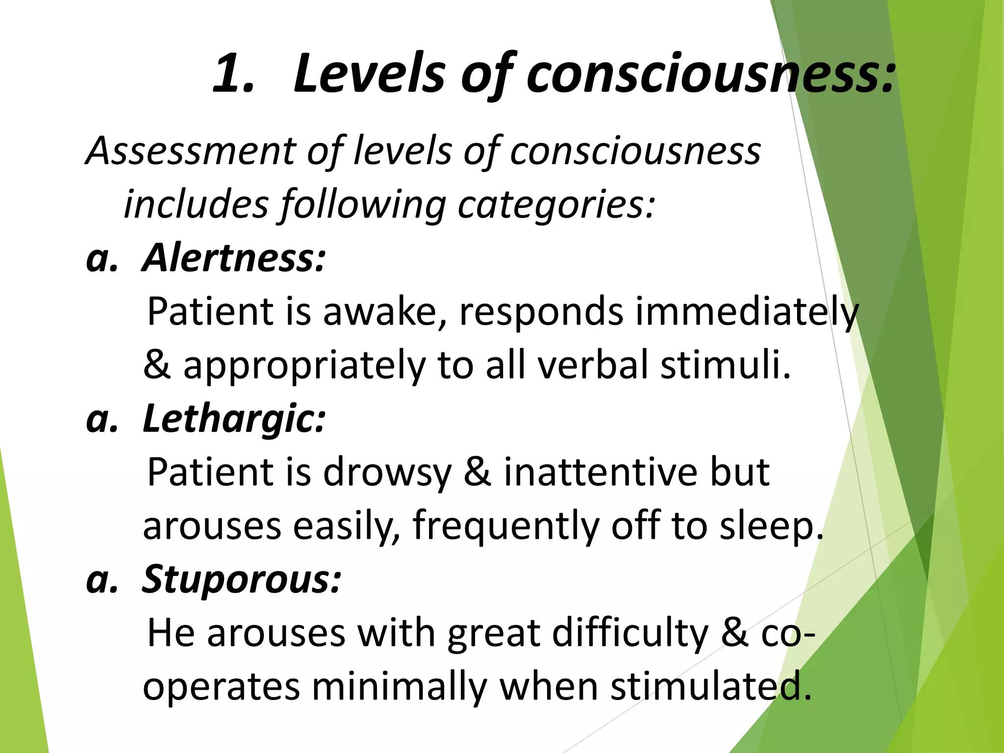
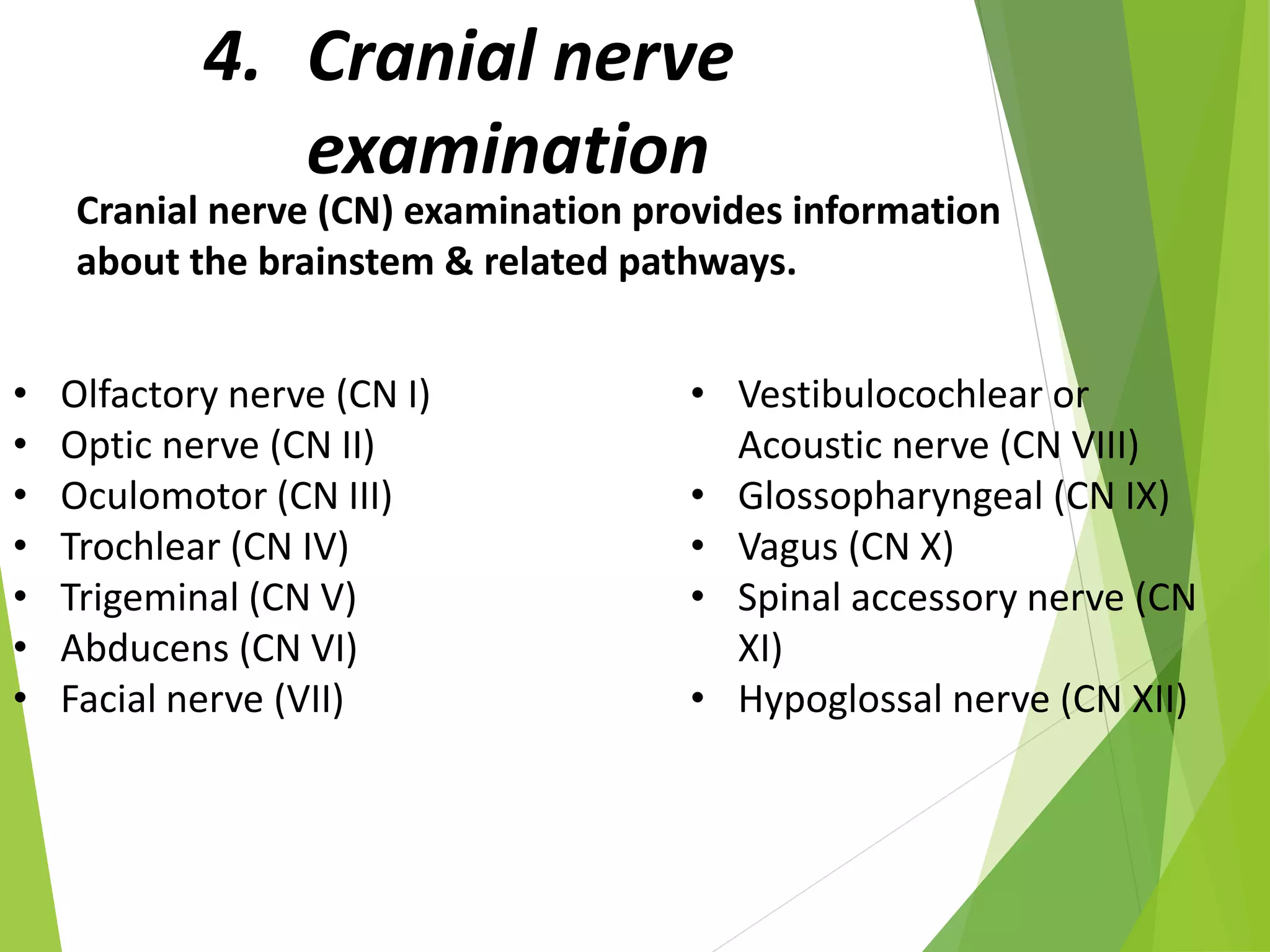
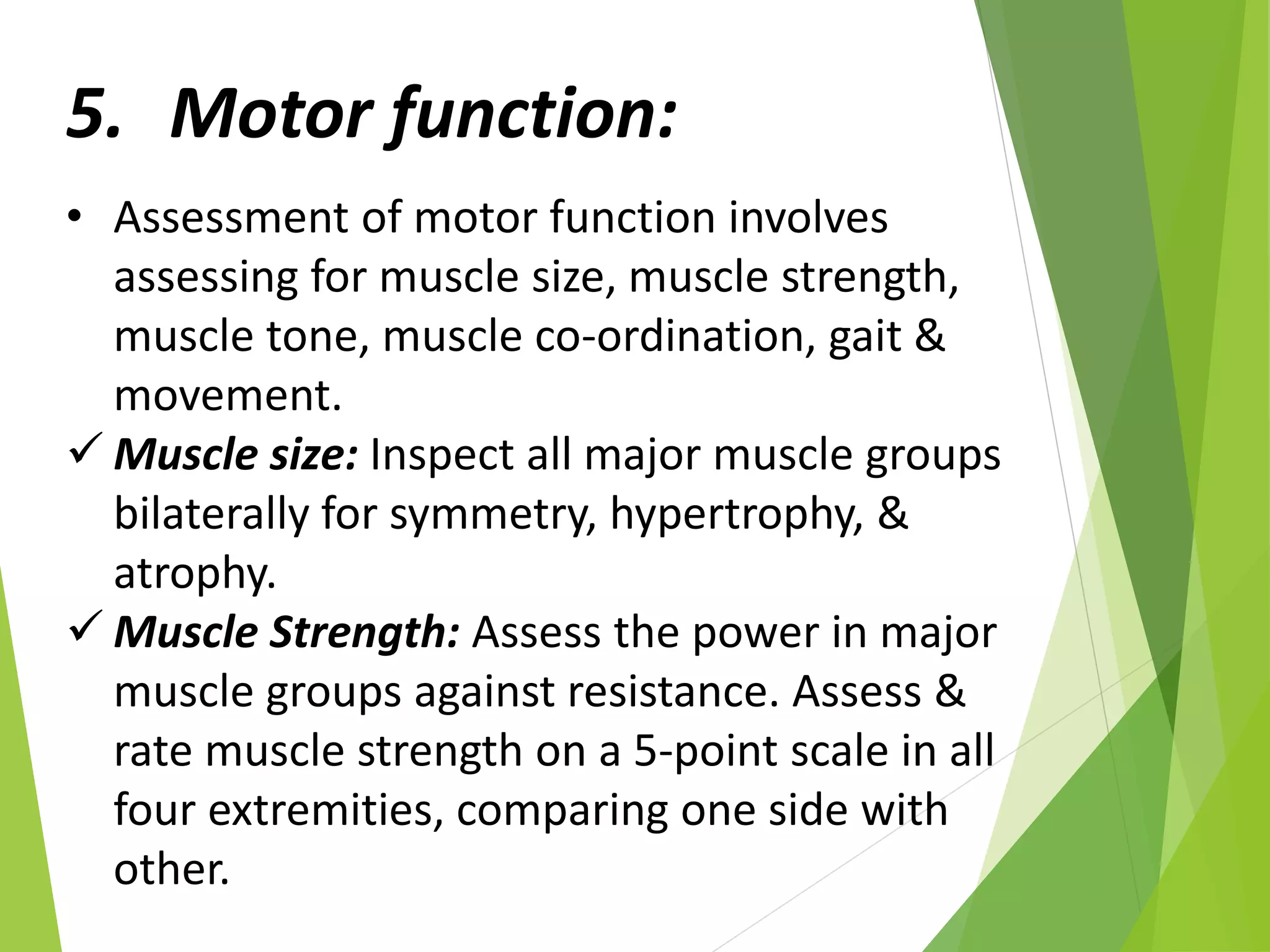
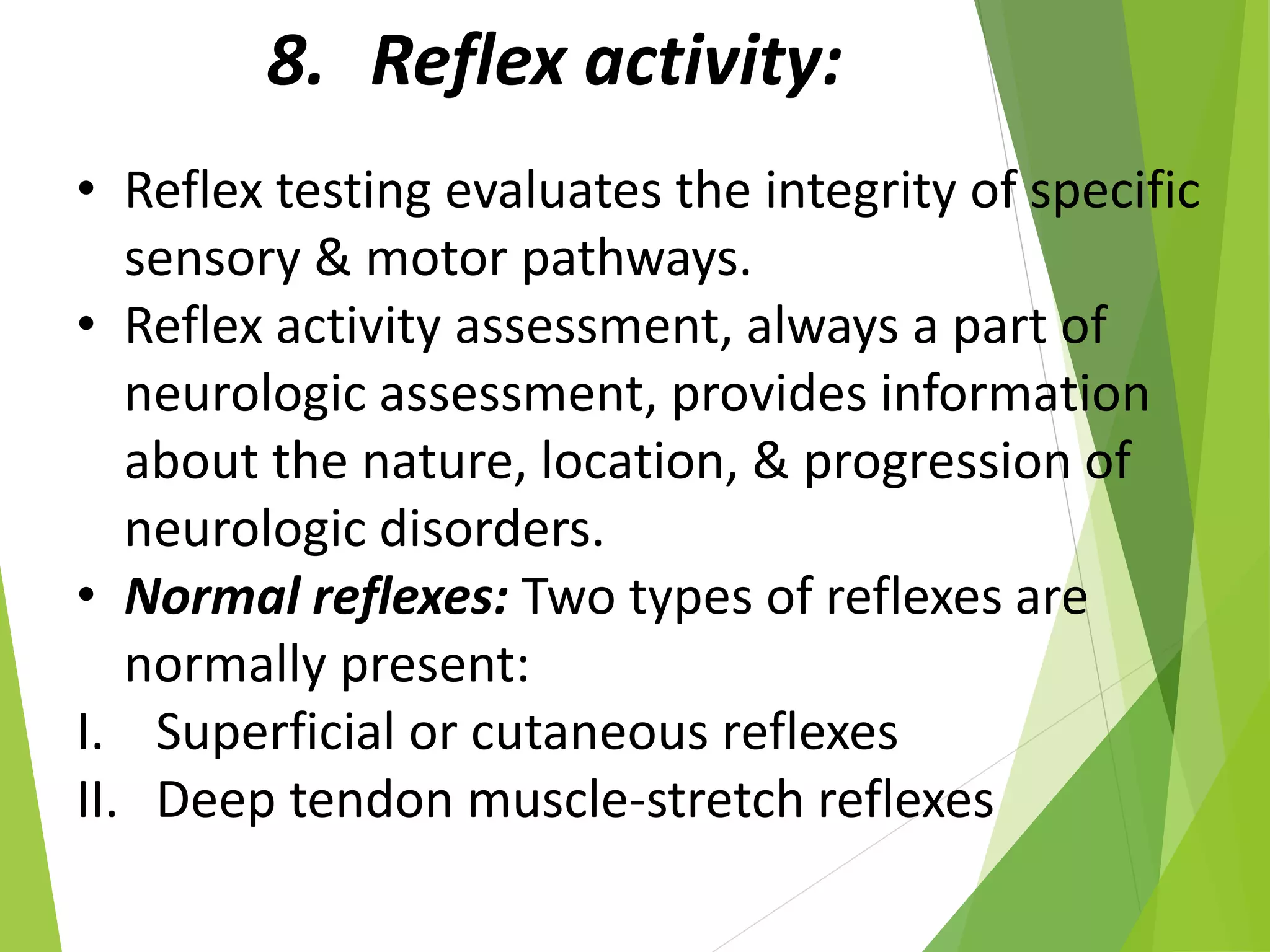
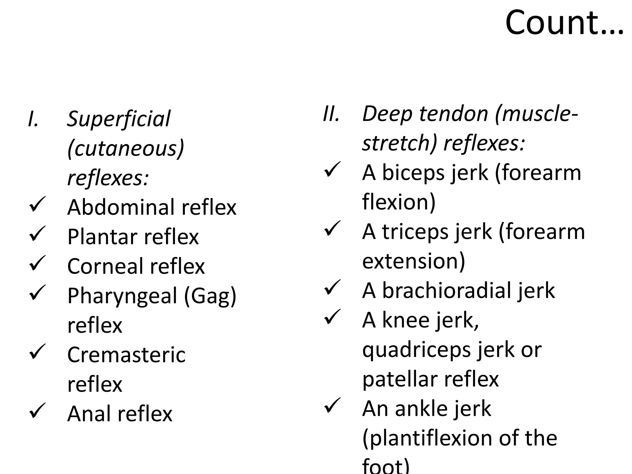
NEUROLOGICAL
EXAMINATION
DR.QURATULAIN MUGHAL
ISRA UNIVERSITY
INTRODUCTION…
• The purpose of neurological
examination is to determine the
presence or absence of disease in the
nervous system.
Aspects of Neurological
Examination…
1. Levels of consciousness
2. Mental status examination
3. Special cerebral functions
4. Cranial nerve function
5. Motor function
6. Sensory function
7. Cerebellar function
8. Reflexes
1. Levels of consciousness:
Assessment of levels of consciousness
includes following categories:
a. Alertness:
Patient is awake, responds immediately
& appropriately to all verbal stimuli.
a. Lethargic:
Patient is drowsy & inattentive but
arouses easily, frequently off to sleep.
a. Stuporous:
He arouses with great difficulty & co-
operates minimally when stimulated.
Count…
d. Semi-comatose:
• The patient has lost his ability to respond to
verbal stimuli.
• There is some response to painful stimuli.
• Little motor function is seen.
d. Comatose:
• When the patient is stimulated there is no
response to verbal or painful stimuli, no
motor activity is seen.
• The Glasgow coma scale is widely used to
measure the patient’s level of consciousness.
2. Mental status
examination:
• The components of mental status
examination include the assessment for
following categories; General
appearance, speech, thought process,
mood, cognitive functions, attention,
concentration, orientation, memory,
general knowledge, abstract reasoning,
judgment & insight.
3. Special cerebral functions:
• Assess for agnosia, apraxia & aphasia.
• Agnosia – inability to recognize
common objects through the senses
• Apraxia – patient cannot carry out
skilled act in the absence of paralysis.
• Aphasia – inability to communicate.
4. Cranial nerve
examination
Cranial nerve (CN) examination provides information
about the brainstem & related pathways.
• Olfactory nerve (CN I)
• Optic nerve (CN II)
• Oculomotor (CN III)
• Trochlear (CN IV)
• Trigeminal (CN V)
• Abducens (CN VI)
• Facial nerve (VII)
• Vestibulocochlear or
Acoustic nerve (CN VIII)
• Glossopharyngeal (CN IX)
• Vagus (CN X)
• Spinal accessory nerve (CN
XI)
• Hypoglossal nerve (CN XII)
5. Motor function:
• Assessment of motor function involves
assessing for muscle size, muscle strength,
muscle tone, muscle co-ordination, gait &
movement.
 Muscle size: Inspect all major muscle groups
bilaterally for symmetry, hypertrophy, &
atrophy.
 Muscle Strength: Assess the power in major
muscle groups against resistance. Assess &
rate muscle strength on a 5-point scale in all
four extremities, comparing one side with
other.
Count…
 Muscle tone:
• Assess muscle tone while moving each
extremity through its range of passive
motion.
• When tone is decreased (hypotonicity), the
muscle are soft, flabby, or flaccid; when tone
is increased (hypertonicity), the muscles are
resistant to movement, rigid, or spastic.
• Note the presence of abnormal flexion or
extension posture.
 Muscle coordination:
• Disorders related to coordination indicate
Cerebellar or posterior column lesions.
Count…
 Gait & station:
• Assess gait station by having the patient stand still,
walk & in tandem (one foot in front of the other in a
straight line).
• Walking involves the functions of motor power,
sensation & coordination.
• The ability to stand quietly with the feet together
requires coordination & intact proprioception (sense of
body position).
 Movement:
• Examine the muscles for fine & gross abnormal
movements.
• Move all the points through a full range of passive
motion.
• Abnormal findings include pain, joint contractures, &
muscle resistance.
6. Sensory function:
• Sensory assessment involves testing for touch,
pain, vibration & discrimination.
• A complete sensory examination is possible
only on a conscious & co-operative patient.
• Always test sensation with patient’s eye
closed.
• Help the patient relax & keep warm.
• Conduct sensory assessment systematically.
• Test a particular area of the body, & then test
the corresponding are on the other side.
7. Assessment of cerebellar
function:
• For evaluation of balance & co-ordination the tests
used are:
a. Finger to finger test: It is performed by
instructing the patient to place her index finger
on the nurse’s index finder. He is asked to repeat
this for several times in succession on both sides.
b. Finger to nose test: Tell the patient to extend his
index finger & then touch the tip of his nose
several times in rapid succession. This test is done
with patient’s eyes both open & closed.
Count…
c. Romberg test: Here the nurse instructs the
patient to stand with his feet together with
arms positioned at his sides. He is told to close
his eyes. This position is maintained for 10
seconds. This test is considered positive only if
there is actual loss of balance.
d. Tandom walking test: This is tested by having
the patient assume a normal standing position.
He is then instructed to walk over heel on a
straight line. Any unsteadiness, lurching or
broadening of the gait base is noted.
8. Reflex activity:
• Reflex testing evaluates the integrity of specific
sensory & motor pathways.
• Reflex activity assessment, always a part of
neurologic assessment, provides information
about the nature, location, & progression of
neurologic disorders.
• Normal reflexes: Two types of reflexes are
normally present:
I. Superficial or cutaneous reflexes
II. Deep tendon muscle-stretch reflexes
Count…
I. Superficial
(cutaneous)
reflexes:
 Abdominal reflex
 Plantar reflex
 Corneal reflex
 Pharyngeal (Gag)
reflex
 Cremasteric
reflex
 Anal reflex
II. Deep tendon (muscle-
stretch) reflexes:
 A biceps jerk (forearm
flexion)
 A triceps jerk (forearm
extension)
 A brachioradial jerk
 A knee jerk,
quadriceps jerk or
patellar reflex
 An ankle jerk
(plantiflexion of the
Count…
Abnormal reflexes:
Babinski’s reflex
Jaw reflex
Palm-chin (Palmomental) reflex
Clonus
Snout reflex
Rooting reflex
Sucking reflex
Glabella reflex
Grasp reflex
Chewing reflex
Neurological examination

Neurological examination

  • 1.
  • 2.
    INTRODUCTION… • The purposeof neurological examination is to determine the presence or absence of disease in the nervous system.
  • 3.
    Aspects of Neurological Examination… 1.Levels of consciousness 2. Mental status examination 3. Special cerebral functions 4. Cranial nerve function 5. Motor function 6. Sensory function 7. Cerebellar function 8. Reflexes
  • 4.
    1. Levels ofconsciousness: Assessment of levels of consciousness includes following categories: a. Alertness: Patient is awake, responds immediately & appropriately to all verbal stimuli. a. Lethargic: Patient is drowsy & inattentive but arouses easily, frequently off to sleep. a. Stuporous: He arouses with great difficulty & co- operates minimally when stimulated.
  • 5.
    Count… d. Semi-comatose: • Thepatient has lost his ability to respond to verbal stimuli. • There is some response to painful stimuli. • Little motor function is seen. d. Comatose: • When the patient is stimulated there is no response to verbal or painful stimuli, no motor activity is seen. • The Glasgow coma scale is widely used to measure the patient’s level of consciousness.
  • 6.
    2. Mental status examination: •The components of mental status examination include the assessment for following categories; General appearance, speech, thought process, mood, cognitive functions, attention, concentration, orientation, memory, general knowledge, abstract reasoning, judgment & insight.
  • 7.
    3. Special cerebralfunctions: • Assess for agnosia, apraxia & aphasia. • Agnosia – inability to recognize common objects through the senses • Apraxia – patient cannot carry out skilled act in the absence of paralysis. • Aphasia – inability to communicate.
  • 8.
    4. Cranial nerve examination Cranialnerve (CN) examination provides information about the brainstem & related pathways. • Olfactory nerve (CN I) • Optic nerve (CN II) • Oculomotor (CN III) • Trochlear (CN IV) • Trigeminal (CN V) • Abducens (CN VI) • Facial nerve (VII) • Vestibulocochlear or Acoustic nerve (CN VIII) • Glossopharyngeal (CN IX) • Vagus (CN X) • Spinal accessory nerve (CN XI) • Hypoglossal nerve (CN XII)
  • 9.
    5. Motor function: •Assessment of motor function involves assessing for muscle size, muscle strength, muscle tone, muscle co-ordination, gait & movement.  Muscle size: Inspect all major muscle groups bilaterally for symmetry, hypertrophy, & atrophy.  Muscle Strength: Assess the power in major muscle groups against resistance. Assess & rate muscle strength on a 5-point scale in all four extremities, comparing one side with other.
  • 10.
    Count…  Muscle tone: •Assess muscle tone while moving each extremity through its range of passive motion. • When tone is decreased (hypotonicity), the muscle are soft, flabby, or flaccid; when tone is increased (hypertonicity), the muscles are resistant to movement, rigid, or spastic. • Note the presence of abnormal flexion or extension posture.  Muscle coordination: • Disorders related to coordination indicate Cerebellar or posterior column lesions.
  • 11.
    Count…  Gait &station: • Assess gait station by having the patient stand still, walk & in tandem (one foot in front of the other in a straight line). • Walking involves the functions of motor power, sensation & coordination. • The ability to stand quietly with the feet together requires coordination & intact proprioception (sense of body position).  Movement: • Examine the muscles for fine & gross abnormal movements. • Move all the points through a full range of passive motion. • Abnormal findings include pain, joint contractures, & muscle resistance.
  • 12.
    6. Sensory function: •Sensory assessment involves testing for touch, pain, vibration & discrimination. • A complete sensory examination is possible only on a conscious & co-operative patient. • Always test sensation with patient’s eye closed. • Help the patient relax & keep warm. • Conduct sensory assessment systematically. • Test a particular area of the body, & then test the corresponding are on the other side.
  • 13.
    7. Assessment ofcerebellar function: • For evaluation of balance & co-ordination the tests used are: a. Finger to finger test: It is performed by instructing the patient to place her index finger on the nurse’s index finder. He is asked to repeat this for several times in succession on both sides. b. Finger to nose test: Tell the patient to extend his index finger & then touch the tip of his nose several times in rapid succession. This test is done with patient’s eyes both open & closed.
  • 14.
    Count… c. Romberg test:Here the nurse instructs the patient to stand with his feet together with arms positioned at his sides. He is told to close his eyes. This position is maintained for 10 seconds. This test is considered positive only if there is actual loss of balance. d. Tandom walking test: This is tested by having the patient assume a normal standing position. He is then instructed to walk over heel on a straight line. Any unsteadiness, lurching or broadening of the gait base is noted.
  • 15.
    8. Reflex activity: •Reflex testing evaluates the integrity of specific sensory & motor pathways. • Reflex activity assessment, always a part of neurologic assessment, provides information about the nature, location, & progression of neurologic disorders. • Normal reflexes: Two types of reflexes are normally present: I. Superficial or cutaneous reflexes II. Deep tendon muscle-stretch reflexes
  • 16.
    Count… I. Superficial (cutaneous) reflexes:  Abdominalreflex  Plantar reflex  Corneal reflex  Pharyngeal (Gag) reflex  Cremasteric reflex  Anal reflex II. Deep tendon (muscle- stretch) reflexes:  A biceps jerk (forearm flexion)  A triceps jerk (forearm extension)  A brachioradial jerk  A knee jerk, quadriceps jerk or patellar reflex  An ankle jerk (plantiflexion of the
  • 17.
    Count… Abnormal reflexes: Babinski’s reflex Jawreflex Palm-chin (Palmomental) reflex Clonus Snout reflex Rooting reflex Sucking reflex Glabella reflex Grasp reflex Chewing reflex