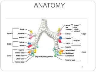
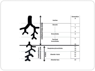
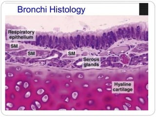
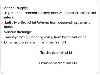
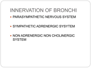
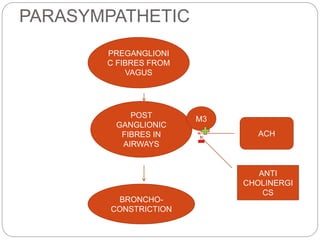
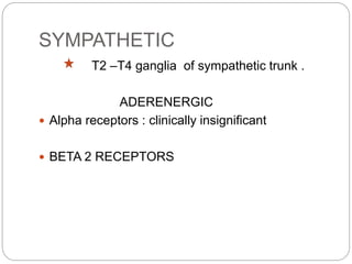
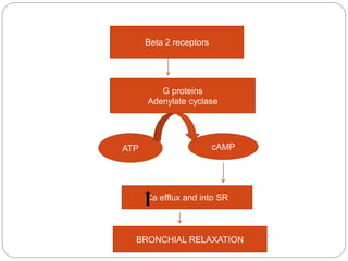
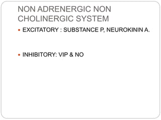
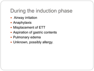
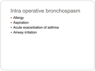
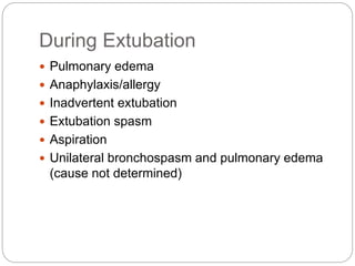
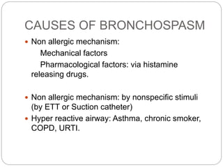
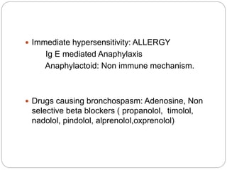
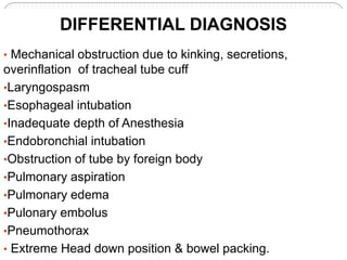
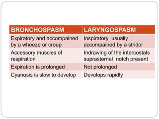
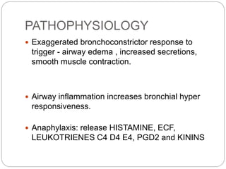
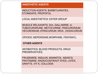
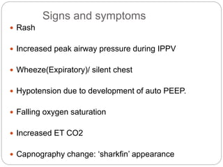
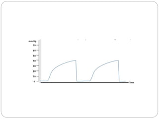
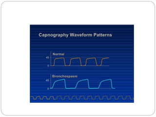
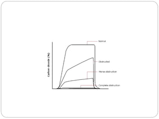
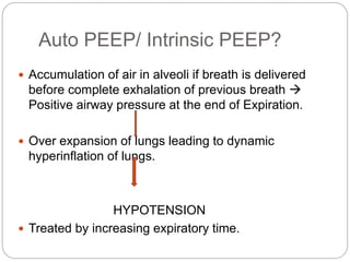
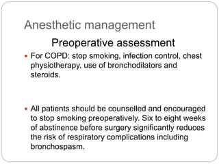
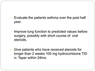
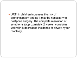
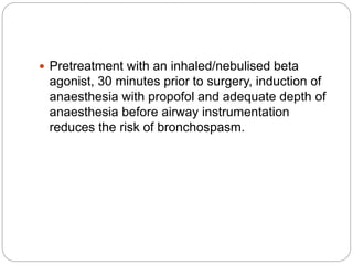
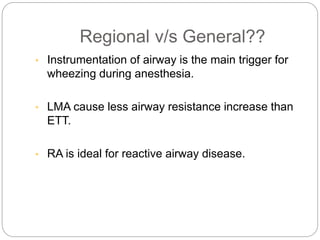
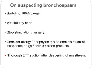
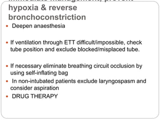
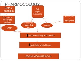
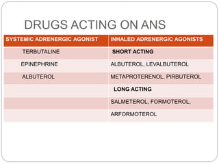
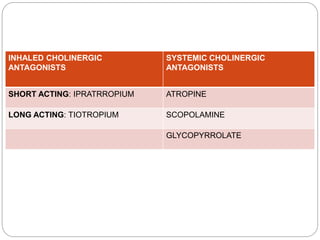
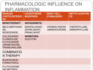
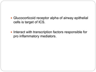
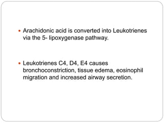
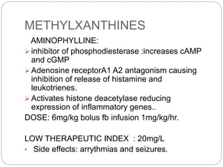
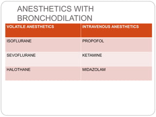
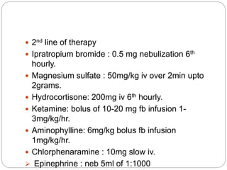
Dr. Imran
MANAGEMENT OF
INTRAOPERATIVE
BRONCHOSPASM
Definition: constriction of smooth muscles of
bronchi & bronchioles.
BRONCHOSPASM
ANATOMY
 Arterial supply:
 Right , one Bronchial Artery from 3rd posterior intercostal
artery.
 Left , two Bronchial Arteries from descending thoracic
aorta.
 Venous drainage:
mostly from pulmonary veins, from bronchial veins.
 Lymphatic drainage : Interbronchial LN
Tracheobronchial LN
Bronchomediastinal LN
INNERVATION OF BRONCHI
 PARASYMPATHETIC NERVOUS SYSTEM
 SYMPATHETIC ADRENERGIC SYSYTEM
 NON ADRENERGIC NON CHOLINERGIC
SYSTEM
PARASYMPATHETIC
PREGANGLIONI
C FIBRES FROM
VAGUS
POST
GANGLIONIC
FIBRES IN
AIRWAYS
ACH
M3
BRONCHO-
CONSTRICTION
ANTI
CHOLINERGI
CS
SYMPATHETIC
T2 –T4 ganglia of sympathetic trunk .
ADERENERGIC
 Alpha receptors : clinically insignificant
 BETA 2 RECEPTORS
Beta 2 receptors
cAMP
Ca efflux and into SR
G proteins
Adenylate cyclase
BRONCHIAL RELAXATION
ATP
NON ADRENERGIC NON
CHOLINERGIC SYSTEM
 EXCITATORY : SUBSTANCE P, NEUROKININ A.
 INHIBITORY: VIP & NO
During the induction phase
 Airway irritation
 Anaphylaxis
 Misplacement of ETT
 Aspiration of gastric contents
 Pulmonary edema
 Unknown, possibly allergy.
Intra operative bronchospasm
 Allergy
 Aspiration
 Acute exacerbation of asthma
 Airway irritation
During Extubation
 Pulmonary edema
 Anaphylaxis/allergy
 Inadvertent extubation
 Extubation spasm
 Aspiration
 Unilateral bronchospasm and pulmonary edema
(cause not determined)
CAUSES OF BRONCHOSPASM
 Non allergic mechanism:
Mechanical factors
Pharmacological factors: via histamine
releasing drugs.
 Non allergic mechanism: by nonspecific stimuli
(by ETT or Suction catheter)
 Hyper reactive airway: Asthma, chronic smoker,
COPD, URTI.
 Immediate hypersensitivity: ALLERGY
Ig E mediated Anaphylaxis
Anaphylactoid: Non immune mechanism.
 Drugs causing bronchospasm: Adenosine, Non
selective beta blockers ( propanolol, timolol,
nadolol, pindolol, alprenolol,oxprenolol)
• Mechanical obstruction due to kinking, secretions,
overinflation of tracheal tube cuff
•Laryngospasm
•Esophageal intubation
•Inadequate depth of Anesthesia
•Endobronchial intubation
•Obstruction of tube by foreign body
•Pulmonary aspiration
•Pulmonary edema
•Pulonary embolus
•Pneumothorax
• Extreme Head down position & bowel packing.
DIFFERENTIAL DIAGNOSIS
BRONCHOSPASM LARYNGOSPASM
Expiratory and accompained
by a wheeze or croup
Inspiratory usually
accompained by a stridor
Accessory muscles of
respiration
Indrawing of the intercostals
suprasternal notch present
Expiration is prolonged Not prolonged
Cyanosis is slow to develop Develops rapidly
PATHOPHYSIOLOGY
 Exaggerated bronchoconstrictor response to
trigger - airway edema , increased secretions,
smooth muscle contraction.
 Airway inflammation increases bronchial hyper
responsiveness.
 Anaphylaxis: release HISTAMINE, ECF,
LEUKOTRIENES C4 D4 E4, PGD2 and KININS
ANESTHETIC AGENTS
INDUCTION AGENTS: BARBITUARATES,
ETOMIDATE, PROPOFOL
LOCAL ANESTHETICS: ESTER GROUP
MUSCLE RELAXANTS: Sch, GALLAMINE, d-
TUBOCURARUNE, METOCURINE, PANCURONIUM,
VECURONIUM, ATRACURIUM, MIVA , DOXACURIUM
OPIODS: MEPERIDINE,MORPHINE, FENTANYL.
OTHER AGENTS
ANTIBIOTICS, BLOOD PRODUCTS, DRUG
PRESERVATIVES,
FRUSEMIDE, INSULIN, MANNITOL, NSAID’S,
PROTAMINE, RADIOCONTRAST DYES, LATEX,
GRAFTS, VIT K, COLLOIDS
Signs and symptoms
 Rash
 Increased peak airway pressure during IPPV
 Wheeze(Expiratory)/ silent chest
 Hypotension due to development of auto PEEP.
 Falling oxygen saturation
 Increased ET CO2
 Capnography change: ‘sharkfin’ appearance
Auto PEEP/ Intrinsic PEEP?
 Accumulation of air in alveoli if breath is delivered
before complete exhalation of previous breath 
Positive airway pressure at the end of Expiration.
 Over expansion of lungs leading to dynamic
hyperinflation of lungs.
HYPOTENSION
 Treated by increasing expiratory time.
Other causes of auto PEEP
 Mucus plugging of airways
 Large Minute Ventilation
 Block in expiratory limb of breathing circuit
 Smaller ETT with inadequate expiratory time.
DIAGNOSIS by APNEA TEST for 30sec.
Anesthetic management
Preoperative assessment
 For COPD: stop smoking, infection control, chest
physiotherapy, use of bronchodilators and
steroids.
 All patients should be counselled and encouraged
to stop smoking preoperatively. Six to eight weeks
of abstinence before surgery significantly reduces
the risk of respiratory complications including
bronchospasm.
• Evaluate the patients asthma over the past half
year.
• Improve lung function to predicted values before
surgery, possibly with short course of oral
steroids.
• Give patients who have received steroids for
longer than 2 weeks 100 mg hydrocortisone TID
iv. Taper within 24hrs.
 URTI in children increases the risk of
bronchospasm and so it may be necessary to
postpone surgery. The complete resolution of
symptoms (approximately 2 weeks) correlates
well with a decreased incidence of airway hyper
reactivity.
 Pretreatment with an inhaled/nebulised beta
agonist, 30 minutes prior to surgery, induction of
anaesthesia with propofol and adequate depth of
anaesthesia before airway instrumentation
reduces the risk of bronchospasm.
Regional v/s General??
• Instrumentation of airway is the main trigger for
wheezing during anesthesia.
• LMA cause less airway resistance increase than
ETT.
• RA is ideal for reactive airway disease.
On suspecting bronchospasm
• Switch to 100% oxygen
• Ventilate by hand
• Stop stimulation / surgery
• Consider allergy / anaphylaxis; stop administration of
suspected drugs / colloid / blood products
 Thorough ETT suction after deepening of anesthesia.
Immediate management; prevent
hypoxia & reverse
bronchoconstriction
 Deepen anaesthesia
 If ventilation through ETT difficult/impossible, check
tube position and exclude blocked/misplaced tube.
 If necessary eliminate breathing circuit occlusion by
using self-inflating bag
 In non-intubated patients exclude laryngospasm and
consider aspiration
 DRUG THERAPY
PHARMOCOLOGY
 Beta 2
agonists
Calcium sensitivity and ca infux
Myosin light chain kinase
BRONCHOCONSTRICTION
Ach
Vagus
histamine
cGMP
G proteins
Adenylate
cyclase
cAMP
Cholinergi
c
antagonist
sPhospholi
pase C &
IP3
DRUGS ACTING ON ANS
SYSTEMIC ADRENERGIC AGONIST INHALED ADRENERGIC AGONISTS
TERBUTALINE SHORT ACTING
EPINEPHRINE ALBUTEROL, LEVALBUTEROL
ALBUTEROL METAPROTERENOL, PIRBUTEROL
LONG ACTING
SALMETEROL, FORMOTEROL,
ARFORMOTEROL
INHALED CHOLINERGIC
ANTAGONISTS
SYSTEMIC CHOLINERGIC
ANTAGONISTS
SHORT ACTING: IPRATRROPIUM ATROPINE
LONG ACTING: TIOTROPIUM SCOPOLAMINE
GLYCOPYRROLATE
PHARMACOLOGIC INFLUENCE ON
INFLAMMATION
INHALED
CORTICOSTEROI
DS
LEUKOTRIENE
MODIFIERS
MAST CELL
STABILIZERS
METHYLXANTHIN
ES
MONOTHERAPY ANTAGONISTS:
BECLOMETHASO
NE,
BUDESONIDE,
MONTELUKAST,
ZAFIRLUKAST,
PRANLUKAST
CROMOLYNSODI
UMNEDOCROMIL
THEOPHYLLINE,
AMINOPHYLLINE
CICLESONIDE,
FLUNISOLIDE,
FLUTICASONE
,MOMETASONE,
TRIAMCINALONE
INHIBITORS:
ZILEUTON
COMBINATIO
N THERAPY
BUDESONIDE/
FORMOTEROL
FLUTICASONE/
 Glucocorticoid receptor alpha of airway epithelial
cells is target of ICS.
 Interact with transcription factors responsible for
pro inflammatory mediators.
 Arachidonic acid is converted into Leukotrienes
via the 5- lipoxygenase pathway.
 Leukotrienes C4, D4, E4 causes
bronchoconstriction, tissue edema, eosinophil
migration and increased airway secretion.
METHYLXANTHINES
AMINOPHYLLINE:
 inhibitor of phosphodiesterase :increases cAMP
and cGMP
 Adenosine receptorA1 A2 antagonism causing
inhibition of release of histamine and
leukotrienes.
 Activates histone deacetylase reducing
expression of inflammatory genes..
DOSE: 6mg/kg bolus fb infusion 1mg/kg/hr.
LOW THERAPEUTIC INDEX : 20mg/L
• Side effects: arrythmias and seizures.
ANESTHETICS WITH
BRONCHODILATION
VOLATILE ANESTHETICS INTRAVENOUS ANESTHETICS
ISOFLURANE PROPOFOL
SEVOFLURANE KETAMINE
HALOTHANE MIDAZOLAM
 Blockade of T-type voltage dependent calcium
channels on distal airway smooth muscles.
 Decreases intracellular calcium by decreasing
sensitivity of calcium mediated by Protein kinase
C and also increase in cAMP.
Other agents
 Antihistamines : allergen induced broncho-
constriction
 Magnesium sulfate.
 1st line Drug therapy
 Salbutamol
 MDI: 6-8 puffs, 100micrograms per puff, repeat
as necessary.
 Nebulisation : 5mg repeat as necessary.
 Intravenous: 250 mcg slow iv followed by
5mcg/min upto 20 mcg/min.
 2nd line of therapy
 Ipratropium bromide : 0.5 mg nebulization 6th
hourly.
 Magnesium sulfate : 50mg/kg iv over 2min upto
2grams.
 Hydrocortisone: 200mg iv 6th hourly.
 Ketamine: bolus of 10-20 mg fb infusion 1-
3mg/kg/hr.
 Aminophylline: 6mg/kg bolus fb infusion
1mg/kg/hr.
 Chlorphenaramine : 10mg slow iv.
 Epinephrine : neb 5ml of 1:1000
REFERENCES
 STOELTING’S PHARMACOLOGY AND
PHYSIOLOGY- 5TH EDITION.
 BARASH CLINICAL ANESTHESIA : 7TH
EDITION.
 MILLERS ANESTHESIA : 7TH EDITION.
THANK YOU

Bronchospasm ppt

  • 1.
  • 2.
    Definition: constriction ofsmooth muscles of bronchi & bronchioles. BRONCHOSPASM
  • 3.
  • 6.
     Arterial supply: Right , one Bronchial Artery from 3rd posterior intercostal artery.  Left , two Bronchial Arteries from descending thoracic aorta.  Venous drainage: mostly from pulmonary veins, from bronchial veins.  Lymphatic drainage : Interbronchial LN Tracheobronchial LN Bronchomediastinal LN
  • 7.
    INNERVATION OF BRONCHI PARASYMPATHETIC NERVOUS SYSTEM  SYMPATHETIC ADRENERGIC SYSYTEM  NON ADRENERGIC NON CHOLINERGIC SYSTEM
  • 8.
    PARASYMPATHETIC PREGANGLIONI C FIBRES FROM VAGUS POST GANGLIONIC FIBRESIN AIRWAYS ACH M3 BRONCHO- CONSTRICTION ANTI CHOLINERGI CS
  • 9.
    SYMPATHETIC T2 –T4 gangliaof sympathetic trunk . ADERENERGIC  Alpha receptors : clinically insignificant  BETA 2 RECEPTORS
  • 10.
    Beta 2 receptors cAMP Caefflux and into SR G proteins Adenylate cyclase BRONCHIAL RELAXATION ATP
  • 11.
    NON ADRENERGIC NON CHOLINERGICSYSTEM  EXCITATORY : SUBSTANCE P, NEUROKININ A.  INHIBITORY: VIP & NO
  • 12.
    During the inductionphase  Airway irritation  Anaphylaxis  Misplacement of ETT  Aspiration of gastric contents  Pulmonary edema  Unknown, possibly allergy.
  • 13.
    Intra operative bronchospasm Allergy  Aspiration  Acute exacerbation of asthma  Airway irritation
  • 14.
    During Extubation  Pulmonaryedema  Anaphylaxis/allergy  Inadvertent extubation  Extubation spasm  Aspiration  Unilateral bronchospasm and pulmonary edema (cause not determined)
  • 15.
    CAUSES OF BRONCHOSPASM Non allergic mechanism: Mechanical factors Pharmacological factors: via histamine releasing drugs.  Non allergic mechanism: by nonspecific stimuli (by ETT or Suction catheter)  Hyper reactive airway: Asthma, chronic smoker, COPD, URTI.
  • 16.
     Immediate hypersensitivity:ALLERGY Ig E mediated Anaphylaxis Anaphylactoid: Non immune mechanism.  Drugs causing bronchospasm: Adenosine, Non selective beta blockers ( propanolol, timolol, nadolol, pindolol, alprenolol,oxprenolol)
  • 17.
    • Mechanical obstructiondue to kinking, secretions, overinflation of tracheal tube cuff •Laryngospasm •Esophageal intubation •Inadequate depth of Anesthesia •Endobronchial intubation •Obstruction of tube by foreign body •Pulmonary aspiration •Pulmonary edema •Pulonary embolus •Pneumothorax • Extreme Head down position & bowel packing. DIFFERENTIAL DIAGNOSIS
  • 18.
    BRONCHOSPASM LARYNGOSPASM Expiratory andaccompained by a wheeze or croup Inspiratory usually accompained by a stridor Accessory muscles of respiration Indrawing of the intercostals suprasternal notch present Expiration is prolonged Not prolonged Cyanosis is slow to develop Develops rapidly
  • 19.
    PATHOPHYSIOLOGY  Exaggerated bronchoconstrictorresponse to trigger - airway edema , increased secretions, smooth muscle contraction.  Airway inflammation increases bronchial hyper responsiveness.  Anaphylaxis: release HISTAMINE, ECF, LEUKOTRIENES C4 D4 E4, PGD2 and KININS
  • 20.
    ANESTHETIC AGENTS INDUCTION AGENTS:BARBITUARATES, ETOMIDATE, PROPOFOL LOCAL ANESTHETICS: ESTER GROUP MUSCLE RELAXANTS: Sch, GALLAMINE, d- TUBOCURARUNE, METOCURINE, PANCURONIUM, VECURONIUM, ATRACURIUM, MIVA , DOXACURIUM OPIODS: MEPERIDINE,MORPHINE, FENTANYL. OTHER AGENTS ANTIBIOTICS, BLOOD PRODUCTS, DRUG PRESERVATIVES, FRUSEMIDE, INSULIN, MANNITOL, NSAID’S, PROTAMINE, RADIOCONTRAST DYES, LATEX, GRAFTS, VIT K, COLLOIDS
  • 21.
    Signs and symptoms Rash  Increased peak airway pressure during IPPV  Wheeze(Expiratory)/ silent chest  Hypotension due to development of auto PEEP.  Falling oxygen saturation  Increased ET CO2  Capnography change: ‘sharkfin’ appearance
  • 25.
    Auto PEEP/ IntrinsicPEEP?  Accumulation of air in alveoli if breath is delivered before complete exhalation of previous breath  Positive airway pressure at the end of Expiration.  Over expansion of lungs leading to dynamic hyperinflation of lungs. HYPOTENSION  Treated by increasing expiratory time.
  • 26.
    Other causes ofauto PEEP  Mucus plugging of airways  Large Minute Ventilation  Block in expiratory limb of breathing circuit  Smaller ETT with inadequate expiratory time. DIAGNOSIS by APNEA TEST for 30sec.
  • 27.
    Anesthetic management Preoperative assessment For COPD: stop smoking, infection control, chest physiotherapy, use of bronchodilators and steroids.  All patients should be counselled and encouraged to stop smoking preoperatively. Six to eight weeks of abstinence before surgery significantly reduces the risk of respiratory complications including bronchospasm.
  • 28.
    • Evaluate thepatients asthma over the past half year. • Improve lung function to predicted values before surgery, possibly with short course of oral steroids. • Give patients who have received steroids for longer than 2 weeks 100 mg hydrocortisone TID iv. Taper within 24hrs.
  • 29.
     URTI inchildren increases the risk of bronchospasm and so it may be necessary to postpone surgery. The complete resolution of symptoms (approximately 2 weeks) correlates well with a decreased incidence of airway hyper reactivity.
  • 30.
     Pretreatment withan inhaled/nebulised beta agonist, 30 minutes prior to surgery, induction of anaesthesia with propofol and adequate depth of anaesthesia before airway instrumentation reduces the risk of bronchospasm.
  • 31.
    Regional v/s General?? •Instrumentation of airway is the main trigger for wheezing during anesthesia. • LMA cause less airway resistance increase than ETT. • RA is ideal for reactive airway disease.
  • 32.
    On suspecting bronchospasm •Switch to 100% oxygen • Ventilate by hand • Stop stimulation / surgery • Consider allergy / anaphylaxis; stop administration of suspected drugs / colloid / blood products  Thorough ETT suction after deepening of anesthesia.
  • 33.
    Immediate management; prevent hypoxia& reverse bronchoconstriction  Deepen anaesthesia  If ventilation through ETT difficult/impossible, check tube position and exclude blocked/misplaced tube.  If necessary eliminate breathing circuit occlusion by using self-inflating bag  In non-intubated patients exclude laryngospasm and consider aspiration  DRUG THERAPY
  • 34.
    PHARMOCOLOGY  Beta 2 agonists Calciumsensitivity and ca infux Myosin light chain kinase BRONCHOCONSTRICTION Ach Vagus histamine cGMP G proteins Adenylate cyclase cAMP Cholinergi c antagonist sPhospholi pase C & IP3
  • 35.
    DRUGS ACTING ONANS SYSTEMIC ADRENERGIC AGONIST INHALED ADRENERGIC AGONISTS TERBUTALINE SHORT ACTING EPINEPHRINE ALBUTEROL, LEVALBUTEROL ALBUTEROL METAPROTERENOL, PIRBUTEROL LONG ACTING SALMETEROL, FORMOTEROL, ARFORMOTEROL
  • 36.
    INHALED CHOLINERGIC ANTAGONISTS SYSTEMIC CHOLINERGIC ANTAGONISTS SHORTACTING: IPRATRROPIUM ATROPINE LONG ACTING: TIOTROPIUM SCOPOLAMINE GLYCOPYRROLATE
  • 37.
    PHARMACOLOGIC INFLUENCE ON INFLAMMATION INHALED CORTICOSTEROI DS LEUKOTRIENE MODIFIERS MASTCELL STABILIZERS METHYLXANTHIN ES MONOTHERAPY ANTAGONISTS: BECLOMETHASO NE, BUDESONIDE, MONTELUKAST, ZAFIRLUKAST, PRANLUKAST CROMOLYNSODI UMNEDOCROMIL THEOPHYLLINE, AMINOPHYLLINE CICLESONIDE, FLUNISOLIDE, FLUTICASONE ,MOMETASONE, TRIAMCINALONE INHIBITORS: ZILEUTON COMBINATIO N THERAPY BUDESONIDE/ FORMOTEROL FLUTICASONE/
  • 38.
     Glucocorticoid receptoralpha of airway epithelial cells is target of ICS.  Interact with transcription factors responsible for pro inflammatory mediators.
  • 39.
     Arachidonic acidis converted into Leukotrienes via the 5- lipoxygenase pathway.  Leukotrienes C4, D4, E4 causes bronchoconstriction, tissue edema, eosinophil migration and increased airway secretion.
  • 40.
    METHYLXANTHINES AMINOPHYLLINE:  inhibitor ofphosphodiesterase :increases cAMP and cGMP  Adenosine receptorA1 A2 antagonism causing inhibition of release of histamine and leukotrienes.  Activates histone deacetylase reducing expression of inflammatory genes.. DOSE: 6mg/kg bolus fb infusion 1mg/kg/hr. LOW THERAPEUTIC INDEX : 20mg/L • Side effects: arrythmias and seizures.
  • 41.
    ANESTHETICS WITH BRONCHODILATION VOLATILE ANESTHETICSINTRAVENOUS ANESTHETICS ISOFLURANE PROPOFOL SEVOFLURANE KETAMINE HALOTHANE MIDAZOLAM
  • 42.
     Blockade ofT-type voltage dependent calcium channels on distal airway smooth muscles.  Decreases intracellular calcium by decreasing sensitivity of calcium mediated by Protein kinase C and also increase in cAMP.
  • 43.
    Other agents  Antihistamines: allergen induced broncho- constriction  Magnesium sulfate.
  • 44.
     1st lineDrug therapy  Salbutamol  MDI: 6-8 puffs, 100micrograms per puff, repeat as necessary.  Nebulisation : 5mg repeat as necessary.  Intravenous: 250 mcg slow iv followed by 5mcg/min upto 20 mcg/min.
  • 47.
     2nd lineof therapy  Ipratropium bromide : 0.5 mg nebulization 6th hourly.  Magnesium sulfate : 50mg/kg iv over 2min upto 2grams.  Hydrocortisone: 200mg iv 6th hourly.  Ketamine: bolus of 10-20 mg fb infusion 1- 3mg/kg/hr.  Aminophylline: 6mg/kg bolus fb infusion 1mg/kg/hr.  Chlorphenaramine : 10mg slow iv.  Epinephrine : neb 5ml of 1:1000
  • 48.
    REFERENCES  STOELTING’S PHARMACOLOGYAND PHYSIOLOGY- 5TH EDITION.  BARASH CLINICAL ANESTHESIA : 7TH EDITION.  MILLERS ANESTHESIA : 7TH EDITION.
  • 49.