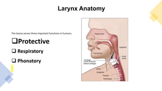
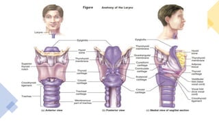
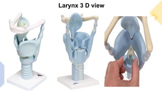
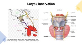
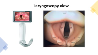
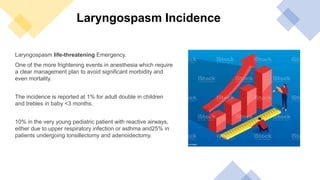
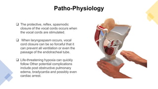
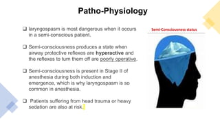
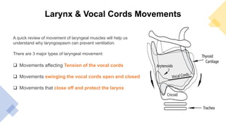
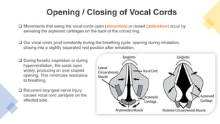
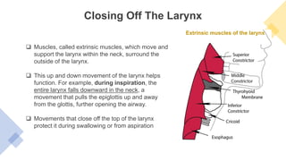
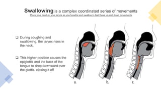
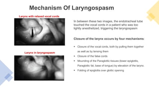
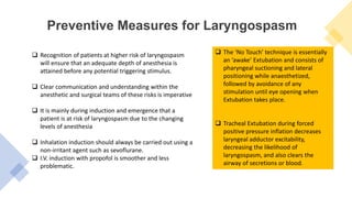
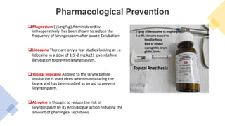
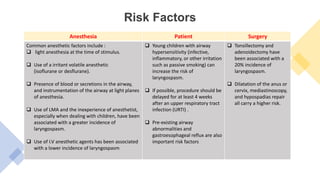
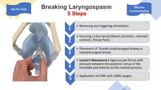
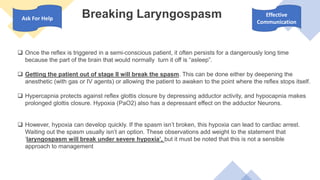
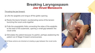
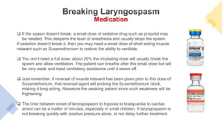
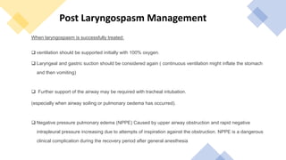
LARYNGOSPASM
No conflict of interest
OBJECTIVES
 Larynx Anatomy
 Definition of Laryngospasm
 Patho-Physiology
 Recognizing Laryngospasm
 Prevention
 Risk Factors
 Treatment And Management
Larynx Anatomy
The larynx serves three important functions in humans.
Protective
 Respiratory
 Phonatory
Larynx 3 D view
Larynx Innervation
The highest receptor density exists posteriorly at the true vocal
cords, where foreign material is most likely to spill into the airway.
Laryngoscopy view
Laryngospasm Definition
Laryngospasm is the sustained closure of
the vocal cords resulting in the partial or
complete loss of the patient’s airway.
Essentially a protective reflex which acts
to prevent foreign material entering
trachea-bronchial tree.
Laryngospasm life-threatening Emergency.
One of the more frightening events in anesthesia which require
a clear management plan to avoid significant morbidity and
even mortality.
The incidence is reported at 1% for adult double in children
and trebles in baby ˂3 months.
10% in the very young pediatric patient with reactive airways,
either due to upper respiratory infection or asthma and25% in
patients undergoing tonsillectomy and adenoidectomy.
Laryngospasm Incidence
Patho-Physiology
 The protective, reflex, spasmodic
closure of the vocal cords occurs when
the vocal cords are stimulated.
 When laryngospasm occurs, vocal
cord closure can be so forceful that it
can prevent all ventilation or even the
passage of the endotracheal tube.
 Life-threatening hypoxia can quickly
follow Other potential complications
include post obstructive pulmonary
edema, bradycardia and possibly even
cardiac arrest.
Patho-Physiology
 laryngospasm is most dangerous when it occurs
in a semi-conscious patient.
 Semi-consciousness produces a state when
airway protective reflexes are hyperactive and
the reflexes to turn them off are poorly operative.
 Semi-consciousness is present in Stage II of
anesthesia during both induction and
emergence, which is why laryngospasm is so
common in anesthesia.
 Patients suffering from head trauma or heavy
sedation are also at risk.
Semi-Consciousness status
Larynx & Vocal Cords Movements
A quick review of movement of laryngeal muscles will help us
understand why laryngospasm can prevent ventilation.
There are 3 major types of laryngeal movement:
 Movements affecting Tension of the vocal cords
 Movements swinging the vocal cords open and closed
 Movements that close off and protect the larynx
 Our vocal cords pivot constantly during the breathing cycle: opening during inhalation,
closing into a slightly separated rest position after exhalation.
Opening / Closing of Vocal Cords
 Movements that swing the vocal cords open (abduction) or closed (adduction) occur by
swiveling the arytenoid cartilages on the back of the cricoid ring.
 During forceful inspiration or during
hyperventilation, the cords open
widely, producing an oval shaped
opening. This minimizes resistance
to breathing.
 Recurrent laryngeal nerve injury
causes vocal cord paralysis on the
affected side.
Closing Off The Larynx
Extrinsic muscles of the larynx
 Muscles, called extrinsic muscles, which move and
support the larynx within the neck, surround the
outside of the larynx.
 This up and down movement of the larynx helps
function. For example, during inspiration, the
entire larynx falls downward in the neck, a
movement that pulls the epiglottis up and away
from the glottis, further opening the airway.
 Movements that close off the top of the larynx
protect it during swallowing or from aspiration
.
Swallowing is a complex coordinated series of movements
Place your hand on your larynx as you breathe and swallow to feel these up and down movements
 During coughing and
swallowing, the larynx rises in
the neck.
 This higher position causes the
epiglottis and the back of the
tongue to drop downward over
the glottis, closing it off
Recognizing Laryngospasm
 Occur when secretions, mucus, blood, or instruments such as a laryngoscope, in
the airway stimulate the vocal cords of a patient who is lightly anesthetized.
 One of the most common times for laryngospasm to occur is after Extubation if the
endotracheal tube is removed during Stage II when reflexes are hyperactive,
instead of while more deeply anesthetized or while awake.
 Avoiding vocal cord stimulation when the patient is lightly anesthetized can usually
prevent laryngospasm. However, laryngospasm can occur even with the best of
care, especially in patients with irritable airways such as those with asthma, COPD,
smokers, and with upper or lower respiratory infection.
Recognizing Laryngospasm
 Laryngospasm can be obvious or subtle, but always has signs of airway obstruction.
 If laryngospasm is partial, there will be chest movement with stridor but very little air
movement. There is very little bag movement with spontaneous ventilation .
 If laryngospasm is complete, then there is chest movement but the airway is silent.
Without air movement there is no stridor. The bag doesn’t move. No ventilation is
possible.
 Signs of airway obstruction include:
1. Rib retraction
2. Tracheal tug
3. Paradoxical breathing movements (chest falls and abdomen rises with inspiration)
4. Possibly stridor.
In between these two images, the endotracheal tube
touched the vocal cords in a patient who was too
lightly anesthetized, triggering the laryngospasm
Mechanism Of Laryngospasm
Closure of the larynx occurs by four mechanisms:
 Closure of the vocal cords, both by pulling them together
as well as by tensing them
 Closure of the false cords
 Mounding of the Paraglottic tissues (lower epiglottis,
Paraglottic fat, base of tongue) by elevation of the larynx.
 Folding of epiglottis over glottic opening
Larynx with relaxed vocal cords
Larynx in laryngospasm
Preventive Measures for Laryngospasm
 Recognition of patients at higher risk of laryngospasm
will ensure that an adequate depth of anesthesia is
attained before any potential triggering stimulus.
 Clear communication and understanding within the
anesthetic and surgical teams of these risks is imperative
 It is mainly during induction and emergence that a
patient is at risk of laryngospasm due to the changing
levels of anesthesia
 Inhalation induction should always be carried out using a
non-irritant agent such as sevoflurane.
 I.V. induction with propofol is smoother and less
problematic.
 The ‘No Touch’ technique is essentially
an ‘awake’ Extubation and consists of
pharyngeal suctioning and lateral
positioning while anaesthetized,
followed by avoidance of any
stimulation until eye opening when
Extubation takes place.
 Tracheal Extubation during forced
positive pressure inflation decreases
laryngeal adductor excitability,
decreasing the likelihood of
laryngospasm, and also clears the
airway of secretions or blood.
Pharmacological Prevention
Magnesium (15mg/kg) Administered i.v
intraoperatively has been shown to reduce the
frequency of laryngospasm after awake Extubation
Lidocaine There are only a few studies looking at i.v.
lidocaine in a dose of 1.5–2 mg kg21 given before
Extubation to prevent laryngospasm.
Topical lidocaine Applied to the larynx before
intubation is used often when manipulating the
larynx and has been studied as an aid to prevent
laryngospasm.
Atropine Is thought to reduce the risk of
laryngospasm by its Antisialogue action reducing the
amount of pharyngeal secretions.
Risk Factors
.
Anesthesia Patient Surgery
Common anesthetic factors include :
 light anesthesia at the time of stimulus.
 Use of a irritant volatile anesthetic
(isoflurane or desflurane).
 Presence of blood or secretions in the airway,
and instrumentation of the airway at light planes
of anesthesia.
 Use of LMA and the inexperience of anesthetist,
especially when dealing with children, have been
associated with a greater incidence of
laryngospasm.
 Use of I.V anesthetic agents has been associated
with a lower incidence of laryngospasm
 Young children with airway
hypersensitivity (infective,
inflammatory, or other irritation
such as passive smoking) can
increase the risk of
laryngospasm.
 If possible, procedure should be
delayed for at least 4 weeks
after an upper respiratory tract
infection (URTI) .
 Pre-existing airway
abnormalities and
gastroesophageal reflux are also
important risk factors
 Tonsillectomy and
adenoidectomy have
been associated with a
20% incidence of
laryngospasm.
 Dilatation of the anus or
cervix, mediastinoscopy,
and hypospadias repair
all carry a higher risk.
Breaking Laryngospasm
5 Steps
1
• Removing any triggering stimulation.
2
• Ensuring a clear larynx (blood ,Secretion , stomach
contents ,Throat Pack).
3
• Placement of Guedel oropharyngeal airway or
nasopharyngeal airway
4
• Larson’s Manoeuvre ( vigorous jaw thrust with
pressure between the posterior ramus of the
mandible and anterior to the mastoid process.
5
• Application of CPAP with 100% oxygen.
Ask For Help
Effective
Communication
Breaking Laryngospasm
 Once the reflex is triggered in a semi-conscious patient, it often persists for a dangerously long time
because the part of the brain that would normally turn it off is “asleep”.
 Getting the patient out of stage II will break the spasm. This can be done either by deepening the
anesthetic (with gas or IV agents) or allowing the patient to awaken to the point where the reflex stops itself.
 Hypercapnia protects against reflex glottis closure by depressing adductor activity, and hypocapnia makes
prolonged glottis closure. Hypoxia (PaO2) also has a depressant effect on the adductor Neurons.
 However, hypoxia can develop quickly. If the spasm isn’t broken, this hypoxia can lead to cardiac arrest.
Waiting out the spasm usually isn’t an option. These observations add weight to the statement that
‘laryngospasm will break under severe hypoxia’, but it must be noted that this is not a sensible
approach to management
Ask For Help
Effective
Communication
Thrusting the jaw forward:
 Lifts the epiglottis and tongue off the glottic opening
 Rocks the larynx forward, counteracting some of the tension
bunching the vocal cords together
 Pulls the aryepiglottic folds connecting the sides of the epiglottis
to the back of the arytenoids, opening a small gap between the
vocal cords
 Stimulates the patient because it’s painful, perhaps awakening the
patient out of Stage II toward consciousness
All of these actions are directed at creating a gap between the vocal cords.
Breaking Laryngospasm
Jaw thrust Manoeuvre
 If the spasm doesn’t break, a small dose of sedative drug such as propofol may
be needed. This deepens the level of anesthesia and usually stops the spasm.
If sedation doesn’t break it, then you may need a small dose of short acting muscle
relaxant such as Suxamethonium to restore the ability to ventilate.
 You don’t need a full dose: about 20% the intubating dose will usually break the
spasm and allow ventilation. The patient can breathe after this small dose but will
be very weak and need ventilatory assistance until it wears off.
 Just remember, if reversal of muscle relaxant has been given prior to this dose of
Suxamethonium, that reversal agent will prolong the Suxamethonium block,
making it long acting. Reassure the awaking patient since such weakness will be
frightening.
 The time between onset of laryngospasm to hypoxia to bradycardia to cardiac
arrest can be a matter of minutes, especially in small children. If laryngospasm is
not breaking quickly with positive pressure alone, to not delay further treatment.
Breaking Laryngospasm
Medication
When laryngospasm is successfully treated:
 ventilation should be supported initially with 100% oxygen.
 Laryngeal and gastric suction should be considered again ( continuous ventilation might inflate the stomach
and then vomiting)
 Further support of the airway may be required with tracheal intubation.
(especially when airway soiling or pulmonary oedema has occurred).
 Negative pressure pulmonary edema (NPPE) Caused by upper airway obstruction and rapid negative
intrapleural pressure increasing due to attempts of inspiration against the obstruction. NPPE is a dangerous
clinical complication during the recovery period after general anesthesia
Post Laryngospasm Management
ETT Cuff Concerns
Cuff Over Pressure
Cuff sensation
Slowly cuff Deflation
Call For Help
Keep Calm
Deep Breathing
Effective Communication
How Stressful Situations Affect the Brain
• Laryngospasm can be prevented by paying attention to
the depth of anesthesia and recognition of risk factors.
• Recognition of at-risk patients will help to prevent
laryngospasm and avoid potential significant morbidity.
• Treatment of laryngospasm should proceed traditionally
by clearing supraglottic airway obstruction and soiling,
CPAP with 100% O2, deepening of anesthesia and/or
paralysis.
• Effective team communication and leadership is
essential during this emergency situation.
Conclusion
• 1. Olsson GL, Hallen B. Laryngospasm during anaesthesia. A computer-
aided incidence study in 136,929 patients. Acta Anaesthesiol Scand 1984;
28: 567–75 2. Gulhas N, Durmus N, Demirbileks S et al. The use of
magnesium to prevent laryngospasm after tonsillectomy and
adenoidectomy: a preliminary study. Paediatr Anaesth 2003; 13: 43– 7 3.
Ikari T, Sasaki CT. Laryngospasm: a neurophysiological definition. Ann Otol
Rhinol Laryngol 1980; 89: 220– 4 4. Visvanathan T, Kluger MT, Webb RK,
Westhorpe RN. Crisis management during anaesthesia: laryngospasm.
Qual Saf Health Care 2005; 14: e3 5. Landsman IS. Mechanisms and
treatment of laryngospasm. Int Anesthesiol Clin 1997; 35: 67– 73 6.
Alalami AA, Ayoub CM, Baraka AS. Laryngospasm: review of different
prevention and treatment modalities. Paediatr Anaesth 2008; 18: 281 –8
7. Hampson-Evans D, Morgan P, Farrar M. Pediatric laryngospasm. Paediatr
Anaesth 2008; 18: 303– 7 8. Walker RWM, Sutton R. Which post in a
storm? Use of suxamethonium without intravenous access for severe
laryngospasm. Anaesthesia 2007; 62: 757– 9 9. Al-Metwalli RR, Mowafi
HA, Ismail SA. Gentle chest compression relieves extubation laryngospasm
in children. J Anesth 2010; 24: 854–7
References
Thank You

Laryngospasm Presentation.pptx By Ak (Anesthesia )

  • 1.
  • 2.
  • 3.
    OBJECTIVES  Larynx Anatomy Definition of Laryngospasm  Patho-Physiology  Recognizing Laryngospasm  Prevention  Risk Factors  Treatment And Management
  • 4.
    Larynx Anatomy The larynxserves three important functions in humans. Protective  Respiratory  Phonatory
  • 6.
  • 7.
    Larynx Innervation The highestreceptor density exists posteriorly at the true vocal cords, where foreign material is most likely to spill into the airway.
  • 8.
  • 9.
    Laryngospasm Definition Laryngospasm isthe sustained closure of the vocal cords resulting in the partial or complete loss of the patient’s airway. Essentially a protective reflex which acts to prevent foreign material entering trachea-bronchial tree.
  • 10.
    Laryngospasm life-threatening Emergency. Oneof the more frightening events in anesthesia which require a clear management plan to avoid significant morbidity and even mortality. The incidence is reported at 1% for adult double in children and trebles in baby ˂3 months. 10% in the very young pediatric patient with reactive airways, either due to upper respiratory infection or asthma and25% in patients undergoing tonsillectomy and adenoidectomy. Laryngospasm Incidence
  • 11.
    Patho-Physiology  The protective,reflex, spasmodic closure of the vocal cords occurs when the vocal cords are stimulated.  When laryngospasm occurs, vocal cord closure can be so forceful that it can prevent all ventilation or even the passage of the endotracheal tube.  Life-threatening hypoxia can quickly follow Other potential complications include post obstructive pulmonary edema, bradycardia and possibly even cardiac arrest.
  • 12.
    Patho-Physiology  laryngospasm ismost dangerous when it occurs in a semi-conscious patient.  Semi-consciousness produces a state when airway protective reflexes are hyperactive and the reflexes to turn them off are poorly operative.  Semi-consciousness is present in Stage II of anesthesia during both induction and emergence, which is why laryngospasm is so common in anesthesia.  Patients suffering from head trauma or heavy sedation are also at risk. Semi-Consciousness status
  • 13.
    Larynx & VocalCords Movements A quick review of movement of laryngeal muscles will help us understand why laryngospasm can prevent ventilation. There are 3 major types of laryngeal movement:  Movements affecting Tension of the vocal cords  Movements swinging the vocal cords open and closed  Movements that close off and protect the larynx
  • 14.
     Our vocalcords pivot constantly during the breathing cycle: opening during inhalation, closing into a slightly separated rest position after exhalation. Opening / Closing of Vocal Cords  Movements that swing the vocal cords open (abduction) or closed (adduction) occur by swiveling the arytenoid cartilages on the back of the cricoid ring.  During forceful inspiration or during hyperventilation, the cords open widely, producing an oval shaped opening. This minimizes resistance to breathing.  Recurrent laryngeal nerve injury causes vocal cord paralysis on the affected side.
  • 15.
    Closing Off TheLarynx Extrinsic muscles of the larynx  Muscles, called extrinsic muscles, which move and support the larynx within the neck, surround the outside of the larynx.  This up and down movement of the larynx helps function. For example, during inspiration, the entire larynx falls downward in the neck, a movement that pulls the epiglottis up and away from the glottis, further opening the airway.  Movements that close off the top of the larynx protect it during swallowing or from aspiration
  • 16.
    . Swallowing is acomplex coordinated series of movements Place your hand on your larynx as you breathe and swallow to feel these up and down movements  During coughing and swallowing, the larynx rises in the neck.  This higher position causes the epiglottis and the back of the tongue to drop downward over the glottis, closing it off
  • 17.
    Recognizing Laryngospasm  Occurwhen secretions, mucus, blood, or instruments such as a laryngoscope, in the airway stimulate the vocal cords of a patient who is lightly anesthetized.  One of the most common times for laryngospasm to occur is after Extubation if the endotracheal tube is removed during Stage II when reflexes are hyperactive, instead of while more deeply anesthetized or while awake.  Avoiding vocal cord stimulation when the patient is lightly anesthetized can usually prevent laryngospasm. However, laryngospasm can occur even with the best of care, especially in patients with irritable airways such as those with asthma, COPD, smokers, and with upper or lower respiratory infection.
  • 18.
    Recognizing Laryngospasm  Laryngospasmcan be obvious or subtle, but always has signs of airway obstruction.  If laryngospasm is partial, there will be chest movement with stridor but very little air movement. There is very little bag movement with spontaneous ventilation .  If laryngospasm is complete, then there is chest movement but the airway is silent. Without air movement there is no stridor. The bag doesn’t move. No ventilation is possible.  Signs of airway obstruction include: 1. Rib retraction 2. Tracheal tug 3. Paradoxical breathing movements (chest falls and abdomen rises with inspiration) 4. Possibly stridor.
  • 19.
    In between thesetwo images, the endotracheal tube touched the vocal cords in a patient who was too lightly anesthetized, triggering the laryngospasm Mechanism Of Laryngospasm Closure of the larynx occurs by four mechanisms:  Closure of the vocal cords, both by pulling them together as well as by tensing them  Closure of the false cords  Mounding of the Paraglottic tissues (lower epiglottis, Paraglottic fat, base of tongue) by elevation of the larynx.  Folding of epiglottis over glottic opening Larynx with relaxed vocal cords Larynx in laryngospasm
  • 20.
    Preventive Measures forLaryngospasm  Recognition of patients at higher risk of laryngospasm will ensure that an adequate depth of anesthesia is attained before any potential triggering stimulus.  Clear communication and understanding within the anesthetic and surgical teams of these risks is imperative  It is mainly during induction and emergence that a patient is at risk of laryngospasm due to the changing levels of anesthesia  Inhalation induction should always be carried out using a non-irritant agent such as sevoflurane.  I.V. induction with propofol is smoother and less problematic.  The ‘No Touch’ technique is essentially an ‘awake’ Extubation and consists of pharyngeal suctioning and lateral positioning while anaesthetized, followed by avoidance of any stimulation until eye opening when Extubation takes place.  Tracheal Extubation during forced positive pressure inflation decreases laryngeal adductor excitability, decreasing the likelihood of laryngospasm, and also clears the airway of secretions or blood.
  • 21.
    Pharmacological Prevention Magnesium (15mg/kg)Administered i.v intraoperatively has been shown to reduce the frequency of laryngospasm after awake Extubation Lidocaine There are only a few studies looking at i.v. lidocaine in a dose of 1.5–2 mg kg21 given before Extubation to prevent laryngospasm. Topical lidocaine Applied to the larynx before intubation is used often when manipulating the larynx and has been studied as an aid to prevent laryngospasm. Atropine Is thought to reduce the risk of laryngospasm by its Antisialogue action reducing the amount of pharyngeal secretions.
  • 22.
    Risk Factors . Anesthesia PatientSurgery Common anesthetic factors include :  light anesthesia at the time of stimulus.  Use of a irritant volatile anesthetic (isoflurane or desflurane).  Presence of blood or secretions in the airway, and instrumentation of the airway at light planes of anesthesia.  Use of LMA and the inexperience of anesthetist, especially when dealing with children, have been associated with a greater incidence of laryngospasm.  Use of I.V anesthetic agents has been associated with a lower incidence of laryngospasm  Young children with airway hypersensitivity (infective, inflammatory, or other irritation such as passive smoking) can increase the risk of laryngospasm.  If possible, procedure should be delayed for at least 4 weeks after an upper respiratory tract infection (URTI) .  Pre-existing airway abnormalities and gastroesophageal reflux are also important risk factors  Tonsillectomy and adenoidectomy have been associated with a 20% incidence of laryngospasm.  Dilatation of the anus or cervix, mediastinoscopy, and hypospadias repair all carry a higher risk.
  • 23.
    Breaking Laryngospasm 5 Steps 1 •Removing any triggering stimulation. 2 • Ensuring a clear larynx (blood ,Secretion , stomach contents ,Throat Pack). 3 • Placement of Guedel oropharyngeal airway or nasopharyngeal airway 4 • Larson’s Manoeuvre ( vigorous jaw thrust with pressure between the posterior ramus of the mandible and anterior to the mastoid process. 5 • Application of CPAP with 100% oxygen. Ask For Help Effective Communication
  • 24.
    Breaking Laryngospasm  Oncethe reflex is triggered in a semi-conscious patient, it often persists for a dangerously long time because the part of the brain that would normally turn it off is “asleep”.  Getting the patient out of stage II will break the spasm. This can be done either by deepening the anesthetic (with gas or IV agents) or allowing the patient to awaken to the point where the reflex stops itself.  Hypercapnia protects against reflex glottis closure by depressing adductor activity, and hypocapnia makes prolonged glottis closure. Hypoxia (PaO2) also has a depressant effect on the adductor Neurons.  However, hypoxia can develop quickly. If the spasm isn’t broken, this hypoxia can lead to cardiac arrest. Waiting out the spasm usually isn’t an option. These observations add weight to the statement that ‘laryngospasm will break under severe hypoxia’, but it must be noted that this is not a sensible approach to management Ask For Help Effective Communication
  • 25.
    Thrusting the jawforward:  Lifts the epiglottis and tongue off the glottic opening  Rocks the larynx forward, counteracting some of the tension bunching the vocal cords together  Pulls the aryepiglottic folds connecting the sides of the epiglottis to the back of the arytenoids, opening a small gap between the vocal cords  Stimulates the patient because it’s painful, perhaps awakening the patient out of Stage II toward consciousness All of these actions are directed at creating a gap between the vocal cords. Breaking Laryngospasm Jaw thrust Manoeuvre
  • 26.
     If thespasm doesn’t break, a small dose of sedative drug such as propofol may be needed. This deepens the level of anesthesia and usually stops the spasm. If sedation doesn’t break it, then you may need a small dose of short acting muscle relaxant such as Suxamethonium to restore the ability to ventilate.  You don’t need a full dose: about 20% the intubating dose will usually break the spasm and allow ventilation. The patient can breathe after this small dose but will be very weak and need ventilatory assistance until it wears off.  Just remember, if reversal of muscle relaxant has been given prior to this dose of Suxamethonium, that reversal agent will prolong the Suxamethonium block, making it long acting. Reassure the awaking patient since such weakness will be frightening.  The time between onset of laryngospasm to hypoxia to bradycardia to cardiac arrest can be a matter of minutes, especially in small children. If laryngospasm is not breaking quickly with positive pressure alone, to not delay further treatment. Breaking Laryngospasm Medication
  • 27.
    When laryngospasm issuccessfully treated:  ventilation should be supported initially with 100% oxygen.  Laryngeal and gastric suction should be considered again ( continuous ventilation might inflate the stomach and then vomiting)  Further support of the airway may be required with tracheal intubation. (especially when airway soiling or pulmonary oedema has occurred).  Negative pressure pulmonary edema (NPPE) Caused by upper airway obstruction and rapid negative intrapleural pressure increasing due to attempts of inspiration against the obstruction. NPPE is a dangerous clinical complication during the recovery period after general anesthesia Post Laryngospasm Management
  • 28.
    ETT Cuff Concerns CuffOver Pressure Cuff sensation Slowly cuff Deflation
  • 29.
    Call For Help KeepCalm Deep Breathing Effective Communication How Stressful Situations Affect the Brain
  • 30.
    • Laryngospasm canbe prevented by paying attention to the depth of anesthesia and recognition of risk factors. • Recognition of at-risk patients will help to prevent laryngospasm and avoid potential significant morbidity. • Treatment of laryngospasm should proceed traditionally by clearing supraglottic airway obstruction and soiling, CPAP with 100% O2, deepening of anesthesia and/or paralysis. • Effective team communication and leadership is essential during this emergency situation. Conclusion
  • 31.
    • 1. OlssonGL, Hallen B. Laryngospasm during anaesthesia. A computer- aided incidence study in 136,929 patients. Acta Anaesthesiol Scand 1984; 28: 567–75 2. Gulhas N, Durmus N, Demirbileks S et al. The use of magnesium to prevent laryngospasm after tonsillectomy and adenoidectomy: a preliminary study. Paediatr Anaesth 2003; 13: 43– 7 3. Ikari T, Sasaki CT. Laryngospasm: a neurophysiological definition. Ann Otol Rhinol Laryngol 1980; 89: 220– 4 4. Visvanathan T, Kluger MT, Webb RK, Westhorpe RN. Crisis management during anaesthesia: laryngospasm. Qual Saf Health Care 2005; 14: e3 5. Landsman IS. Mechanisms and treatment of laryngospasm. Int Anesthesiol Clin 1997; 35: 67– 73 6. Alalami AA, Ayoub CM, Baraka AS. Laryngospasm: review of different prevention and treatment modalities. Paediatr Anaesth 2008; 18: 281 –8 7. Hampson-Evans D, Morgan P, Farrar M. Pediatric laryngospasm. Paediatr Anaesth 2008; 18: 303– 7 8. Walker RWM, Sutton R. Which post in a storm? Use of suxamethonium without intravenous access for severe laryngospasm. Anaesthesia 2007; 62: 757– 9 9. Al-Metwalli RR, Mowafi HA, Ismail SA. Gentle chest compression relieves extubation laryngospasm in children. J Anesth 2010; 24: 854–7 References
  • 32.

Editor's Notes

  • #2 I selected this topic because the tech can show a good talent in the management of such critical anesthesia moment.
  • #7 The larynx consists of the cartilaginous-osseous framework, elastic membranes and ligaments, joints, muscles, nerves, vessels, and interior cavity. The laryngeal functions are the protection of the airway during swallowing, respiration (i.e. maintainance of the airway) and phonation.
  • #8 Right & Left superior laryngeal nerve /Right & Left Recurrent laryngeal nerves They are all supplied by the vagus nerve
  • #14 Movement affecting the tension of the vocal cords, like the pitch of the voice, occurs by rocking the thyroid cartilage backward and forward on the cricoid cartilage at the cricothyroid joint.////One only has to listen to a singer to realize the dynamic control of pitch that changes in vocal cord tension produce.
  • #21 Right & Left superior laryngeal nerve /Right & Left Recurrent laryngeal nerves They are all supplied by the vagus nerve The highest receptor density exists posteriorly at the true vocal cords, where foreign material is most likely to spill into the airway.
  • #22 The only available study shows a slight decrease in the incidence of laryngospasm.
  • #24 Initial treatment of laryngospasm classically consists of:
  • #25 After exclusion of other obvious causes of airway obstruction, this plan should be put into action An oxygen saturation which continues to decrease below 80% with or without an accompanying bradycardia should prompt the anaesthetist to act quickly to regain oxygenation of the patient Initial treatment of laryngospasm classically consists of: