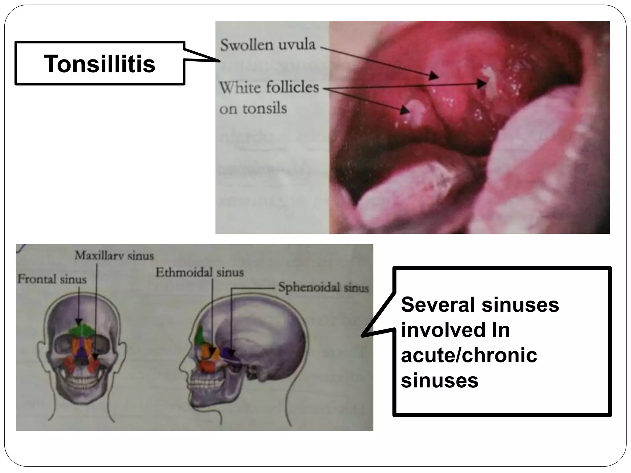
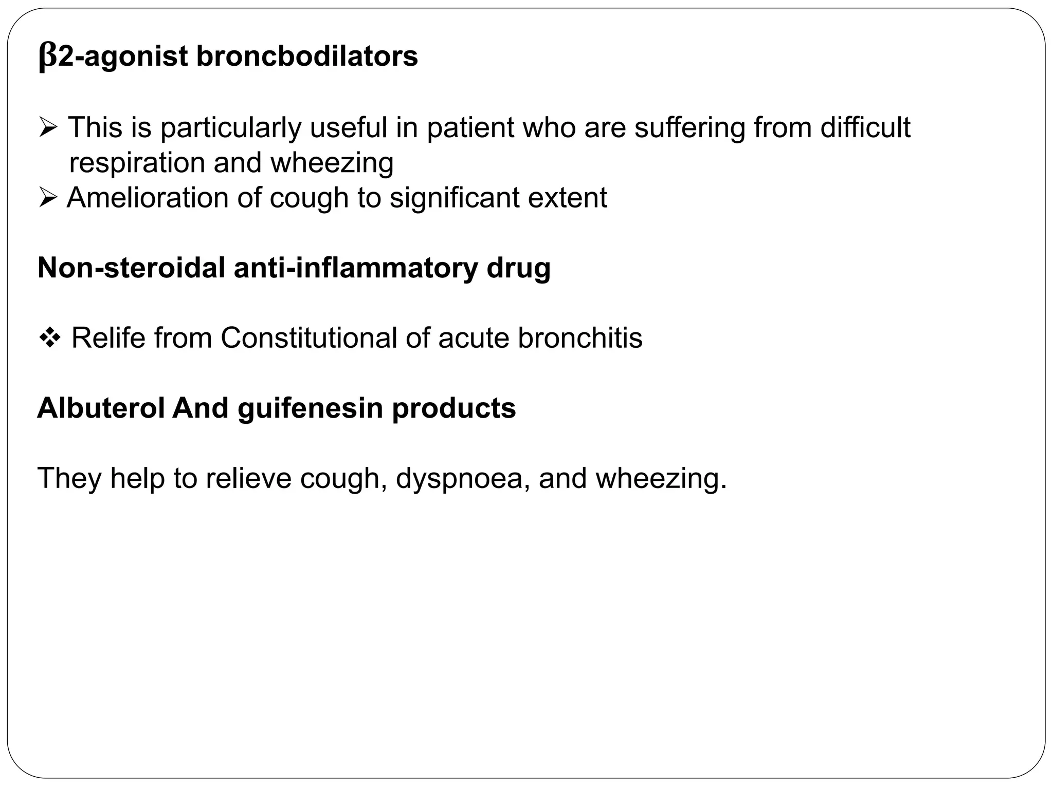
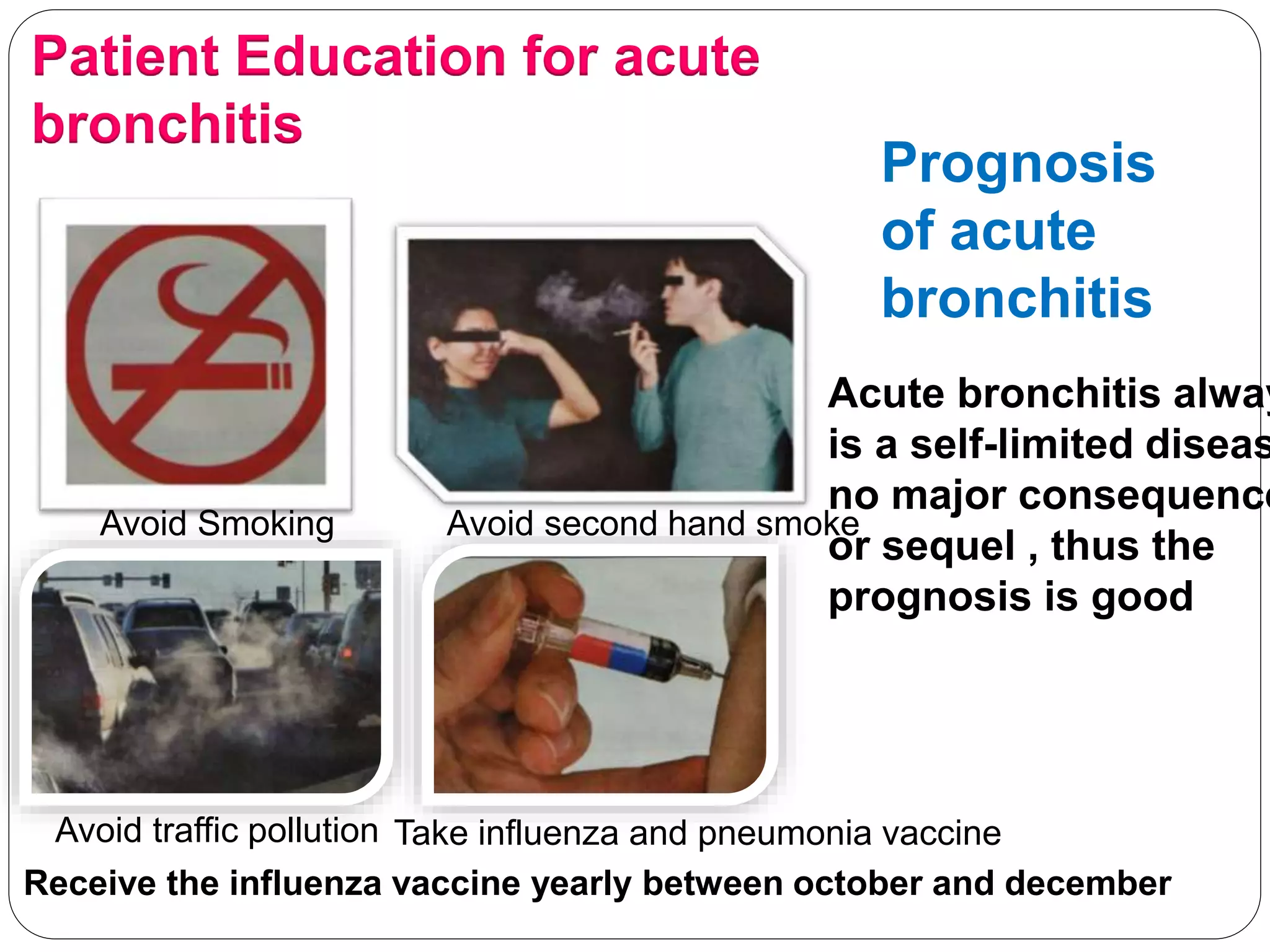
22/04/2018 Respiratory Diseases Management Course 2017-18
Respiratory Diseases
Management Course
2017-18
22/04/2018 Ahmedabad
22/04/2018 Respiratory Diseases Management Course 2017-18
Acute & Chronic
Bronchitis
Dr. Meghanaben Mehta M.B.B.S.
Senior Family Physician
Jafrabad, Gujarat
Defenation
The involvement of the large bronchi i.e. the air passages
from the trachea Into the small airways and alveoli, in term
of inflammation when infected either By becteria or virus i
called as acute bronchitis(fig.).
Acute bronchitis and Chronic bronchitis
Difference between acute bronchitis and chronic
bronchitis
Parameters Acute Bronchitis Chronic Bronchitis
Pathogens involved Mainly virus Both Bacteria and virus
Onset of the disease Sudden Gradual
Duration Of the
Disease
Short not more than 3
weeks
Long Lasting, for atleast 2
consecutive years
Age group Affected More Commonly seen in
children
More Commonly seen in
elderly population
Clinical Presentation Cough and occasionally,
sputum production
Cough with significant
amount of sputum
expectroration for al least 3
months a year for two
consecutive years.
 All over the world, all age group
 Most common cause of visit to a paediatrician in child
 Low Socioeconomic status
 people who live in urban and highly industrialised areas
 Mainly Children Below 2 years, Most commonly affected, another Peak
seen in children aged 9-15 years
Epidemiology
Risk Factors of acute bronchitis
Acute bronchitis can be classified , infectious and non infectious
A) Infectious causative agent
Viruses
Typical Viruses which cause acute
bronchitis include
 Influenza A and B
 parainfluenza
 respiratory syncytial virus
 coronavirus
 Rhinovirus
 Human bocavirus (parvovirus)
 Coxsackievirus
 Herpes simplex virus
Bacteria
The most common bacteria affecting the
bronchi are
 Mycoplasma species
 Cblamydia pneumoniae
 Moraxella catarrbalis
 Haemopbilus influenzae
Influenza and moraxella catarrbalis are
Found to be significant causative
pathogens in preschoolers(age <5 years
 Mycoplasma pneumoniae is observed t
be a causative agent in school-aged
children (age 6-18 years)
 Bordetella Pertussis generally affects
the children who are incompletely
vaccinated
B) Non-infectious causative agents
 Allergens or irritants: Mainly air pollutants (fig.3)
 Cigarette Smoke
 Carbon and nitrogen dioxide airborne particulates
 chemicals
 Tobacco smoke
Fig. 3: Air pollutants responsible for causing acute bronchitis
C) Other causes include the following
 Allergies
 Chronic aspiration or gastro-oesophageal reflux
 Fungal infection
Pathophysiology of acute bronchitis
Irritation of airways due to any aetiological stimulant
Infiltration of neutrophils in the lung tissue
Release of inflammatory substance by neutrophils
Hypersecretion of goblet
cells
Hyperemic and oedematous mucus
lining
Diminished bronchial mucociliary
function
Clogging of air passages
Acute bronchitis with predominant cough
Normal and inflamed bronchial tube
Complications of acute bronchitis
Various Complications of acute bronchitis are:
 Bacteria superinfection
 Heamoptysis
 Pneumonia
 Reactive airway disease which occur as a result of acute bronchitis
 Chronic bronchitis
Clinical Presentation of acute bronchitis
 Running nose with watery coryza
 Body pain malaise
 Slight fever with or without chills
 Sore throat
 Back Pain
 Whole body muscle pain
Nasal Discharge: Initially water gradually become thick. 7 -10 days to resolve.
when nasal Discharge in purulent, it is Mainly viral in origin, purulent discharge
does not imply Bacteria involvement
Cough: The cough is usually accompanied by nasal discharge, initially is dry and
Harsh and then over a period of the time becomes loose and more productive
Dyspnoea: Difficulty in breathing, due to decreased elastic recoil of the alveolus
And the narrowed bronchioles
Clinical Presentation of dyspnoea in acute bronchitis
Tachypnoea: Increase in respiratory rate
Heamoptysis: Blood streaked sputum or frank blood with the co
Fever: Fever is not Commonly present ,fever along with a cough
influenza or pneumonia
Chest Pain: Chest pain accompanying the cough
Other Symptoms: burning with substernal chest pain
3 Major parameters:
I. Detailed and precise history taking
II. Physical examination
III. Laboratory Investigation
History Taking:
 History of repeated exposure to second hand smoke
 Personal history of smoking
 duration of the complaint
 Type of onset of the symptoms, sudden or gradual.
 Inquire about the duration from which cough is present, the cough
lasts for more than 5 days, then is usually indicates acute bronchitis.
 Type of sputum, in acute bronchitis can be clear, yellow, green or even
blood-tinged. Purulent sputum is reported in 50% of persons.
Physical Examination
 Presence of crackles
 Presence of rhonchi
 At time large airway wheezing, it is usually scattered and bilateral
 Decreased intensity of breath sounds
 Prolonged Expiration
 The presence of inspiratory stridor indicates obstruction of a major
bronchus of the trachea
 At times, phyaryngeal erythema is present
 Localised Lymphadenopathy is occasionally present
Phyaryngeal Erythema Presence of localised cervical
lymphadenopathy in acute bronchitis
Note:
 The Change of color of the sputum, dose not indicate secondary bacteria
infection
 The Presence of inspiratory stridor indicates obstruction of a major
bronchus or the treachea, and it is a medical emergency tackled by
insertion indotracheal intubation
 The combination of conjunctivitis, adenopathy, and rginorrhoea suggest
adenovirus infection
Laboratory investigation
Complete blood count
• increase in eosinophils count ,denotes allergic reaction of respiratory tract
• Presence of leucocytosis along with fever is indicative of secondary bacteria
infection
C-reactive protein
• C-reactive protein which is indicative of infective pathology.
Sputum Microcopy
• Show the presence of neutrophil granulocytes
Sputum Culture
• I show the presence of pathogenic microorganisms such a s streptococcus
species
• Culture of respiratory secretions is obtain for certain viruses like influenza
virus, Mycoplasma pneumoniae, and bordetella pertusis.
Blood Culture
• Beneficial only if bacteria superinfection is suspected
Procalcitonin levels
• Distinguishing between the bacterial infections from non-bacterial
infections
• physician as it my guide therapy and it also helps in reducing the use of
antibiotics.
Imaging Studies
• Chest X-ray PA view
• It is usually usefull in the diagnosis of associated underlying condition of
the lung e.g.hyperinflation,collapse,pneumonia
• It is done if the clinical findings are suspicious of pneumonia
• Elderly population it is mandatory to do Chest X-ray if signs of cold cough
and sneezing are present.
• some conditions that predispose to bronchitis can be well seen with help of
chest radiography
X-ray Chest showing
consolidation patch in left
middle lobe
Bronchoscopy
It is not needed for diagnosis of acute bronchitis, But it helps in ruling out
the presence of any other underlying chronic disease like foreign body
aspiration, tuberculosis, tumors, etc
Bronchoscopy
Spirometry
 bronchospasm is seen as the underlying pathophysiology in acute
bronchitis.
 A large reduction in forced expiratory volume in one second(FEV)
 This reduction Generally resolve over 4-6 weeks.
Spirometry use in acute bronchitis
Differential diagnosis of acute bronchitis
Streptococcal pbaryngitis
 Caused by Group A streptococci(45%) and anaerobes (18%)
Appearance of throat on physcal examination of streptococcal pharyngitis.
Other differential diagnosis to be considered are
 Exercise induced asthma
 bacterial tracheitis
 influenza
 Hyperreactive airway disease
 Tonsillitis
 Viral pharyngitis
 Acute and Chroonic sinusitis
Bacterial
tracheitis
Tonsillitis
Several sinuses
involved In
acute/chronic
sinuses
The treatment is divided into pharmacological treatment
1) Pharmacological treatment approacb
Antibiotics
 the bacterial pathogens are responsible only in about 5-10% of bronchitis
most cases are caused by viral infection, self-limited.
 Antibiotics are useually not recommended ,Unless microscopic
examination of the sputum reveals large numbers of bacteria.
 The use of antibiotic has not shown any consistent benefit in the
symptomatology or natural history of acute bronochits
 Do not treat acute bronochitis with antibiotic unless a risk serious
complication exists because of co-morbid conditions. Older than 65 years
with acute cough had a hospitalisation in the past year, have diabetes
mellitus or congestive heart failure or are on steroids
β2-agonist broncbodilators
 This is particularly useful in patient who are suffering from difficult
respiration and wheezing
 Amelioration of cough to significant extent
Non-steroidal anti-inflammatory drug
 Relife from Constitutional of acute bronchitis
Albuterol And guifenesin products
They help to relieve cough, dyspnoea, and wheezing.
Caution
DO NOT USE ANTIHISTAMINE IN ACUTE BRONCHITIS
Increase in thinkness of mucous and thus hinder its expulsion.
 it also helps the bacteria to persist
 antihistamine along with an axpectorant cough syrup may be doubly
harmful, it would lead to increased production of mucous
2) Non-pharmacological approacb
Stop Smoking
 Quitting Smoking bronchial tree to heal faster
Influenza Vaccination
 Influenza vaccination helps in reducing – incidence of upper respiratory
Zinc
Intranasal zinc products help in reducing duration & severity of cold symptoms
Patient Education for acute
bronchitis
Avoid Smoking Avoid second hand smoke
Avoid traffic pollution Take influenza and pneumonia vaccine
Receive the influenza vaccine yearly between october and december
Prognosis
of acute
bronchitis
Acute bronchitis alway
is a self-limited diseas
no major consequence
or sequel , thus the
prognosis is good
22/04/2018 Respiratory Diseases Management Course 2017-18
Module Coordinator

Bronchitis afpa rdmc_06_dr meghanaben mehta_20180422

  • 1.
    22/04/2018 Respiratory DiseasesManagement Course 2017-18 Respiratory Diseases Management Course 2017-18 22/04/2018 Ahmedabad
  • 2.
    22/04/2018 Respiratory DiseasesManagement Course 2017-18 Acute & Chronic Bronchitis Dr. Meghanaben Mehta M.B.B.S. Senior Family Physician Jafrabad, Gujarat
  • 3.
    Defenation The involvement ofthe large bronchi i.e. the air passages from the trachea Into the small airways and alveoli, in term of inflammation when infected either By becteria or virus i called as acute bronchitis(fig.).
  • 4.
    Acute bronchitis andChronic bronchitis Difference between acute bronchitis and chronic bronchitis Parameters Acute Bronchitis Chronic Bronchitis Pathogens involved Mainly virus Both Bacteria and virus Onset of the disease Sudden Gradual Duration Of the Disease Short not more than 3 weeks Long Lasting, for atleast 2 consecutive years Age group Affected More Commonly seen in children More Commonly seen in elderly population Clinical Presentation Cough and occasionally, sputum production Cough with significant amount of sputum expectroration for al least 3 months a year for two consecutive years.
  • 5.
     All overthe world, all age group  Most common cause of visit to a paediatrician in child  Low Socioeconomic status  people who live in urban and highly industrialised areas  Mainly Children Below 2 years, Most commonly affected, another Peak seen in children aged 9-15 years Epidemiology Risk Factors of acute bronchitis
  • 6.
    Acute bronchitis canbe classified , infectious and non infectious A) Infectious causative agent Viruses Typical Viruses which cause acute bronchitis include  Influenza A and B  parainfluenza  respiratory syncytial virus  coronavirus  Rhinovirus  Human bocavirus (parvovirus)  Coxsackievirus  Herpes simplex virus Bacteria The most common bacteria affecting the bronchi are  Mycoplasma species  Cblamydia pneumoniae  Moraxella catarrbalis  Haemopbilus influenzae Influenza and moraxella catarrbalis are Found to be significant causative pathogens in preschoolers(age <5 years  Mycoplasma pneumoniae is observed t be a causative agent in school-aged children (age 6-18 years)  Bordetella Pertussis generally affects the children who are incompletely vaccinated
  • 7.
    B) Non-infectious causativeagents  Allergens or irritants: Mainly air pollutants (fig.3)  Cigarette Smoke  Carbon and nitrogen dioxide airborne particulates  chemicals  Tobacco smoke Fig. 3: Air pollutants responsible for causing acute bronchitis C) Other causes include the following  Allergies  Chronic aspiration or gastro-oesophageal reflux  Fungal infection
  • 8.
    Pathophysiology of acutebronchitis Irritation of airways due to any aetiological stimulant Infiltration of neutrophils in the lung tissue Release of inflammatory substance by neutrophils Hypersecretion of goblet cells Hyperemic and oedematous mucus lining Diminished bronchial mucociliary function Clogging of air passages Acute bronchitis with predominant cough
  • 9.
    Normal and inflamedbronchial tube Complications of acute bronchitis Various Complications of acute bronchitis are:  Bacteria superinfection  Heamoptysis  Pneumonia  Reactive airway disease which occur as a result of acute bronchitis  Chronic bronchitis
  • 10.
    Clinical Presentation ofacute bronchitis  Running nose with watery coryza  Body pain malaise  Slight fever with or without chills  Sore throat  Back Pain  Whole body muscle pain Nasal Discharge: Initially water gradually become thick. 7 -10 days to resolve. when nasal Discharge in purulent, it is Mainly viral in origin, purulent discharge does not imply Bacteria involvement Cough: The cough is usually accompanied by nasal discharge, initially is dry and Harsh and then over a period of the time becomes loose and more productive Dyspnoea: Difficulty in breathing, due to decreased elastic recoil of the alveolus And the narrowed bronchioles
  • 11.
    Clinical Presentation ofdyspnoea in acute bronchitis Tachypnoea: Increase in respiratory rate Heamoptysis: Blood streaked sputum or frank blood with the co Fever: Fever is not Commonly present ,fever along with a cough influenza or pneumonia Chest Pain: Chest pain accompanying the cough Other Symptoms: burning with substernal chest pain
  • 12.
    3 Major parameters: I.Detailed and precise history taking II. Physical examination III. Laboratory Investigation History Taking:  History of repeated exposure to second hand smoke  Personal history of smoking  duration of the complaint  Type of onset of the symptoms, sudden or gradual.  Inquire about the duration from which cough is present, the cough lasts for more than 5 days, then is usually indicates acute bronchitis.  Type of sputum, in acute bronchitis can be clear, yellow, green or even blood-tinged. Purulent sputum is reported in 50% of persons.
  • 13.
    Physical Examination  Presenceof crackles  Presence of rhonchi  At time large airway wheezing, it is usually scattered and bilateral  Decreased intensity of breath sounds  Prolonged Expiration  The presence of inspiratory stridor indicates obstruction of a major bronchus of the trachea  At times, phyaryngeal erythema is present  Localised Lymphadenopathy is occasionally present Phyaryngeal Erythema Presence of localised cervical lymphadenopathy in acute bronchitis
  • 14.
    Note:  The Changeof color of the sputum, dose not indicate secondary bacteria infection  The Presence of inspiratory stridor indicates obstruction of a major bronchus or the treachea, and it is a medical emergency tackled by insertion indotracheal intubation  The combination of conjunctivitis, adenopathy, and rginorrhoea suggest adenovirus infection
  • 15.
    Laboratory investigation Complete bloodcount • increase in eosinophils count ,denotes allergic reaction of respiratory tract • Presence of leucocytosis along with fever is indicative of secondary bacteria infection C-reactive protein • C-reactive protein which is indicative of infective pathology. Sputum Microcopy • Show the presence of neutrophil granulocytes Sputum Culture • I show the presence of pathogenic microorganisms such a s streptococcus species • Culture of respiratory secretions is obtain for certain viruses like influenza virus, Mycoplasma pneumoniae, and bordetella pertusis. Blood Culture • Beneficial only if bacteria superinfection is suspected
  • 16.
    Procalcitonin levels • Distinguishingbetween the bacterial infections from non-bacterial infections • physician as it my guide therapy and it also helps in reducing the use of antibiotics. Imaging Studies • Chest X-ray PA view • It is usually usefull in the diagnosis of associated underlying condition of the lung e.g.hyperinflation,collapse,pneumonia • It is done if the clinical findings are suspicious of pneumonia • Elderly population it is mandatory to do Chest X-ray if signs of cold cough and sneezing are present. • some conditions that predispose to bronchitis can be well seen with help of chest radiography X-ray Chest showing consolidation patch in left middle lobe
  • 17.
    Bronchoscopy It is notneeded for diagnosis of acute bronchitis, But it helps in ruling out the presence of any other underlying chronic disease like foreign body aspiration, tuberculosis, tumors, etc Bronchoscopy
  • 18.
    Spirometry  bronchospasm isseen as the underlying pathophysiology in acute bronchitis.  A large reduction in forced expiratory volume in one second(FEV)  This reduction Generally resolve over 4-6 weeks. Spirometry use in acute bronchitis
  • 19.
    Differential diagnosis ofacute bronchitis Streptococcal pbaryngitis  Caused by Group A streptococci(45%) and anaerobes (18%) Appearance of throat on physcal examination of streptococcal pharyngitis.
  • 20.
    Other differential diagnosisto be considered are  Exercise induced asthma  bacterial tracheitis  influenza  Hyperreactive airway disease  Tonsillitis  Viral pharyngitis  Acute and Chroonic sinusitis Bacterial tracheitis
  • 21.
  • 22.
    The treatment isdivided into pharmacological treatment 1) Pharmacological treatment approacb Antibiotics  the bacterial pathogens are responsible only in about 5-10% of bronchitis most cases are caused by viral infection, self-limited.  Antibiotics are useually not recommended ,Unless microscopic examination of the sputum reveals large numbers of bacteria.  The use of antibiotic has not shown any consistent benefit in the symptomatology or natural history of acute bronochits  Do not treat acute bronochitis with antibiotic unless a risk serious complication exists because of co-morbid conditions. Older than 65 years with acute cough had a hospitalisation in the past year, have diabetes mellitus or congestive heart failure or are on steroids
  • 23.
    β2-agonist broncbodilators  Thisis particularly useful in patient who are suffering from difficult respiration and wheezing  Amelioration of cough to significant extent Non-steroidal anti-inflammatory drug  Relife from Constitutional of acute bronchitis Albuterol And guifenesin products They help to relieve cough, dyspnoea, and wheezing.
  • 24.
    Caution DO NOT USEANTIHISTAMINE IN ACUTE BRONCHITIS Increase in thinkness of mucous and thus hinder its expulsion.  it also helps the bacteria to persist  antihistamine along with an axpectorant cough syrup may be doubly harmful, it would lead to increased production of mucous 2) Non-pharmacological approacb Stop Smoking  Quitting Smoking bronchial tree to heal faster Influenza Vaccination  Influenza vaccination helps in reducing – incidence of upper respiratory Zinc Intranasal zinc products help in reducing duration & severity of cold symptoms
  • 25.
    Patient Education foracute bronchitis Avoid Smoking Avoid second hand smoke Avoid traffic pollution Take influenza and pneumonia vaccine Receive the influenza vaccine yearly between october and december Prognosis of acute bronchitis Acute bronchitis alway is a self-limited diseas no major consequence or sequel , thus the prognosis is good
  • 26.
    22/04/2018 Respiratory DiseasesManagement Course 2017-18 Module Coordinator