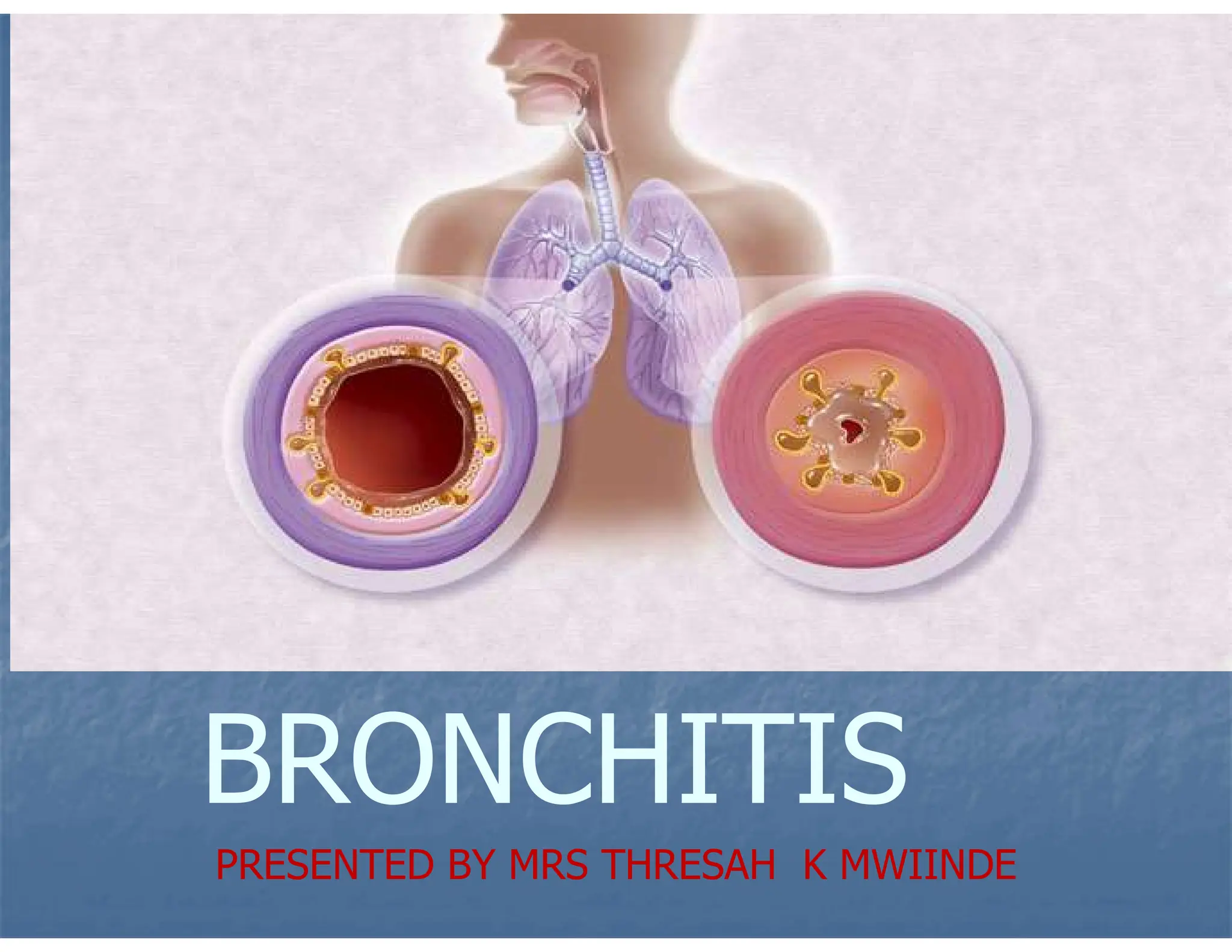
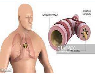
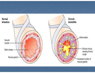
PRESENTED BY MRS THRESAH K MWIINDE
GENERAL OBJECTIVES
At the end of the lecture
Student nurses should
understand Bronchitis
and its management.
SPECIFIC OBJECTIVES
 Define Bronchitis
 Mention the two types of Bronchitis
 List the predisposition factors of Bronchitis
 Describe the pathophysiology of Acute
and Chronic Bronchitis
 State the signs and symptom of Bronchitis
 Describe the management of Bronchitis
 State the complications of Bronchitis
DEFINITION OF ACUTE
BRONCHITIS
 Acute Bronchitis is an inflammation of
the lower respiratory tract
accompanied by coughing and
coughing up of phlegm. It can be
caused by exposure to a cold or the
flu, infection, or irritants. It takes less
than two weeks
DEFINITION
 Bronchitis is an inflammation
of the bronchi in the lower
respiratory tract usually due
to infection, characterized by
cough, wheezing and Dyspnea.
TYPES OF BRONCHITIS
There are 2 types of bronchitis:
• Acute bronchitis
• Chronic bronchitis
ACUTE BRONCHITIS
 Acute bronchitis is usually a self
limiting and treatment is supportive.
 Upto 90% of acute bronchial
infections are viral by origin
 Cough which is the most common
sign lasts for upto 3 weeks
ACUTE BRONCHITIS
When bronchitis is mild and
brief in duration, it is called
acute. It usually takes less
than 2 weeks
Acute Bronchitis
 This is the inflammation of the bronchial tree with minimal changes
in the parts affected.
 Acute bronchitis is characterised by the development of a cough or
small sensation in the back of the throat, with or without the production
of sputum.
 Acute bronchitis is most often caused by viruses that infect the
epithelium of the bronchi, resulting in inflammation and increased
mucus secretion.
Predisposing factors:
• Exposure to cold
• Irritants like cigarettes
smoke
• Very young and very old
• Dust
• Toxic gases
10
Causes of Bronchitis
• Microorganisms: viruses and bacteria
• Extension of infection from the
trachea
• Traumatic injuries
• Over exposure to industrial fumes.
Causes of Acute Bronchitis
 It is caused by both viruses and bacteria.
The main viruses are:
 Rhinoviruses
 Coronaviruses
 Adenoviruses
Causes of Acute Bronchitis
 Metapneumovirus
 Parainfluenza virus
 Respiratory syncytial virus
 Influenza.
Causes of Acute Bronchitis
 The main bacteria is:
 Mycoplasma pneumoniae,
 Chlamydophila pneumoniae,
 Bordetella pertussis,
 Streptococcus pneumoniae,
15
PATHOPHYSIOLOGY
-The presence of an irritant like smoke or
infection will lead to inflammation which narrows
the air way.
-There will also be increased production of
secretions which will further impair air passage.
-Irritation of the respiratory tract will lead to
cough in an attempt to clear the air way.
-There may also be pain behind the sternum due
to the inflammation of the airways.
INCIDENCE
 Anyone can get acute bronchitis, but
infants, young children, and the elderly
are more likely to come down with the
disease. Smokers and people with heart or
lung diseases are at a higher risk of
developing acute bronchitis. Individuals
exposed to chemical fumes or high levels
of air pollution also have a greater chance
of developing acute bronchitis.
Signs and symptoms
 Cough due to irritation in the air way.
 Dyspnoea ( shortness of breath) due
to inflammation of the bronchial
airways.
 Soreness behind the sternum due to
the inflammatory processes.
Signs and symptoms
 Wheezing as air passes through the
fluid filled air way.
 Fever due to infection.
 In the initial stage sign of flu eg
running nose, sore throat, nasal
congestion, etc
Diagnosis
 History may reveal a predisposing factor
like Flu
 On Auscultation there will sign of
crepitations.
 Sputum for culture and sensitivity will
show the causative agent
 Bronchoscopy will show inflamed mucosal
membranes
Treatment
 If there is no bacterial infection treat
like flu
 If bacterial infection is present give
antibiotics like Amoxyl 500mg tds for
5/7
 Analgesic and antipyretics for pain
e.g. Panadol 1g tds 3/7
Treatment
 If dyspnea is present oxygen therapy may
be given
 Rest should be ensured
 Copious fluids to prevent dehydration
 Cough expectorants may be given.
 Broncho dilators may be given eg ventolin
4mg tid 3/7
Complications of acute
Bronchitis
 Chronic bronchitis
 Pneumonia
 Bronchiectasis; This is chronic
irreversible dilation of the bronchi and
bronchioles that results from the
destruction of the muscles and elastic
connective tissue
Complications of acute
Bronchitis
 Atelectasis:This is partial collapse or
incomplete inflation of the lungs
 Pnuemothorax;This is the
accumulation of air in the pleural
space through breach of either
Parietal or Visceral pleura
Complications of acute
Bronchitis
 Emphysema; Is a pathological term that
describes an abnormal distention of the air
spaces beyond the terminal bronchioles
and destruction of the walls of the alveoli
 Empyema;An accumulation of thick,
purulent fluid within the pleural space
often with fibrin development and loculated (
walled - off) area where infection is located.
INTRODUCTION
 Chronic bronchitis is caused by
inhaling respiratory tract irritants; it
may also be due to recurrent bouts of
acute bronchitis. The most common
cause, however, is the irritation of
cigarette smoke.
Chronic Bronchitis
 Chronic bronchitis is the
chronic inflammation of the
bronchi and fall under a group
of diseases that are collectively
known as chronic obstructive
pulmonary disease (COPD).
Chronic Bronchitis
Chronic Bronchitis is the
presence of cough and
sputum production for
atleast 3 months.
Chronic bronchitis is
recurrent, has a
prolonged course, and is
often a sign of a serious
underlying disease
Chronic Bronchitis
 Chronic bronchitis usually develops due to
recurrent injury to the airways caused by
inhaled irritants. Cigarette smoking is the
most common cause, followed by
exposure to air pollutants such as sulphur
dioxide or nitrogen dioxide, and
occupational exposure to respiratory
irritants.
Chronic Bronchitis
 Individuals exposed to
cigarette smoke, chemical lung
irritants, or who are
immunocompromised have an
increased risk of developing
bronchitis
Incidence continues
 Because this disease progresses slowly,
middle-aged and older people are more
likely to be diagnosed with chronic
bronchitis
 Chronic bronchitis shows symptoms similar
to acute bronchitis, but it recurs and is
present for at least three months a year
Causes of Chronic Bronchitis
Most cases of chronic bronchitis are caused
by:
 Smoking cigarettes or other forms of
tobacco.
 Chronic inhalation of air pollutants,
irritating fumes or dust from hazardous
exposures in occupations such as coal
mining.
Causes of Chronic Bronchitis
• Grain handling.
• Textile manufacturing.
• Livestock farming.
• Metal moulding may also be a risk factor
for the development of chronic bronchitis.
Pathophysiology
 In many cases smoke or other
environmental pullutants irritate the
airways resulting in inflammation.
 Smoking will cause the destruction of
the cillia by shortening and making
them to be less motile
Pathophysiology
 Constant irritatation will stimulate the
mucus glands to hypersecrete the mucus
to coat the area
 Persistent irritation causes hypertrophy
and hyperplasia of the mucus secreting
glands and the goblet cells.
 Increase in number leading to increased
mucus production
Pathophysiology
 Mucus plugging of the airway reduces
cilliary function, air is trapped leading
to hypoxia and hypercapnia
(hypercarbia)
 Bronchial walls also become
thickened further narrowing the
bronchial lumen.
Pathophysiology
 Alveoli adjacent to the bronchioles
may become damaged ,fibrosed and
develop scars resulting in altered
function of the alveolar macrophages
making the patient susceptible to
other infections
47
Signs and Symptoms of
Chronic Bronchitis
 The main signs and symptoms
are:
 Chronic cough due to repeated
irritation.
Signs and Symptoms of
Chronic Bronchitis
 Copious sputum that is thick and
brown in colour resulting from
increased production of mucus from
the goblet cells.
 Haemoptysis due to bleeding in some
parts of the bronchi.
Signs and Symptoms of
Chronic Bronchitis
 Dyspnoea due to constriction of the
bronchiole tree resulting from inflammation
and presence of mucus.
 Chest pains due to reduced air spaces.
 Cyanosis due to hypoxia
 Fever due to fever.
 Wheezing due to accumulation of mucus
in the airway
Treatment
 Antibiotics if the cause is bacterial, e.g.,
Ampicillin.
 Anti-inflammatory drugs such a
prednisolone.
 Cough expectorants, e.g., Ammonium
chloride.
Treatment
 Steam inhalations to liquefy
secretions.
 A warm, well ventilated room.
 Light diet with plenty of fluids.
Prevention
 Stopping smoking
 Prompt treatment of respiratory tract
infections
 Avoiding respiratory irritants
NURSING CARE
 Aims;
 To relieve Dyspnea
 To promote quick recovery
 To educate the patient on the need to
stop smocking
 To prevent complications
 To maintain a good nutritional status
Environment
 Patient will be nursed in a well ventilated
room to prevent other respiratory tract
infection
 Patient will be nursed in a well lit room
for easy observation and for orientation to
time and place
 Patient will be nursed in a general ward
but reverse barrier nursing will be used to
prevent acquisition of nosocomial
infections
Environment
 Will include the oxygen giving
apparatus for use because patient is
dyspneic
 will include the drip stand in the room
for IVI when need arises
Position
 Patient will be nursed in fowlers position
to promote lung expansion and relieve
dyspnea
 I will change the patient’s position two
hourly to prevent development of pressure
sores
 As the condition improves I will let the
patient adopt any position of comfort to
promote rest
Rest
 I will nurse the patient in a quiet room to
promote rest
 I will play the radio at low volume to
promote rest
 I will answer all phone calls promptly to
prevent disturbing the patient there by
promote rest
Rest
 I will do related procedures in blocks to
promote rest
 I will administer prescribed analgesics in
order to promote rest
 I will ensure that squeaking trolleys a
oiled to prevent noise and there by
promote rest
observations
 I will do vital sign and BP to act as the
base line data in order to know if the
condition is improving or deteriorating
 I will observe for cyanosis if improving or
getting worse and give oxygen therapy
when necessary
 I will observe Dyspnea if present will prop
up the patient to promote lung expansion
and there by relieve dyspnea
observations
 I will observe the pressure area to
detect on set of pressure sore
development
 I will observe the sputum for colour
amount and consistency to detect
haemoptysis and report the physician
observations
 I will observe the patient’s facial
expressions to detect pain and administer
prescribed analgesics like panadol
 I will observe the feeding pattern of my
patient and take measures like giving
small frequent meals to promote appetite
 I will observe the respirations to detect
tachycardia and report accordingly
Psychological care
 I will explain the disease process in order
to raise the knowledge levels and thereby
alley anxiety
 I will encourage the patient to ask
question and I will answer accordingly
those I cant answer I will refer to the
physician
 I will explain all procedures to my patient
in order to allay anxiety
Psychological care
 I will involve a successfully managed case
to come and talk to my patient in order to
allow the patient ask pressing question
and get answer this will improve the
patients out look on his condition
 I will involve the loved ones in his care in
order for him not to feel neglected.
 I will provide diversional therapy in order
to shift the patient’s mind from the
hospital routine and his condition
Psychological care
 I will involve him in planning his own care
in order for him not to feel left out
 I will explain to him that as the health
care team we are doing everything
possible to ensure that he get better in
order to promote co-operation.
Hygiene
 I will encourage the patient to take plunge baths
in order to remove dead epithelium and promote
comfort
 I will do hair care to promote self esteem and
also prevent pediculosis
 I will do nail care to prevent auto infection
 I will do mouth care to prevent halitosis
 Any soiled linen and clothes will be changed to
promote comfort
Elimination
 I will provide copious fluids in order to
promote renal wash out and there by
prevent renal problems
 If my patient is confined to bed I will give
a bed pan to promote bowel movement
 I will provide a lot of fluids and roughage
to prevent constipation
Nutrition
 I will provide energy giving foods like
inshima to provide the energy needed for
the metabolic processes
 I will provide protein foods like fish and
beans to promote replacement of worn
out tissues
 Vegetables and fruits will be provided to
raise the immunity and promote skin and
mucous membrane integrity
Nutrition
 I will provide a lot of oral fluids to prevent
dehydration due to fever and vomiting
 I will serve small frequent meals to
promote appetite
 I will allow visitors to bring food preferred
by the patient in order to promote
appetite
 I will do regular mouth washes in order to
promote appetite
Exercises
 I will encourage the patient to do deep
breathing exercises in order to promote lung
expansion
 I will encourage early ambulation as soon as the
condition permits in order to prevent deep vein
thrombosis and other complications of
immobility
 If my patient is confined to bed i will do passive
exercises like limb movement and massage in
order to prevent muscle atrophy and promote
blood circulation
medication
 I will administer prescribed analgesic like
panadol at the right time to promote rest
 I will give prescribed antibiotics like amoxil
to promote quick recovery
 I will ensure that the drugs are swallowed
in my presence to promote recovery.
Information, Education and Communication
 Advise the patient to avoid crowds
and people with known infections,
and to obtain influenza and
pneumococcus immunizations.
 .
Information, Education and Communication
 Teach the patient and family how to
perform postural drainage and chest
percussion. Instruct the patient to maintain
each position for 10 minutes before a
caregiver performs percussion and the
patient coughs. Also teach the patient
coughing and deep-breathing techniques
to promote good ventilation and to remove
secretions
Information, Education and Communication
 Review all medications, including dosage,
adverse effects, and purposes for the
prescriptions. Advise them to report any
adverse reactions to the doctor
immediately.
Information, Education and Communication
 Encourage the patient to eat high-calorie,
protein-rich meals and to drink plenty of
fluids to prevent dehydration and help
loosen secretions
 If the patient smokes, advise them to stop.
Provide them with lessons on how to stop
smoking and counselling if necessary.
Information, Education and Communication
 Urge the patient to avoid irritants such as
automobile exhausts fumes, aerosol sprays, and
industrial pollutants.
 Warn the patient that exposure to blasts of cold
air may precipitate bronchospasms. Suggest
that they avoid cold, windy weather by covering
their mouth and nose with a scarf or mask if they
must go outdoors.
Information, Education and Communication
 If the patient takes theophylline
(bronchodilator), warn them that smoking
cigarettes or marijuana significantly
increases plasma clearance of
theophylline. Also, patients who quit
smoking should notify the doctor because
they may experience the onset of the
adverse effects of higher blood levels of
theophylline
Information education and
communication
 I will talk to the patient about the
need to take a balanced diet
using locally available foods in
order to boost the immunity
 I will educate the patient about
the need keep the review dates
so that his progress is monitored
to ensure full recovery
Complications
 Acute respiratory failure.
 Pneumonia due to spread of infection.
 Right side heart failure due to
increased pressure on the right side
of the heart in order to push blood
into the lungs.
Complications
 Bronchopneumonia with repeated
infections of the lower respiratory tract.
 Cor –pulmonale (pulmonary heart disease)
comes as a result of obstruction in the
lungs.
Complications
• Atelectasis, This is the
complete collapse of a lung.
• Bronchiectasis due to chronic
dilatation of the bronchi and
destruction of the bronchial
walls
BRONCHITIS.pdfdfvvvvdddgnnnnhhreerffssdf

BRONCHITIS.pdfdfvvvvdddgnnnnhhreerffssdf

  • 1.
    PRESENTED BY MRSTHRESAH K MWIINDE
  • 2.
    GENERAL OBJECTIVES At theend of the lecture Student nurses should understand Bronchitis and its management.
  • 3.
    SPECIFIC OBJECTIVES  DefineBronchitis  Mention the two types of Bronchitis  List the predisposition factors of Bronchitis  Describe the pathophysiology of Acute and Chronic Bronchitis  State the signs and symptom of Bronchitis  Describe the management of Bronchitis  State the complications of Bronchitis
  • 4.
    DEFINITION OF ACUTE BRONCHITIS Acute Bronchitis is an inflammation of the lower respiratory tract accompanied by coughing and coughing up of phlegm. It can be caused by exposure to a cold or the flu, infection, or irritants. It takes less than two weeks
  • 5.
    DEFINITION  Bronchitis isan inflammation of the bronchi in the lower respiratory tract usually due to infection, characterized by cough, wheezing and Dyspnea.
  • 6.
    TYPES OF BRONCHITIS Thereare 2 types of bronchitis: • Acute bronchitis • Chronic bronchitis
  • 7.
    ACUTE BRONCHITIS  Acutebronchitis is usually a self limiting and treatment is supportive.  Upto 90% of acute bronchial infections are viral by origin  Cough which is the most common sign lasts for upto 3 weeks
  • 8.
    ACUTE BRONCHITIS When bronchitisis mild and brief in duration, it is called acute. It usually takes less than 2 weeks
  • 9.
    Acute Bronchitis  Thisis the inflammation of the bronchial tree with minimal changes in the parts affected.  Acute bronchitis is characterised by the development of a cough or small sensation in the back of the throat, with or without the production of sputum.  Acute bronchitis is most often caused by viruses that infect the epithelium of the bronchi, resulting in inflammation and increased mucus secretion.
  • 10.
    Predisposing factors: • Exposureto cold • Irritants like cigarettes smoke • Very young and very old • Dust • Toxic gases 10
  • 11.
    Causes of Bronchitis •Microorganisms: viruses and bacteria • Extension of infection from the trachea • Traumatic injuries • Over exposure to industrial fumes.
  • 12.
    Causes of AcuteBronchitis  It is caused by both viruses and bacteria. The main viruses are:  Rhinoviruses  Coronaviruses  Adenoviruses
  • 13.
    Causes of AcuteBronchitis  Metapneumovirus  Parainfluenza virus  Respiratory syncytial virus  Influenza.
  • 14.
    Causes of AcuteBronchitis  The main bacteria is:  Mycoplasma pneumoniae,  Chlamydophila pneumoniae,  Bordetella pertussis,  Streptococcus pneumoniae,
  • 15.
  • 21.
    PATHOPHYSIOLOGY -The presence ofan irritant like smoke or infection will lead to inflammation which narrows the air way. -There will also be increased production of secretions which will further impair air passage. -Irritation of the respiratory tract will lead to cough in an attempt to clear the air way. -There may also be pain behind the sternum due to the inflammation of the airways.
  • 22.
    INCIDENCE  Anyone canget acute bronchitis, but infants, young children, and the elderly are more likely to come down with the disease. Smokers and people with heart or lung diseases are at a higher risk of developing acute bronchitis. Individuals exposed to chemical fumes or high levels of air pollution also have a greater chance of developing acute bronchitis.
  • 23.
    Signs and symptoms Cough due to irritation in the air way.  Dyspnoea ( shortness of breath) due to inflammation of the bronchial airways.  Soreness behind the sternum due to the inflammatory processes.
  • 24.
    Signs and symptoms Wheezing as air passes through the fluid filled air way.  Fever due to infection.  In the initial stage sign of flu eg running nose, sore throat, nasal congestion, etc
  • 25.
    Diagnosis  History mayreveal a predisposing factor like Flu  On Auscultation there will sign of crepitations.  Sputum for culture and sensitivity will show the causative agent  Bronchoscopy will show inflamed mucosal membranes
  • 26.
    Treatment  If thereis no bacterial infection treat like flu  If bacterial infection is present give antibiotics like Amoxyl 500mg tds for 5/7  Analgesic and antipyretics for pain e.g. Panadol 1g tds 3/7
  • 27.
    Treatment  If dyspneais present oxygen therapy may be given  Rest should be ensured  Copious fluids to prevent dehydration  Cough expectorants may be given.  Broncho dilators may be given eg ventolin 4mg tid 3/7
  • 28.
    Complications of acute Bronchitis Chronic bronchitis  Pneumonia  Bronchiectasis; This is chronic irreversible dilation of the bronchi and bronchioles that results from the destruction of the muscles and elastic connective tissue
  • 29.
    Complications of acute Bronchitis Atelectasis:This is partial collapse or incomplete inflation of the lungs  Pnuemothorax;This is the accumulation of air in the pleural space through breach of either Parietal or Visceral pleura
  • 30.
    Complications of acute Bronchitis Emphysema; Is a pathological term that describes an abnormal distention of the air spaces beyond the terminal bronchioles and destruction of the walls of the alveoli  Empyema;An accumulation of thick, purulent fluid within the pleural space often with fibrin development and loculated ( walled - off) area where infection is located.
  • 32.
    INTRODUCTION  Chronic bronchitisis caused by inhaling respiratory tract irritants; it may also be due to recurrent bouts of acute bronchitis. The most common cause, however, is the irritation of cigarette smoke.
  • 33.
    Chronic Bronchitis  Chronicbronchitis is the chronic inflammation of the bronchi and fall under a group of diseases that are collectively known as chronic obstructive pulmonary disease (COPD).
  • 34.
    Chronic Bronchitis Chronic Bronchitisis the presence of cough and sputum production for atleast 3 months.
  • 35.
    Chronic bronchitis is recurrent,has a prolonged course, and is often a sign of a serious underlying disease
  • 36.
    Chronic Bronchitis  Chronicbronchitis usually develops due to recurrent injury to the airways caused by inhaled irritants. Cigarette smoking is the most common cause, followed by exposure to air pollutants such as sulphur dioxide or nitrogen dioxide, and occupational exposure to respiratory irritants.
  • 37.
    Chronic Bronchitis  Individualsexposed to cigarette smoke, chemical lung irritants, or who are immunocompromised have an increased risk of developing bronchitis
  • 40.
    Incidence continues  Becausethis disease progresses slowly, middle-aged and older people are more likely to be diagnosed with chronic bronchitis  Chronic bronchitis shows symptoms similar to acute bronchitis, but it recurs and is present for at least three months a year
  • 41.
    Causes of ChronicBronchitis Most cases of chronic bronchitis are caused by:  Smoking cigarettes or other forms of tobacco.  Chronic inhalation of air pollutants, irritating fumes or dust from hazardous exposures in occupations such as coal mining.
  • 42.
    Causes of ChronicBronchitis • Grain handling. • Textile manufacturing. • Livestock farming. • Metal moulding may also be a risk factor for the development of chronic bronchitis.
  • 43.
    Pathophysiology  In manycases smoke or other environmental pullutants irritate the airways resulting in inflammation.  Smoking will cause the destruction of the cillia by shortening and making them to be less motile
  • 44.
    Pathophysiology  Constant irritatationwill stimulate the mucus glands to hypersecrete the mucus to coat the area  Persistent irritation causes hypertrophy and hyperplasia of the mucus secreting glands and the goblet cells.  Increase in number leading to increased mucus production
  • 45.
    Pathophysiology  Mucus pluggingof the airway reduces cilliary function, air is trapped leading to hypoxia and hypercapnia (hypercarbia)  Bronchial walls also become thickened further narrowing the bronchial lumen.
  • 46.
    Pathophysiology  Alveoli adjacentto the bronchioles may become damaged ,fibrosed and develop scars resulting in altered function of the alveolar macrophages making the patient susceptible to other infections
  • 47.
  • 48.
    Signs and Symptomsof Chronic Bronchitis  The main signs and symptoms are:  Chronic cough due to repeated irritation.
  • 49.
    Signs and Symptomsof Chronic Bronchitis  Copious sputum that is thick and brown in colour resulting from increased production of mucus from the goblet cells.  Haemoptysis due to bleeding in some parts of the bronchi.
  • 50.
    Signs and Symptomsof Chronic Bronchitis  Dyspnoea due to constriction of the bronchiole tree resulting from inflammation and presence of mucus.  Chest pains due to reduced air spaces.  Cyanosis due to hypoxia  Fever due to fever.  Wheezing due to accumulation of mucus in the airway
  • 51.
    Treatment  Antibiotics ifthe cause is bacterial, e.g., Ampicillin.  Anti-inflammatory drugs such a prednisolone.  Cough expectorants, e.g., Ammonium chloride.
  • 52.
    Treatment  Steam inhalationsto liquefy secretions.  A warm, well ventilated room.  Light diet with plenty of fluids.
  • 53.
    Prevention  Stopping smoking Prompt treatment of respiratory tract infections  Avoiding respiratory irritants
  • 54.
    NURSING CARE  Aims; To relieve Dyspnea  To promote quick recovery  To educate the patient on the need to stop smocking  To prevent complications  To maintain a good nutritional status
  • 55.
    Environment  Patient willbe nursed in a well ventilated room to prevent other respiratory tract infection  Patient will be nursed in a well lit room for easy observation and for orientation to time and place  Patient will be nursed in a general ward but reverse barrier nursing will be used to prevent acquisition of nosocomial infections
  • 56.
    Environment  Will includethe oxygen giving apparatus for use because patient is dyspneic  will include the drip stand in the room for IVI when need arises
  • 57.
    Position  Patient willbe nursed in fowlers position to promote lung expansion and relieve dyspnea  I will change the patient’s position two hourly to prevent development of pressure sores  As the condition improves I will let the patient adopt any position of comfort to promote rest
  • 58.
    Rest  I willnurse the patient in a quiet room to promote rest  I will play the radio at low volume to promote rest  I will answer all phone calls promptly to prevent disturbing the patient there by promote rest
  • 59.
    Rest  I willdo related procedures in blocks to promote rest  I will administer prescribed analgesics in order to promote rest  I will ensure that squeaking trolleys a oiled to prevent noise and there by promote rest
  • 60.
    observations  I willdo vital sign and BP to act as the base line data in order to know if the condition is improving or deteriorating  I will observe for cyanosis if improving or getting worse and give oxygen therapy when necessary  I will observe Dyspnea if present will prop up the patient to promote lung expansion and there by relieve dyspnea
  • 61.
    observations  I willobserve the pressure area to detect on set of pressure sore development  I will observe the sputum for colour amount and consistency to detect haemoptysis and report the physician
  • 62.
    observations  I willobserve the patient’s facial expressions to detect pain and administer prescribed analgesics like panadol  I will observe the feeding pattern of my patient and take measures like giving small frequent meals to promote appetite  I will observe the respirations to detect tachycardia and report accordingly
  • 63.
    Psychological care  Iwill explain the disease process in order to raise the knowledge levels and thereby alley anxiety  I will encourage the patient to ask question and I will answer accordingly those I cant answer I will refer to the physician  I will explain all procedures to my patient in order to allay anxiety
  • 64.
    Psychological care  Iwill involve a successfully managed case to come and talk to my patient in order to allow the patient ask pressing question and get answer this will improve the patients out look on his condition  I will involve the loved ones in his care in order for him not to feel neglected.  I will provide diversional therapy in order to shift the patient’s mind from the hospital routine and his condition
  • 65.
    Psychological care  Iwill involve him in planning his own care in order for him not to feel left out  I will explain to him that as the health care team we are doing everything possible to ensure that he get better in order to promote co-operation.
  • 66.
    Hygiene  I willencourage the patient to take plunge baths in order to remove dead epithelium and promote comfort  I will do hair care to promote self esteem and also prevent pediculosis  I will do nail care to prevent auto infection  I will do mouth care to prevent halitosis  Any soiled linen and clothes will be changed to promote comfort
  • 67.
    Elimination  I willprovide copious fluids in order to promote renal wash out and there by prevent renal problems  If my patient is confined to bed I will give a bed pan to promote bowel movement  I will provide a lot of fluids and roughage to prevent constipation
  • 68.
    Nutrition  I willprovide energy giving foods like inshima to provide the energy needed for the metabolic processes  I will provide protein foods like fish and beans to promote replacement of worn out tissues  Vegetables and fruits will be provided to raise the immunity and promote skin and mucous membrane integrity
  • 69.
    Nutrition  I willprovide a lot of oral fluids to prevent dehydration due to fever and vomiting  I will serve small frequent meals to promote appetite  I will allow visitors to bring food preferred by the patient in order to promote appetite  I will do regular mouth washes in order to promote appetite
  • 70.
    Exercises  I willencourage the patient to do deep breathing exercises in order to promote lung expansion  I will encourage early ambulation as soon as the condition permits in order to prevent deep vein thrombosis and other complications of immobility  If my patient is confined to bed i will do passive exercises like limb movement and massage in order to prevent muscle atrophy and promote blood circulation
  • 71.
    medication  I willadminister prescribed analgesic like panadol at the right time to promote rest  I will give prescribed antibiotics like amoxil to promote quick recovery  I will ensure that the drugs are swallowed in my presence to promote recovery.
  • 72.
    Information, Education andCommunication  Advise the patient to avoid crowds and people with known infections, and to obtain influenza and pneumococcus immunizations.  .
  • 73.
    Information, Education andCommunication  Teach the patient and family how to perform postural drainage and chest percussion. Instruct the patient to maintain each position for 10 minutes before a caregiver performs percussion and the patient coughs. Also teach the patient coughing and deep-breathing techniques to promote good ventilation and to remove secretions
  • 74.
    Information, Education andCommunication  Review all medications, including dosage, adverse effects, and purposes for the prescriptions. Advise them to report any adverse reactions to the doctor immediately.
  • 75.
    Information, Education andCommunication  Encourage the patient to eat high-calorie, protein-rich meals and to drink plenty of fluids to prevent dehydration and help loosen secretions  If the patient smokes, advise them to stop. Provide them with lessons on how to stop smoking and counselling if necessary.
  • 76.
    Information, Education andCommunication  Urge the patient to avoid irritants such as automobile exhausts fumes, aerosol sprays, and industrial pollutants.  Warn the patient that exposure to blasts of cold air may precipitate bronchospasms. Suggest that they avoid cold, windy weather by covering their mouth and nose with a scarf or mask if they must go outdoors.
  • 77.
    Information, Education andCommunication  If the patient takes theophylline (bronchodilator), warn them that smoking cigarettes or marijuana significantly increases plasma clearance of theophylline. Also, patients who quit smoking should notify the doctor because they may experience the onset of the adverse effects of higher blood levels of theophylline
  • 78.
    Information education and communication I will talk to the patient about the need to take a balanced diet using locally available foods in order to boost the immunity  I will educate the patient about the need keep the review dates so that his progress is monitored to ensure full recovery
  • 79.
    Complications  Acute respiratoryfailure.  Pneumonia due to spread of infection.  Right side heart failure due to increased pressure on the right side of the heart in order to push blood into the lungs.
  • 80.
    Complications  Bronchopneumonia withrepeated infections of the lower respiratory tract.  Cor –pulmonale (pulmonary heart disease) comes as a result of obstruction in the lungs.
  • 81.
    Complications • Atelectasis, Thisis the complete collapse of a lung. • Bronchiectasis due to chronic dilatation of the bronchi and destruction of the bronchial walls