DR. SIDDHARTHA SHARMA
Moderator : DR. REEMA MEENA
INTRODUCTION

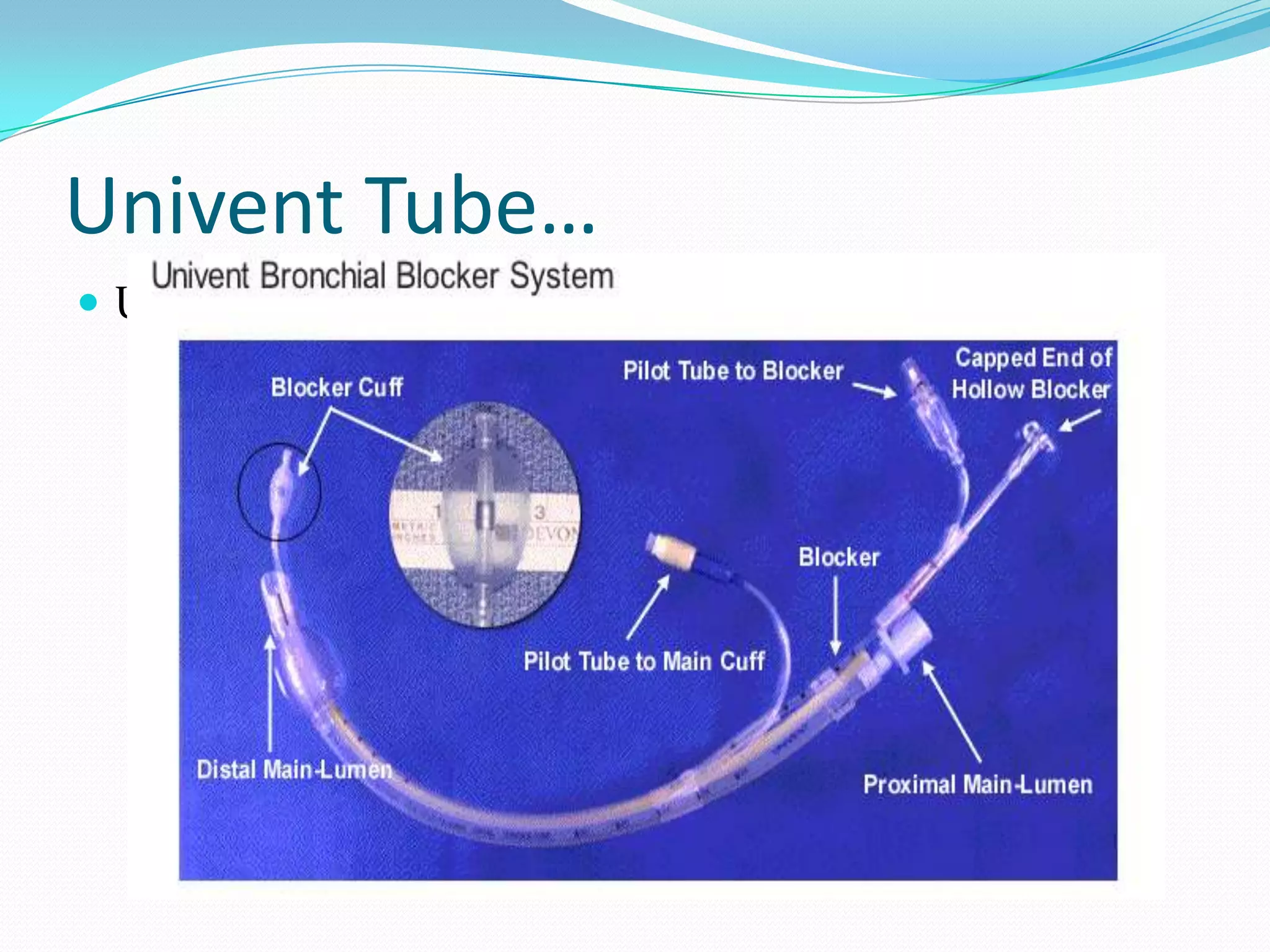
 Bronchial blockers are inflatable devices that are
  placed along side or through a single lumen tracheal
  tube to selectively occlude a bronchial orifice.
 Separation of two lungs is essential for a variety of
  thoracic surgical procedures and can be life-saving in
  certain clinical situations.
 A variety of methods have been described and used to
  isolate one lung.
 These methods includs
       Double-lumen ETT (DLT)
       Bronchial blockers.
       Single lumen bronchial tube
Indications For Separation Of Two Lungs and/or
OLV


    ABSOLUTE
 1. To avoid contamination of a non-diseased lung
      A. Infection (e.g. unilateral pulmonary abscess)
      B. Massive pulmonary hemorrhage
      C. Unilateral pulmonary lavage (e.g. for pulmonary
      alveolar proteinosis)
 2.Control of distribution of ventilation
      A. Bronchopleural fistula
      B. Bronchopleural cutaneous fistula
      C. Surgical opening of a major conducting airway
      D. Giant unilateral lung cyst or bulla
      E. Tracheobronchial tree disruption
      F. Life-threatening hypoxemia due to unilateral
  lung disease

 3. Video assisted thoracoscopy (VATS)
Relative indications
 Thoracic aortic aneurysm repair
 Pneumonectomy
 Pulmonary resection via median sternotomy
 Upper lobectomy
 Lung transplantation
 Unilateral lung disease causing severe hypoxemia
Indication for bronchial blockers
Bronchial blockers are indicated where DLT insertion is not
  possible or advisable as in patients with
 Difficult airway
 Lesions with in the trachea
 Severely distorted tracheobronchial anatomy
 Cervical spine injuries
 Who cannot tolerate period of apnoea
 on anticoagulants
 Endotracheal tube insitu
 When only a lobe is needed to be blocked rather than entire lung
 When sequential blockage of both lungs is needed
 Patients requiring Mechanical ventilation postoperatively
 To apply CPAP
LIMITATIONS
Slow collapse of the desired lung.
Small lumen of bronchial blockers can be connected to
  suction to facilitate more rapid deflation of the lung.
Small lumen of bronchial blockers allow suctioning of
  air but secretions, blood, and pus cannot be
  eliminated through them and cause obstruction of the
  lumen preventing the application of CPAP.
This is remedied by injecting saline or by placing suction
  and/or an appropriate sized wire down the lumen.
LIMITATIONS..
 Bronchial blockers are more easily dislodged during
  patient positioning and surgical manipulation of the
  lung.
 Elliptical-shaped balloons, versus spherical, help to
  prevent dislodgment.
 Whenever, patient position is changed, correct
  bronchial blocker placement needs to be confirmed
  with bronchoscopy.
 Bronchial blockers present the potential risk of
  perforating a bronchus or lung parenchyma causing a
  pneumothorax.
Types of bronchial blockers
Univent bronchial blocker system
Arndt endobronchial blocker
Cohen Flexitip Endobronchial Blocker
Fogarty embolectomy catheters
Foley catheters,
 Balloon-tipped pulmonary artery catheters,
Univent Tube
 Developed in 1982 by Dr. Inoue
 It is a single-lumen silicone tube with a small
  separate lumen along the anterior concave wall.
 This separate lumen contains the small hollow
  nonlatex bronchial blocker that can extend about
  8-10 cm beyond the tip and it has a blue colored
  high pressure and low volume cuff.
 The lumen of the bronchial blocker is 2 mm in
  internal diameter. The Univent is supplied in sizes
  6.0-9.0 mm internal diameter
Univent Tube…
   After inserting the univent tube the blocker is visualized by using a flexible fiberoptic bronchoscope through
    an airway adapter having a port for bronchoscope.

   The blocker is placed in the desired bronchus under vision.

   When the bronchus needs to be blocked the lung is deflated with the blocker open to atmosphere.

   Cuff should be inflated with minimum amount of ait that would provide seal. This can be achived by
    attaching by sample line from Co2 analysizer to the proximal end of the blocker and noting when the
    waveform disappears

   Typical cuff inflation volume is 5-6cc.

   Univent blocker can also be used with normal tracheal tubes and placed coaxially or in parallel.
Univent Tube…
 Univent Tube
Univent Tube…
Advantage
 Difficult airways requiring OLV
 • Selective lobar blockade
 • Tracheostomized patients requiring OLV
 • Rapid sequence induction that requires OLV
 • Robotic (cardiac, thoracic, or esophageal surgery)
Univent Tube…
Disadvantage
 #8.5–9.0 tied fit to pass through vocal cords
 • Enclosed channel of 2.0 mm (not enough lumen
to aspirate secretions)
 • More expensive ($137.00)
 • Potential for inclusion in the stapling line
The Cohen Bronchial Blocker
• It has 9F external diameter, 1.4mm inner lumen
  and length of 65cm with angle tip
• High volume low pressure blue spherical balloon at
  the tip
• Murphy eyes in the distal tip
• A proximal control wheel to adjust the tip
 deflection . An arrow on the wheel indicates the
 direction to which the tip deflects
The Cohen Bronchial Blocker
Fogarty Embolectomy Catheter
 Single-lumen balloon tipped catheter with a removable
    stylet
   In the parallel fashion, the Fogarty catheter is inserted
    prior to intubation
   In the co-axial fashion, the Fogarty catheter is placed
    through the endotracheal tube
   Both techniques require fiberoptic bronchoscopy to direct
    the Fogarty catheter into the correct pulmonary segment
   Once the catheter is in place, the balloon is
    inflated, sealing the airway
Clinical limitations of the
Fogarty catheter
 No accessory lumen
 So Suction , Oxygen insuffulation or applying
 CPAP to the blocked lung is not possible not
 possible

 Latex allergy
 Low volume high pressure cuff
20
Arndt Endobronchial Blocker set
 Invented by Dr. Arndt, an anesthesiologist
 Is available as a 7 or 9 French, wire-guided, yellow
  catheter, 65 and 78cm lengths, with centimeter markings
  from 10-60.
 Ideal for diff intubation, pre-existing ETT and when postop
  ventilation needed or in pediatric patients
 It consists of
       blocking catheter
       airway adaptor
Arndt Endobronchial Blocker set..
 Blocking catheter: it has a blue colored high-
    volume, low-pressure balloon, which is elliptical or
    spherical in shape.
   A flexible nylon wire passes through the proximal end
    of catheter and extends to the distal end then exits as a
    small loop
   Air way adapter: have 4 ports
   1    15 mm port that attaches to the ETT
   2     ventilation port that connects to the breathing
    circuit
   3     port for a flexible fiberoptic scope
   4     port angled approximately 30º for the blocker
23
Arndt Endobronchial Blocker set..
 The fiberoptic scope (a pediatric scope is most easily used) and
    the blocker are placed through their specific ports in the adapter.
   Use an adequate amount of lubricant on the scope and the
    blocker.
   The scope is threaded through the wire loop at the end of the
    blocker and the wire loop should remain loose.
    The entire unit is placed on the ETT and the circuit connected to
    the ventilation port, allowing continuous ventilation during
    placement.
   The fiberoptic scope is “driven” into the left mainstem bronchus
    and the blocker is gently passed down over the scope until
    resistance is encountered.
   The scope is then gently withdrawn until the carina and the
    blocker are in view.
Single lumen bronchial tube
 Rarely used now
 Gordon Green tube is a right sided Single lumen bronchial
    tube that can be used for left thoracotomies
   It has both tracheal and bronchial cuffs
   Used in pediatric patients or patients with massive
    hemoptysis
   Elastic bougie can be inserted into the chosen bronchus by
    using a bronchoscope and bronchial tube can be railroaded
    over the bougie
   Neither suctioning nor application of CPAP to the
    nonventilated lung is possible

Bronchial blockers & endobronchial tubes

  • 1.
  • 2.
    INTRODUCTION  Bronchial blockersare inflatable devices that are placed along side or through a single lumen tracheal tube to selectively occlude a bronchial orifice.  Separation of two lungs is essential for a variety of thoracic surgical procedures and can be life-saving in certain clinical situations.
  • 3.
     A varietyof methods have been described and used to isolate one lung.  These methods includs Double-lumen ETT (DLT) Bronchial blockers. Single lumen bronchial tube
  • 4.
    Indications For SeparationOf Two Lungs and/or OLV  ABSOLUTE 1. To avoid contamination of a non-diseased lung  A. Infection (e.g. unilateral pulmonary abscess)  B. Massive pulmonary hemorrhage  C. Unilateral pulmonary lavage (e.g. for pulmonary alveolar proteinosis)
  • 5.
     2.Control ofdistribution of ventilation A. Bronchopleural fistula B. Bronchopleural cutaneous fistula C. Surgical opening of a major conducting airway D. Giant unilateral lung cyst or bulla E. Tracheobronchial tree disruption F. Life-threatening hypoxemia due to unilateral lung disease  3. Video assisted thoracoscopy (VATS)
  • 6.
    Relative indications  Thoracicaortic aneurysm repair  Pneumonectomy  Pulmonary resection via median sternotomy  Upper lobectomy  Lung transplantation  Unilateral lung disease causing severe hypoxemia
  • 7.
    Indication for bronchialblockers Bronchial blockers are indicated where DLT insertion is not possible or advisable as in patients with  Difficult airway  Lesions with in the trachea  Severely distorted tracheobronchial anatomy  Cervical spine injuries  Who cannot tolerate period of apnoea  on anticoagulants  Endotracheal tube insitu  When only a lobe is needed to be blocked rather than entire lung  When sequential blockage of both lungs is needed  Patients requiring Mechanical ventilation postoperatively  To apply CPAP
  • 8.
    LIMITATIONS Slow collapse ofthe desired lung. Small lumen of bronchial blockers can be connected to suction to facilitate more rapid deflation of the lung. Small lumen of bronchial blockers allow suctioning of air but secretions, blood, and pus cannot be eliminated through them and cause obstruction of the lumen preventing the application of CPAP. This is remedied by injecting saline or by placing suction and/or an appropriate sized wire down the lumen.
  • 9.
    LIMITATIONS..  Bronchial blockersare more easily dislodged during patient positioning and surgical manipulation of the lung.  Elliptical-shaped balloons, versus spherical, help to prevent dislodgment.  Whenever, patient position is changed, correct bronchial blocker placement needs to be confirmed with bronchoscopy.  Bronchial blockers present the potential risk of perforating a bronchus or lung parenchyma causing a pneumothorax.
  • 10.
    Types of bronchialblockers Univent bronchial blocker system Arndt endobronchial blocker Cohen Flexitip Endobronchial Blocker Fogarty embolectomy catheters Foley catheters,  Balloon-tipped pulmonary artery catheters,
  • 11.
    Univent Tube  Developedin 1982 by Dr. Inoue  It is a single-lumen silicone tube with a small separate lumen along the anterior concave wall.  This separate lumen contains the small hollow nonlatex bronchial blocker that can extend about 8-10 cm beyond the tip and it has a blue colored high pressure and low volume cuff.  The lumen of the bronchial blocker is 2 mm in internal diameter. The Univent is supplied in sizes 6.0-9.0 mm internal diameter
  • 12.
    Univent Tube…  After inserting the univent tube the blocker is visualized by using a flexible fiberoptic bronchoscope through an airway adapter having a port for bronchoscope.  The blocker is placed in the desired bronchus under vision.  When the bronchus needs to be blocked the lung is deflated with the blocker open to atmosphere.  Cuff should be inflated with minimum amount of ait that would provide seal. This can be achived by attaching by sample line from Co2 analysizer to the proximal end of the blocker and noting when the waveform disappears  Typical cuff inflation volume is 5-6cc.  Univent blocker can also be used with normal tracheal tubes and placed coaxially or in parallel.
  • 13.
  • 14.
    Univent Tube… Advantage  Difficultairways requiring OLV  • Selective lobar blockade  • Tracheostomized patients requiring OLV  • Rapid sequence induction that requires OLV  • Robotic (cardiac, thoracic, or esophageal surgery)
  • 15.
    Univent Tube… Disadvantage  #8.5–9.0tied fit to pass through vocal cords  • Enclosed channel of 2.0 mm (not enough lumen to aspirate secretions)  • More expensive ($137.00)  • Potential for inclusion in the stapling line
  • 16.
    The Cohen BronchialBlocker • It has 9F external diameter, 1.4mm inner lumen and length of 65cm with angle tip • High volume low pressure blue spherical balloon at the tip • Murphy eyes in the distal tip • A proximal control wheel to adjust the tip deflection . An arrow on the wheel indicates the direction to which the tip deflects
  • 17.
  • 18.
    Fogarty Embolectomy Catheter Single-lumen balloon tipped catheter with a removable stylet  In the parallel fashion, the Fogarty catheter is inserted prior to intubation  In the co-axial fashion, the Fogarty catheter is placed through the endotracheal tube  Both techniques require fiberoptic bronchoscopy to direct the Fogarty catheter into the correct pulmonary segment  Once the catheter is in place, the balloon is inflated, sealing the airway
  • 19.
    Clinical limitations ofthe Fogarty catheter  No accessory lumen So Suction , Oxygen insuffulation or applying CPAP to the blocked lung is not possible not possible  Latex allergy  Low volume high pressure cuff
  • 20.
  • 21.
    Arndt Endobronchial Blockerset  Invented by Dr. Arndt, an anesthesiologist  Is available as a 7 or 9 French, wire-guided, yellow catheter, 65 and 78cm lengths, with centimeter markings from 10-60.  Ideal for diff intubation, pre-existing ETT and when postop ventilation needed or in pediatric patients  It consists of blocking catheter airway adaptor
  • 22.
    Arndt Endobronchial Blockerset..  Blocking catheter: it has a blue colored high- volume, low-pressure balloon, which is elliptical or spherical in shape.  A flexible nylon wire passes through the proximal end of catheter and extends to the distal end then exits as a small loop  Air way adapter: have 4 ports  1 15 mm port that attaches to the ETT  2 ventilation port that connects to the breathing circuit  3 port for a flexible fiberoptic scope  4 port angled approximately 30º for the blocker
  • 23.
  • 24.
    Arndt Endobronchial Blockerset..  The fiberoptic scope (a pediatric scope is most easily used) and the blocker are placed through their specific ports in the adapter.  Use an adequate amount of lubricant on the scope and the blocker.  The scope is threaded through the wire loop at the end of the blocker and the wire loop should remain loose.  The entire unit is placed on the ETT and the circuit connected to the ventilation port, allowing continuous ventilation during placement.  The fiberoptic scope is “driven” into the left mainstem bronchus and the blocker is gently passed down over the scope until resistance is encountered.  The scope is then gently withdrawn until the carina and the blocker are in view.
  • 25.
    Single lumen bronchialtube  Rarely used now  Gordon Green tube is a right sided Single lumen bronchial tube that can be used for left thoracotomies  It has both tracheal and bronchial cuffs  Used in pediatric patients or patients with massive hemoptysis  Elastic bougie can be inserted into the chosen bronchus by using a bronchoscope and bronchial tube can be railroaded over the bougie  Neither suctioning nor application of CPAP to the nonventilated lung is possible