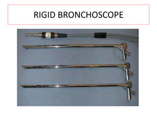
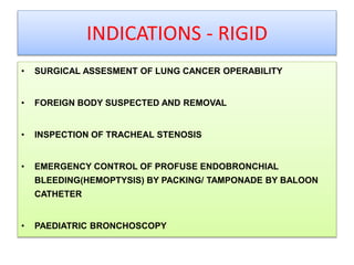
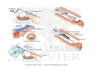
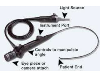
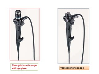
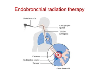
BRONCHOSCOPY BASICS
Dr. RAJASEKHAR THOTTADI
Pulmonary medicine
Rmc ,kakinada
• INTRODUCTION ,HISTORY & EVOLUTION ,
CURRENT APPLICATIONS
• PATIENT PREPARATION, INDICATIONS &
CONSENT
INTRODUCTION
• Bronchoscopy is a technique of visualizing the inside of
the airways for diagnostic and therapeutic purposes
• Flexible bronchoscopy since its inception has had an
exponential growth in pulmonology and clinical medicine. The
advantages of maneuverability, feasibility of wide spectrum of
diagnostics and therapeutics, patient comfort and ease of
conscious sedation has established flexible bronchoscopy as
the most important weapon in a pulmonologists'
armamentarium.
HISTORY & EVALUATION
A German, Gustav Killian, performed the
first bronchoscopy in 1897. From then until
the 1970s, rigid bronchoscopes were used
exclusively. Killian used rigid bronchoscopy
to remove a pork bone. The procedure was
done in an awake patient using topical
cocaine as a local anesthetic.
• Killian demonstrates
the rigid bronchoscopy
in a cadaver specimen
• An American, Chevalier
Jackson, refined the rigid
bronchoscope in the 1920s,
using this rigid tube to visually
inspect the trachea and
mainstem bronchi. The British
laryngologist Victor Negus,
who worked with Jackson,
improved the design of his
endoscopes, including what
came to be called the 'Negus
bronchoscope
• He is sometimes known as the
"father of endoscopy",
although Philipp
Bozzini (1773–1809) is also
often given this sobriquet.
• A Japanese, Shigeto Ikeda, invented
the flexible bronchoscope in 1966. The
flexible scope initially employed
fiberoptic bundles requiring an external
light source for illumination. These
scopes had outside diameters of
approximately 5 mm to 6 mm, with an
ability to flex 180 degrees and to extend
120 degrees, allowing entry into lobar
and segmental bronchi. More recently,
fiberoptic scopes have been replaced
by bronchoscopes with a charge
coupled device (CCD) video chip
located at their distal extremity.
• Shigeto Ikeda
• he developed the first flexible bronchoscope
in conjunction with Machida Endoscope Co.
Ltd (later taken over by Pentax)
and Olympus Optical Co., Ltd. This allowed
better visualisation of the upper
lobe bronchi than is possible with the rigid
bronchoscope. Successive improvements
under his supervision included the
development of video-bronchoscopy.
• His motto was "there is more hope with the
bronchoscope".
TYPES
• RIGID
• FLEXIBLE FIBEROPTIC AND VIDEO
BRONCHOSCOPY
RIGID BRONCHOSCOPE
INDICATIONS - RIGID
• SURGICAL ASSESMENT OF LUNG CANCER OPERABILITY
• FOREIGN BODY SUSPECTED AND REMOVAL
• INSPECTION OF TRACHEAL STENOSIS
• EMERGENCY CONTROL OF PROFUSE ENDOBRONCHIAL
BLEEDING(HEMOPTYSIS) BY PACKING/ TAMPONADE BY BALOON
CATHETER
• PAEDIATRIC BRONCHOSCOPY
• IN TREATMENT OF LARGE AIRWAY TUMOURS BY LASER
• INSPECTION OF SERIOUS CAUSES OF TRACHEAL
NARROWING, WHEREIN A TRACHEAL DILATOR OR STENTING
CAN BE DONE
ADVANTAGES
• wide channel through which large biopsies
and forign bodies can be more easily grasped
and removed
• superior suction capability
DISADVANTAGES
• lack of manoeuvrability
• requirement of a anaesthetist
FIBREOPTIC BRONCHOSCOPY
• The bronchoscope is longer and thinner than a rigid bronchoscope.
It contains a fiberoptic system that transmits an image from the tip of
the instrument to an eyepiece or video camera at the opposite end.
Using Bowden cables connected to a lever at the hand piece, the tip
of the instrument can be oriented, allowing the practitioner to
navigate the instrument into individual lobe or segment bronchi.
Most flexible bronchoscopes also include a channel for suctioning or
instrumentation, but these are significantly smaller than those in a
rigid bronchoscope.
Fiberoptic bronchoscope
with eye piece vediobronchoscope
Current applications
THERAPEUTIC BRONCHOSCOPY
• laser bronchoscopy
– Benign airway stenosis
– Malignant airway obstruction
• Photodynamic therapy
• Endobronchial radiation therapy
• Cryotherapy
• Stenting of airways
• Removal of foreign body
• In hemoptysis
– Lung isolation airway control
– Double-lumen Endotracheal Tube
– Direct Tamponade with Bronchoscope
– Endobronchial Balloon Tamponade
– Use of Topical Vasoconstrictors as Coagulants
– Selective Bronchial Intubation
– Cold Saline Lavage Through Bronchoscope
– Endobronchial Thrombolysis
• laser bronchoscopy
• Laser bronchoscopy uses the thermal and
photochemical effects of laser radiation. Medical
laser machines incorporate a red light producing
helium-neon laser to act as aiming light.
• Unlike ordinary light, laser light waves travel in
phase with each other (coherence), in a single
direction without divergence (collimation) and
represent a single wavelength (monochromatic).
• Application of lasers in pulmonary medicine
• Thermal effects
• Photoresection of airway lesions
Malignant: Bronchogenic carcinoma, metastatic lesions
Benign : Tracheal stenosis, broncholith, suture grartuloma, carcinoid
amyloidosis .
• Photocoagulation
Management of haemoptysis
Bleeding cavitary lesions
• Photochemical effects
• Diagnostic usage (Kr laser)
Carcinoma in situ
Superficial bronchogenic carcinoma
• Therapeutic usage (rhodamine B dye laser)
Palliative resection of malignant airway lesions
Curative therapy for carcinoma in situ,
superficial bronchogenic carcinoma
juvenile laryngo-tracheobronchial papillomatosis
• Stenting of airways
• Endobronchial or endotracheal stents are used where
airway obstruction is caused by extrinsic compression
from a tumour. They may also be required after
endobronchial tumour debulking if the airway has lost its
support structure and also if the tumour is prolapsing
through a lobar bronchus and occluding the main
bronchus
• variety of different stents exist, but the main
group are metallic or non-metallic stents.
• Metallic stents
• subdivided into covered and uncovered
stents.
• usually made from nitinol (a nickel titanium
alloy)
Self-expanding
uncovered nitinol stent.
Nitinol stent,
laser-cut from a single piece
and covered with silicon.
Airway stenting
• Removal of foreign body
• Rigid bronchoscopy has been extensively used in foreign body
removal and is currently recommended as the procedure of choice .
• Rigid bronchoscopy, used in foreign body removal in children, and is
carried out under general anaesthesia.
•
• Several extraction instruments aid in foreign body removal through
bronchoscope include
FB
grasping jaws,
alligator forceps,
four-prong flexible grasping hooks,
Dormia-basket
Fogarty balloons.
alligator forceps
Dormia-basket
four-prong flexible grasping hooks
• For organic foreign bodies which may fracture, either
basket or Fogarty catheters may be used.
• Fogarty ballon catheters
• passing a balloon distal to the foreign body, withdrawal
of inner sheath and telescope 8-10 mm within the outer
sheath, thus creating a hollow space at the tip of the
bronchoscope, inflation of balloon and subsequent
retraction of the foreign body on to the tip of the
bronchoscope.
Fogarty ballon catheters
ADVANTAGES
• Increased experience
• Improved instruments
• Small objects/peripheral
• Use of local anaesthesia and preservation of
cough reflex
DISADVANTAGES
• Narrow channel
• Small extraction instruments
• Potential for distal displacement airways of
foreign body
• Loss of objects in subglottic area
Photodynamic therapy
• Photodynamic therapy involves a photosensitizing agent
which, when exposed to light of proper wavelength, forms
toxic oxygen radicals that result in cell death.
• There are several photosensitizing agents . The most
commonly used ones are porphyrin-based agents and include
haemato-porphyrin derivative named dihaematopor phyrin
ether/ester (DHE, Photofrin II)
• Photodynamic therapy has the potential to cure carcinoma in situ, or
unresectable yet early stage lung carcinoma, where infiltration is
limited to the bronchial mucosa without lymphatic or hematogenous
spread.
• The main limitation is depth of penetration of red light which extends
5 to 15 mm below the tumour surface; once the lesion has extended
deeper into the muscle or cartilage of the bronchial wall, PDT is
solely palliative and other modalities should be included in the
patient's management.
ENDOBRONCHIAL RADIATION THERAPY
• Endobronchial brachytherapy (EBBT) is the placement of
encapsulated radioactive sources within or in close
proximity to a tumour through a bronchoscope used in
the palliative treatment of airway obstruction caused by
bronchogenic carcinoma.
• Low dose brachytherapy
• High dose brachytherapy
Endobronchial radiation therapy
• Low dose brachytherapy
• Low-dose brachytherapy, defined as less than 1 Gy/h to
the point of reference, is effective in about 60%-80% of
patients.
• major disadvantages of low-dose EBBT include the need
for 30-72 hour hospitalization, catheter intolerance and
potential radiation-hazard to hospital workers.
• High dose brachytherapy
• High-dose EBBT is defined as 10 Gy/h to the point of
reference. This is an outpatient procedure requiring
only few minutes per dose.
• Disadvantages
• multiple bronchoscopies are required
• it may be associated with acute side effects.
CRYOTHERAPY
• Cryotherapy refers to the application of extreme cold for local
destruction of lining tissue .
• Cooling can be achieved at different rates : slow (-10°C per
min), rapid (-100°C per sec).
• Cell damage of freezing occurs either as a result of
– fluid shifts between intracellular and extracellular
compartments or
– directly by mechanical damage from ice-crystal formation
• Nitrous oxide is the commonest cooling agent
used in cryotherapy .
• Cryotherapy has recently been combined with
chemotherapy and radiotherapy in the
management of endobronchial tumours.
• In hemoptysis
– Lung isolation airway control
– Double-lumen Endotracheal Tube
– Direct Tamponade with Bronchoscope
– Endobronchial Balloon Tamponade
– Use of Topical Vasoconstrictors as Coagulants
– Selective Bronchial Intubation
– Cold Saline Lavage Through Bronchoscope
– Endobronchial Thrombolysis
Isolation of bleeding site
Double lumen endo-tracheal tube
Selective Bronchial Intubation

Bronchoscopy basics history current applications

  • 1.
    BRONCHOSCOPY BASICS Dr. RAJASEKHARTHOTTADI Pulmonary medicine Rmc ,kakinada
  • 2.
    • INTRODUCTION ,HISTORY& EVOLUTION , CURRENT APPLICATIONS • PATIENT PREPARATION, INDICATIONS & CONSENT
  • 3.
    INTRODUCTION • Bronchoscopy isa technique of visualizing the inside of the airways for diagnostic and therapeutic purposes • Flexible bronchoscopy since its inception has had an exponential growth in pulmonology and clinical medicine. The advantages of maneuverability, feasibility of wide spectrum of diagnostics and therapeutics, patient comfort and ease of conscious sedation has established flexible bronchoscopy as the most important weapon in a pulmonologists' armamentarium.
  • 4.
    HISTORY & EVALUATION AGerman, Gustav Killian, performed the first bronchoscopy in 1897. From then until the 1970s, rigid bronchoscopes were used exclusively. Killian used rigid bronchoscopy to remove a pork bone. The procedure was done in an awake patient using topical cocaine as a local anesthetic.
  • 5.
    • Killian demonstrates therigid bronchoscopy in a cadaver specimen
  • 6.
    • An American,Chevalier Jackson, refined the rigid bronchoscope in the 1920s, using this rigid tube to visually inspect the trachea and mainstem bronchi. The British laryngologist Victor Negus, who worked with Jackson, improved the design of his endoscopes, including what came to be called the 'Negus bronchoscope • He is sometimes known as the "father of endoscopy", although Philipp Bozzini (1773–1809) is also often given this sobriquet.
  • 7.
    • A Japanese,Shigeto Ikeda, invented the flexible bronchoscope in 1966. The flexible scope initially employed fiberoptic bundles requiring an external light source for illumination. These scopes had outside diameters of approximately 5 mm to 6 mm, with an ability to flex 180 degrees and to extend 120 degrees, allowing entry into lobar and segmental bronchi. More recently, fiberoptic scopes have been replaced by bronchoscopes with a charge coupled device (CCD) video chip located at their distal extremity.
  • 8.
    • Shigeto Ikeda •he developed the first flexible bronchoscope in conjunction with Machida Endoscope Co. Ltd (later taken over by Pentax) and Olympus Optical Co., Ltd. This allowed better visualisation of the upper lobe bronchi than is possible with the rigid bronchoscope. Successive improvements under his supervision included the development of video-bronchoscopy. • His motto was "there is more hope with the bronchoscope".
  • 9.
    TYPES • RIGID • FLEXIBLEFIBEROPTIC AND VIDEO BRONCHOSCOPY
  • 10.
  • 11.
    INDICATIONS - RIGID •SURGICAL ASSESMENT OF LUNG CANCER OPERABILITY • FOREIGN BODY SUSPECTED AND REMOVAL • INSPECTION OF TRACHEAL STENOSIS • EMERGENCY CONTROL OF PROFUSE ENDOBRONCHIAL BLEEDING(HEMOPTYSIS) BY PACKING/ TAMPONADE BY BALOON CATHETER • PAEDIATRIC BRONCHOSCOPY
  • 12.
    • IN TREATMENTOF LARGE AIRWAY TUMOURS BY LASER • INSPECTION OF SERIOUS CAUSES OF TRACHEAL NARROWING, WHEREIN A TRACHEAL DILATOR OR STENTING CAN BE DONE
  • 14.
    ADVANTAGES • wide channelthrough which large biopsies and forign bodies can be more easily grasped and removed • superior suction capability
  • 15.
    DISADVANTAGES • lack ofmanoeuvrability • requirement of a anaesthetist
  • 16.
    FIBREOPTIC BRONCHOSCOPY • Thebronchoscope is longer and thinner than a rigid bronchoscope. It contains a fiberoptic system that transmits an image from the tip of the instrument to an eyepiece or video camera at the opposite end. Using Bowden cables connected to a lever at the hand piece, the tip of the instrument can be oriented, allowing the practitioner to navigate the instrument into individual lobe or segment bronchi. Most flexible bronchoscopes also include a channel for suctioning or instrumentation, but these are significantly smaller than those in a rigid bronchoscope.
  • 19.
    Fiberoptic bronchoscope with eyepiece vediobronchoscope
  • 20.
    Current applications THERAPEUTIC BRONCHOSCOPY •laser bronchoscopy – Benign airway stenosis – Malignant airway obstruction • Photodynamic therapy • Endobronchial radiation therapy • Cryotherapy • Stenting of airways • Removal of foreign body
  • 21.
    • In hemoptysis –Lung isolation airway control – Double-lumen Endotracheal Tube – Direct Tamponade with Bronchoscope – Endobronchial Balloon Tamponade – Use of Topical Vasoconstrictors as Coagulants – Selective Bronchial Intubation – Cold Saline Lavage Through Bronchoscope – Endobronchial Thrombolysis
  • 22.
    • laser bronchoscopy •Laser bronchoscopy uses the thermal and photochemical effects of laser radiation. Medical laser machines incorporate a red light producing helium-neon laser to act as aiming light. • Unlike ordinary light, laser light waves travel in phase with each other (coherence), in a single direction without divergence (collimation) and represent a single wavelength (monochromatic).
  • 23.
    • Application oflasers in pulmonary medicine • Thermal effects • Photoresection of airway lesions Malignant: Bronchogenic carcinoma, metastatic lesions Benign : Tracheal stenosis, broncholith, suture grartuloma, carcinoid amyloidosis . • Photocoagulation Management of haemoptysis Bleeding cavitary lesions
  • 24.
    • Photochemical effects •Diagnostic usage (Kr laser) Carcinoma in situ Superficial bronchogenic carcinoma • Therapeutic usage (rhodamine B dye laser) Palliative resection of malignant airway lesions Curative therapy for carcinoma in situ, superficial bronchogenic carcinoma juvenile laryngo-tracheobronchial papillomatosis
  • 25.
    • Stenting ofairways • Endobronchial or endotracheal stents are used where airway obstruction is caused by extrinsic compression from a tumour. They may also be required after endobronchial tumour debulking if the airway has lost its support structure and also if the tumour is prolapsing through a lobar bronchus and occluding the main bronchus
  • 26.
    • variety ofdifferent stents exist, but the main group are metallic or non-metallic stents. • Metallic stents • subdivided into covered and uncovered stents. • usually made from nitinol (a nickel titanium alloy)
  • 27.
    Self-expanding uncovered nitinol stent. Nitinolstent, laser-cut from a single piece and covered with silicon.
  • 28.
  • 29.
    • Removal offoreign body • Rigid bronchoscopy has been extensively used in foreign body removal and is currently recommended as the procedure of choice . • Rigid bronchoscopy, used in foreign body removal in children, and is carried out under general anaesthesia. • • Several extraction instruments aid in foreign body removal through bronchoscope include FB grasping jaws, alligator forceps, four-prong flexible grasping hooks, Dormia-basket Fogarty balloons.
  • 30.
  • 31.
  • 33.
  • 34.
    • For organicforeign bodies which may fracture, either basket or Fogarty catheters may be used. • Fogarty ballon catheters • passing a balloon distal to the foreign body, withdrawal of inner sheath and telescope 8-10 mm within the outer sheath, thus creating a hollow space at the tip of the bronchoscope, inflation of balloon and subsequent retraction of the foreign body on to the tip of the bronchoscope.
  • 35.
  • 36.
    ADVANTAGES • Increased experience •Improved instruments • Small objects/peripheral • Use of local anaesthesia and preservation of cough reflex
  • 37.
    DISADVANTAGES • Narrow channel •Small extraction instruments • Potential for distal displacement airways of foreign body • Loss of objects in subglottic area
  • 38.
    Photodynamic therapy • Photodynamictherapy involves a photosensitizing agent which, when exposed to light of proper wavelength, forms toxic oxygen radicals that result in cell death. • There are several photosensitizing agents . The most commonly used ones are porphyrin-based agents and include haemato-porphyrin derivative named dihaematopor phyrin ether/ester (DHE, Photofrin II)
  • 39.
    • Photodynamic therapyhas the potential to cure carcinoma in situ, or unresectable yet early stage lung carcinoma, where infiltration is limited to the bronchial mucosa without lymphatic or hematogenous spread. • The main limitation is depth of penetration of red light which extends 5 to 15 mm below the tumour surface; once the lesion has extended deeper into the muscle or cartilage of the bronchial wall, PDT is solely palliative and other modalities should be included in the patient's management.
  • 40.
    ENDOBRONCHIAL RADIATION THERAPY •Endobronchial brachytherapy (EBBT) is the placement of encapsulated radioactive sources within or in close proximity to a tumour through a bronchoscope used in the palliative treatment of airway obstruction caused by bronchogenic carcinoma. • Low dose brachytherapy • High dose brachytherapy
  • 41.
  • 42.
    • Low dosebrachytherapy • Low-dose brachytherapy, defined as less than 1 Gy/h to the point of reference, is effective in about 60%-80% of patients. • major disadvantages of low-dose EBBT include the need for 30-72 hour hospitalization, catheter intolerance and potential radiation-hazard to hospital workers.
  • 43.
    • High dosebrachytherapy • High-dose EBBT is defined as 10 Gy/h to the point of reference. This is an outpatient procedure requiring only few minutes per dose. • Disadvantages • multiple bronchoscopies are required • it may be associated with acute side effects.
  • 44.
    CRYOTHERAPY • Cryotherapy refersto the application of extreme cold for local destruction of lining tissue . • Cooling can be achieved at different rates : slow (-10°C per min), rapid (-100°C per sec). • Cell damage of freezing occurs either as a result of – fluid shifts between intracellular and extracellular compartments or – directly by mechanical damage from ice-crystal formation
  • 46.
    • Nitrous oxideis the commonest cooling agent used in cryotherapy . • Cryotherapy has recently been combined with chemotherapy and radiotherapy in the management of endobronchial tumours.
  • 47.
    • In hemoptysis –Lung isolation airway control – Double-lumen Endotracheal Tube – Direct Tamponade with Bronchoscope – Endobronchial Balloon Tamponade – Use of Topical Vasoconstrictors as Coagulants – Selective Bronchial Intubation – Cold Saline Lavage Through Bronchoscope – Endobronchial Thrombolysis
  • 48.
  • 49.
  • 50.