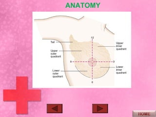
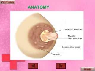
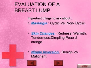
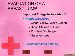
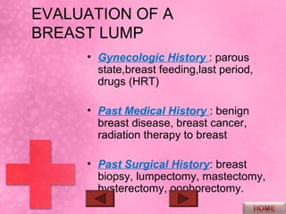
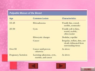
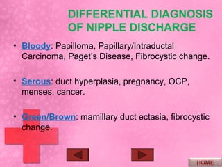
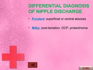
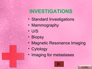
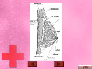
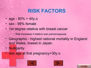
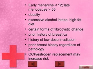
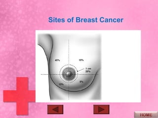
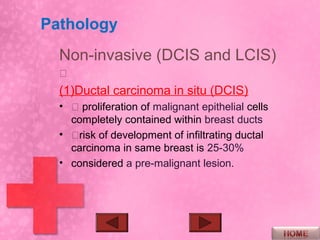
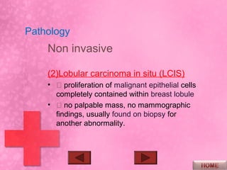
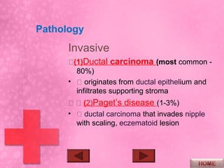
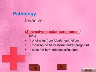
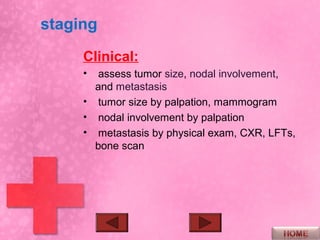
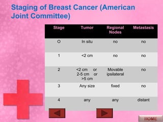
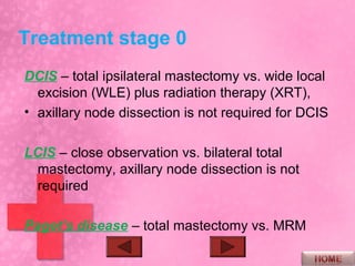
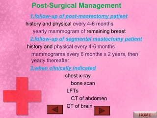
Approaches of
a Patient With a
Breast Lump &
Nipple
Discharge
OBJECTIVES
•   Identify & Discuss the Differential Diagnosis of a
    Breast Lump

•   Present and Interpret a Good History & physical
    Examination for a Patient with a Breast Lump.

•   Discuss the Possible Causes of Nipple Discharge
    According to the Color & Nature of the Discharge.

•   Present a sound Approach to Investigate a Patient
    with a Breast Lump & Nipple Discharge.

•   Present a Possible Approach for Treatment.
ANATOMY
ANATOMY
ANATOMY
ANATOMY
BLOOD SUPPLY
LYMPHATIC DRAINAGE
HISTORY

• Personal Data

• Chief Complaint

• H.C.C : when and how first noticed,
  Pain, tenderness, change in size
  over time and with menstruation.
EVALUATION OF A
BREAST LUMP
    Important things to ask about :
    • Mastalgia : Cyclic Vs. Non- Cyclic

    • Skin Changes : Redness, Warmth,
      Tenderness,Dimpling,Peau d’
      orange

    • Nipple Inversion : Benign Vs.
      Malignant
EVALUATION OF A
BREAST LUMP
      Important Things to Ask About :
      • Nipple Discharge
          - Clear, Yellow, White, Green
          - Blood Stained or Dark
          - Purulent Discharge
          - Galactorrhoea

      • Gynaecomastia ( Males )
EVALUATION OF A
BREAST LUMP
     • Gynecologic History : parous
       state,breast feeding,last period,
       drugs (HRT)

     • Past Medical History : benign
       breast disease, breast cancer,
       radiation therapy to breast

     • Past Surgical History: breast
       biopsy, lumpectomy, mastectomy,
       hysterectomy, oophorectomy.
EVALUATION OF A
BREAST LUMP
• Family History : Especially in first
  degree relatives.

• Constitutional Features :
   - Anorexia
   - weight loss
   - Respiratory Symptoms
   - Bone Pain
PHYSICAL EXAM
 – Inspection, Palpation
• Skin changes: Edema, Dimpling, Redness,
  Retraction, Ulceration, Peau d’orande
• Nipple: Bloody Discharge, Crusting, Ulceration,
  Inversion.
• Prominent Veins
• palpable axillary/supraclavicular lymph nodes
• Arm Edema
EVALUATION OF A
BREAST LUMP
Characteristics of a Breast mass
• Size
• Position
• Mobility
• Composition ( Fluctuant, Hard,
  Rubbery )
• Fixation to underlying tissue or skin
Differential Diagnosis of
a Breast Lump
•   FIBROCYSTIC DISEASE
•   CYSTS
•   ADENOMAS
•   FIBROADENOMA
•   FAT NECROSIS
•   PAPILLOMA
•   BREAST CANCER
DIFFERENTIAL DIAGNOSIS
             OF NIPPLE DISCHARGE
• Bloody: Papilloma, Papillary/Intraductal
  Carcinoma, Paget’s Disease, Fibrocystic change.

• Serous: duct hyperplasia, pregnancy, OCP,
  menses, cancer.

• Green/Brown: mamillary duct ectasia, fibrocystic
  change.
DIFFERENTIAL DIAGNOSIS
OF NIPPLE DISCHARGE
• Purulent: superficial or central abscess

• Milky: post-lactation, OCP, prolactinoma
INVESTIGATIONS
•   Standard Investigations
•   Mammography
•   U/S
•   Biopsy
•   Magnetic Resonance Imaging
•   Cytology
•   Imaging for metastases
INVESTIGATIONS
•   Standard Investigations
•   Mammography
•   U/S
•   Biopsy
•   Magnetic Resonance Imaging
•   Cytology
•   Imaging for metastases
Mammography
  Indications
  • screening – every 1-2 years for
    women ages 50-69.

  • metastatic adenocarcinoma of
    unknown primary.

  • nipple discharge without palpable
    mass.
Mammography
Mammogram findings indicative of
 malignancy

• stellate appearance and spiculated
  border - pathognomonic of breast
  cancer.
• microcalcifications, ill-defined lesion
  border.
• lobulation, architectural distortion
Mammography
NOTE:


• normal mammogram does not rule
  out suspicion of cancer, based on
  clinical findings.
Ultrasonography


• Performed primarily to differentiate
  cystic from solid lesions.
• Not diagnostic
Biopsy
  • The diagnosis of breast cancer
    depends upon examination of tissue
    or cells removed by biopsy.
  • The safest course is biopsy
    examination of all suspicious
    masses found on physical
    examination and of suspicious
    lesions demonstrated by
    mammography.
Biopsy
• The simplest method is needle
  biopsy, either by aspiration of tumor
  cells ( fine – needle aspiration
  cytology) or by obtaining a small
  core of tissue with a hollow needle.
• Open biopsy… ( incisional or
  excisional )
Magnetic Resonance Imaging
       • High Sensitivity for breast cancer
       • Can demonstrate the extent of both
         invasive & non-invasive disease.
       • Determines weather a
         mammographic lesion at the site of
         previous surgery is due to scar or
         recurrence.
       • The optimum method for imaging
         breast implants and detecting
         implant leakage or rupture.
cytology
   • Cytologic examination of nipple
     discharge or cyst fluid may be
     helpful on rare occasions.
   • As a rule, mammography and breast
     biopsy are required when nipple
     discharge or cyst fluid is bloody or
     cytologically questionable.
Imaging for metastases
     • Chest x-ray may show pulmonary
       metastases.
     • CT scanning of liver and brain is of
       value only when metastases are
       suspected in these areas.
Benign Breast Lumps
  •   FIBROCYSTIC DISEASE
  •   FIBROADENOMA
  •   FAT NECROSIS
  •   PAPILLOMA
  •   FIBROADENOSIS-focal/diffuse

                      nodularity
  • GALACTOCOELE
  • ABSCESS
  • PERIDUCTAL MASTITIS-secondary to
                        duct ectasia
FIBROCYSTIC DISEASE
  • Benign breast condition consisting
    of fibrous and cystic changes in
    breast.
  • Age : 30-50 years
  • Clinical Features
     - breast pain
     - swelling with focal areas of
    nodularity or cysts
     - Frequently bilateral
     - varies with menstrual cycle
FIBROCYSTIC DISEASE
    Treatment
    • If no dominant mass, observe to
      ensure no mass dominates.
    • For a dominant mass, FNA
    • If > 40 years, mammography every
      3 years
    • Avoid xanthine-containing products
      (coffee, tea, chocolate, cola drinks)
      and nicotine.
    • For severe symptoms – danozol (2-
      3 months), or tamoxifen (4-6 weeks)
FIBROADENOMA
    • Most common benign breast tumour
      in women under age 30.
    • No malignant potential
    • Clinical features – smooth, rubbery,
      discrete, well circumscribed nodule,
      non-tender, mobile, hormonally
      dependant.
    • Management – usually excised to
      confirm diagnosis
FAT NECROSIS
• Due to trauma (although positive history in only
  50%).

• Clinical features – firm, ill-defined mass with skin
  or nipple retraction, +/– tenderness.

• Management – will regress spontaneously but
  complete excisional biopsy is the safest approach
  to rule out carcinoma.
PAPILLOMA
    • Solitary intraductal benign polyp.

    • Most common cause of bloody nipple
      discharge.

    • Management – excision of involved duct
Breast Cancer
   •   Epidemiology
   •   Risk factors
   •   Pathology
   •   Staging (clinical & pathological)
   •   Metastasis
   •   Treatment
   •   Local/Regional Recurrence
   •   Prognosis
EPIDEMIOLOGY
   • Most common cancer in women.

   • Second leading cause of cancer
     mortality in women.

   • Most common cause of death in 5th
     decade.

   • Lifetime risk of 1/9
RISK FACTORS
• age - 80% > 40y.o
• sex - 99% female
• 1st degree relative with breast cancer
     - Risk increases if relative was premenopausal.
• Geographic - highest national mortality in England
  and Wales, lowest in Japan.
• Nulliparity
• late age at first pregnancy>30y.o
• Early menarche < 12; late
  menopause > 55
• obesity
• excessive alcohol intake, high fat
  diet
• certain forms of fibrocystic change
• prior history of breast ca
• history of low-dose irradiation
• prior breast biopsy regardless of
  pathology
• OCP/estrogen replacement may
  increase risk
Sites of Breast Cancer
Pathology
  Non-invasive (DCIS and LCIS)
  
  (1)Ductal carcinoma in situ (DCIS)
  •  proliferation of malignant epithelial cells
    completely contained within breast ducts
  • risk of development of infiltrating ductal
    carcinoma in same breast is 25-30%
  • considered a pre-malignant lesion.
Pathology
    Non invasive

    (2)Lobular carcinoma in situ (LCIS)
    •  proliferation of malignant epithelial cells
      completely contained within breast lobule
    •  no palpable mass, no mammographic
      findings, usually found on biopsy for
      another abnormality.
Pathology
      Invasive
      (1)Ductal carcinoma (most common -
        80%)
      •  originates from ductal epithelium and
        infiltrates supporting stroma
        (2)Paget’s disease (1-3%)
      •  ductal carcinoma that invades nipple
        with scaling, eczematoid lesion
Pathology
     Invasive

     (3)Invasive lobular carcinoma (8-
       10%)
     •  originates from lobular epithelium
     •  more apt to be bilateral, better prognosis
     •  does not form microcalcifications
     
Pathology
       Invasive
       (4)Inflammatory carcinoma (1-4%)
       •  ductal carcinoma that involves dermal
          lymphatics
       •  most aggressive form of breast cancer
       •  presents with erythema, skin edema,
          warm swollen tender breast, +/– lump
       •  peau d’orange indicates advanced
          disease (IIIb-IV)
staging
     Clinical:
     •  assess tumor size, nodal involvement,
       and metastasis
     • tumor size by palpation, mammogram
     • nodal involvement by palpation
     • metastasis by physical exam, CXR, LFTs,
       bone scan
staging
Pathological
• Histology
• axillary dissection should be performed for accurate
   staging and to reduce risk of axillary recurrence
• estrogen/progesterone receptor testing
  presence or absence of estrogen and progesterone
   receptors,
Staging of Breast Cancer (American
Joint Committee)
           Stage    Tumor      Regional      Metastasis
                                Nodes

            O        In situ       no            no


            1        <2 cm         no            no


            2      <2 cm or     Movable          no
                   2-5 cm or   ipsilateral
                      >5 cm
            3       Any size     fixed           no


            4         any         any          distant
Treatment
    • Primary Treatment of Breast Cancer
    • total mastectomy – removes breast tissue,
       nipple-areolar complex and skin
    • modified radical mastectomy (MRM) –
       removes breast tissue, pectorali fascia,
    nipple-areolar complex, skin and axillary
       lymph nodes
Treatment stage 0
DCIS – total ipsilateral mastectomy vs. wide local
  excision (WLE) plus radiation therapy (XRT),
• axillary node dissection is not required for DCIS

LCIS – close observation vs. bilateral total
 mastectomy, axillary node dissection is not
 required

Paget’s disease – total mastectomy vs. MRM
Treatment stages I,II
     surgery for cure

     MRM vs. WLE with axillary node
      dissection plus XRT

     adjuvant chemotherapy in node-
       positive patients and high risk node-
       negative patient
Treatment Stages III,IV
 operate for local control
 includes surgery, radiation and systemic therapy
   individualized, but mastectomy is most common
   procedure
• even with aggressive therapy most patients die as a result of
  distant metastasis

Indication of chemotherapy
                       tumors > 5 cm
                       inflammatory carcinomas
                       chest wall or skin extention
• Adjuvant Therapy –
  Combination Chemotherapy
• indications – node-positive patients, high
  risk node-negative patients, and palliation
  for metastatic disease,
• ER negative patients
• CMF (cyclophosphamide, methotrexate, 5-
  flurouracil) x 6 months
Adjuvant Therapy -
 Hormonal
• indications – ER positive, pre- or
  post-menopausal, node-positive or
  high risk node-negative patients.
• • adjuvant or palliative therapy
• Tamoxifen (anti-estrogen) – is agent
  of choice, continue for 5 years
• alternatives to tamoxifen
• Adjuvant Therapy -
  Radiation
• with breast-conserving surgery
• those with high-risk of local
  recurrence
• adjuvant radiation to breast
  decreases local recurrence,
  increases disease free survival
• (no change in overall survival)
Post-Surgical Management
  1.follow-up of post-mastectomy patient
history and physical every 4-6 months
    yearly mammogram of remaining breast
  2.follow-up of segmental mastectomy patient
 history and physical every 4-6 months
   mammograms every 6 months x 2 years, then
   yearly thereafter
  3.when clinically indicated
                  chest x-ray
                     bone scan
                   LFTs
                     CT of abdomen
                   CT of brain
CHEMOTHERAPY
Breast lump

Breast lump

  • 2.
    Approaches of a PatientWith a Breast Lump & Nipple Discharge
  • 3.
    OBJECTIVES • Identify & Discuss the Differential Diagnosis of a Breast Lump • Present and Interpret a Good History & physical Examination for a Patient with a Breast Lump. • Discuss the Possible Causes of Nipple Discharge According to the Color & Nature of the Discharge. • Present a sound Approach to Investigate a Patient with a Breast Lump & Nipple Discharge. • Present a Possible Approach for Treatment.
  • 4.
  • 5.
  • 6.
  • 7.
  • 8.
  • 10.
  • 11.
    HISTORY • Personal Data •Chief Complaint • H.C.C : when and how first noticed, Pain, tenderness, change in size over time and with menstruation.
  • 12.
    EVALUATION OF A BREASTLUMP Important things to ask about : • Mastalgia : Cyclic Vs. Non- Cyclic • Skin Changes : Redness, Warmth, Tenderness,Dimpling,Peau d’ orange • Nipple Inversion : Benign Vs. Malignant
  • 13.
    EVALUATION OF A BREASTLUMP Important Things to Ask About : • Nipple Discharge - Clear, Yellow, White, Green - Blood Stained or Dark - Purulent Discharge - Galactorrhoea • Gynaecomastia ( Males )
  • 14.
    EVALUATION OF A BREASTLUMP • Gynecologic History : parous state,breast feeding,last period, drugs (HRT) • Past Medical History : benign breast disease, breast cancer, radiation therapy to breast • Past Surgical History: breast biopsy, lumpectomy, mastectomy, hysterectomy, oophorectomy.
  • 15.
    EVALUATION OF A BREASTLUMP • Family History : Especially in first degree relatives. • Constitutional Features : - Anorexia - weight loss - Respiratory Symptoms - Bone Pain
  • 16.
    PHYSICAL EXAM –Inspection, Palpation • Skin changes: Edema, Dimpling, Redness, Retraction, Ulceration, Peau d’orande • Nipple: Bloody Discharge, Crusting, Ulceration, Inversion. • Prominent Veins • palpable axillary/supraclavicular lymph nodes • Arm Edema
  • 17.
    EVALUATION OF A BREASTLUMP Characteristics of a Breast mass • Size • Position • Mobility • Composition ( Fluctuant, Hard, Rubbery ) • Fixation to underlying tissue or skin
  • 18.
    Differential Diagnosis of aBreast Lump • FIBROCYSTIC DISEASE • CYSTS • ADENOMAS • FIBROADENOMA • FAT NECROSIS • PAPILLOMA • BREAST CANCER
  • 20.
    DIFFERENTIAL DIAGNOSIS OF NIPPLE DISCHARGE • Bloody: Papilloma, Papillary/Intraductal Carcinoma, Paget’s Disease, Fibrocystic change. • Serous: duct hyperplasia, pregnancy, OCP, menses, cancer. • Green/Brown: mamillary duct ectasia, fibrocystic change.
  • 21.
    DIFFERENTIAL DIAGNOSIS OF NIPPLEDISCHARGE • Purulent: superficial or central abscess • Milky: post-lactation, OCP, prolactinoma
  • 22.
    INVESTIGATIONS • Standard Investigations • Mammography • U/S • Biopsy • Magnetic Resonance Imaging • Cytology • Imaging for metastases
  • 23.
    INVESTIGATIONS • Standard Investigations • Mammography • U/S • Biopsy • Magnetic Resonance Imaging • Cytology • Imaging for metastases
  • 24.
    Mammography Indications • screening – every 1-2 years for women ages 50-69. • metastatic adenocarcinoma of unknown primary. • nipple discharge without palpable mass.
  • 25.
    Mammography Mammogram findings indicativeof malignancy • stellate appearance and spiculated border - pathognomonic of breast cancer. • microcalcifications, ill-defined lesion border. • lobulation, architectural distortion
  • 26.
    Mammography NOTE: • normal mammogramdoes not rule out suspicion of cancer, based on clinical findings.
  • 27.
    Ultrasonography • Performed primarilyto differentiate cystic from solid lesions. • Not diagnostic
  • 28.
    Biopsy •The diagnosis of breast cancer depends upon examination of tissue or cells removed by biopsy. • The safest course is biopsy examination of all suspicious masses found on physical examination and of suspicious lesions demonstrated by mammography.
  • 29.
    Biopsy • The simplestmethod is needle biopsy, either by aspiration of tumor cells ( fine – needle aspiration cytology) or by obtaining a small core of tissue with a hollow needle. • Open biopsy… ( incisional or excisional )
  • 30.
    Magnetic Resonance Imaging • High Sensitivity for breast cancer • Can demonstrate the extent of both invasive & non-invasive disease. • Determines weather a mammographic lesion at the site of previous surgery is due to scar or recurrence. • The optimum method for imaging breast implants and detecting implant leakage or rupture.
  • 31.
    cytology • Cytologic examination of nipple discharge or cyst fluid may be helpful on rare occasions. • As a rule, mammography and breast biopsy are required when nipple discharge or cyst fluid is bloody or cytologically questionable.
  • 32.
    Imaging for metastases • Chest x-ray may show pulmonary metastases. • CT scanning of liver and brain is of value only when metastases are suspected in these areas.
  • 33.
    Benign Breast Lumps • FIBROCYSTIC DISEASE • FIBROADENOMA • FAT NECROSIS • PAPILLOMA • FIBROADENOSIS-focal/diffuse nodularity • GALACTOCOELE • ABSCESS • PERIDUCTAL MASTITIS-secondary to duct ectasia
  • 35.
    FIBROCYSTIC DISEASE • Benign breast condition consisting of fibrous and cystic changes in breast. • Age : 30-50 years • Clinical Features - breast pain - swelling with focal areas of nodularity or cysts - Frequently bilateral - varies with menstrual cycle
  • 36.
    FIBROCYSTIC DISEASE Treatment • If no dominant mass, observe to ensure no mass dominates. • For a dominant mass, FNA • If > 40 years, mammography every 3 years • Avoid xanthine-containing products (coffee, tea, chocolate, cola drinks) and nicotine. • For severe symptoms – danozol (2- 3 months), or tamoxifen (4-6 weeks)
  • 37.
    FIBROADENOMA • Most common benign breast tumour in women under age 30. • No malignant potential • Clinical features – smooth, rubbery, discrete, well circumscribed nodule, non-tender, mobile, hormonally dependant. • Management – usually excised to confirm diagnosis
  • 38.
    FAT NECROSIS • Dueto trauma (although positive history in only 50%). • Clinical features – firm, ill-defined mass with skin or nipple retraction, +/– tenderness. • Management – will regress spontaneously but complete excisional biopsy is the safest approach to rule out carcinoma.
  • 39.
    PAPILLOMA • Solitary intraductal benign polyp. • Most common cause of bloody nipple discharge. • Management – excision of involved duct
  • 40.
    Breast Cancer • Epidemiology • Risk factors • Pathology • Staging (clinical & pathological) • Metastasis • Treatment • Local/Regional Recurrence • Prognosis
  • 41.
    EPIDEMIOLOGY • Most common cancer in women. • Second leading cause of cancer mortality in women. • Most common cause of death in 5th decade. • Lifetime risk of 1/9
  • 42.
    RISK FACTORS • age- 80% > 40y.o • sex - 99% female • 1st degree relative with breast cancer - Risk increases if relative was premenopausal. • Geographic - highest national mortality in England and Wales, lowest in Japan. • Nulliparity • late age at first pregnancy>30y.o
  • 43.
    • Early menarche< 12; late menopause > 55 • obesity • excessive alcohol intake, high fat diet • certain forms of fibrocystic change • prior history of breast ca • history of low-dose irradiation • prior breast biopsy regardless of pathology • OCP/estrogen replacement may increase risk
  • 44.
  • 45.
    Pathology Non-invasive(DCIS and LCIS)  (1)Ductal carcinoma in situ (DCIS) •  proliferation of malignant epithelial cells completely contained within breast ducts • risk of development of infiltrating ductal carcinoma in same breast is 25-30% • considered a pre-malignant lesion.
  • 46.
    Pathology Non invasive (2)Lobular carcinoma in situ (LCIS) •  proliferation of malignant epithelial cells completely contained within breast lobule •  no palpable mass, no mammographic findings, usually found on biopsy for another abnormality.
  • 47.
    Pathology Invasive (1)Ductal carcinoma (most common - 80%) •  originates from ductal epithelium and infiltrates supporting stroma   (2)Paget’s disease (1-3%) •  ductal carcinoma that invades nipple with scaling, eczematoid lesion
  • 48.
    Pathology Invasive (3)Invasive lobular carcinoma (8- 10%) •  originates from lobular epithelium •  more apt to be bilateral, better prognosis •  does not form microcalcifications 
  • 49.
    Pathology Invasive (4)Inflammatory carcinoma (1-4%) •  ductal carcinoma that involves dermal lymphatics •  most aggressive form of breast cancer •  presents with erythema, skin edema, warm swollen tender breast, +/– lump •  peau d’orange indicates advanced disease (IIIb-IV)
  • 50.
    staging Clinical: • assess tumor size, nodal involvement, and metastasis • tumor size by palpation, mammogram • nodal involvement by palpation • metastasis by physical exam, CXR, LFTs, bone scan
  • 51.
    staging Pathological • Histology • axillarydissection should be performed for accurate staging and to reduce risk of axillary recurrence • estrogen/progesterone receptor testing presence or absence of estrogen and progesterone receptors,
  • 52.
    Staging of BreastCancer (American Joint Committee) Stage Tumor Regional Metastasis Nodes O In situ no no 1 <2 cm no no 2 <2 cm or Movable no 2-5 cm or ipsilateral >5 cm 3 Any size fixed no 4 any any distant
  • 53.
    Treatment • Primary Treatment of Breast Cancer • total mastectomy – removes breast tissue, nipple-areolar complex and skin • modified radical mastectomy (MRM) – removes breast tissue, pectorali fascia, nipple-areolar complex, skin and axillary lymph nodes
  • 54.
    Treatment stage 0 DCIS– total ipsilateral mastectomy vs. wide local excision (WLE) plus radiation therapy (XRT), • axillary node dissection is not required for DCIS LCIS – close observation vs. bilateral total mastectomy, axillary node dissection is not required Paget’s disease – total mastectomy vs. MRM
  • 55.
    Treatment stages I,II surgery for cure MRM vs. WLE with axillary node dissection plus XRT adjuvant chemotherapy in node- positive patients and high risk node- negative patient
  • 56.
    Treatment Stages III,IV operate for local control includes surgery, radiation and systemic therapy individualized, but mastectomy is most common procedure • even with aggressive therapy most patients die as a result of distant metastasis Indication of chemotherapy tumors > 5 cm inflammatory carcinomas chest wall or skin extention
  • 57.
    • Adjuvant Therapy– Combination Chemotherapy • indications – node-positive patients, high risk node-negative patients, and palliation for metastatic disease, • ER negative patients • CMF (cyclophosphamide, methotrexate, 5- flurouracil) x 6 months
  • 58.
    Adjuvant Therapy - Hormonal • indications – ER positive, pre- or post-menopausal, node-positive or high risk node-negative patients. • • adjuvant or palliative therapy • Tamoxifen (anti-estrogen) – is agent of choice, continue for 5 years • alternatives to tamoxifen
  • 59.
    • Adjuvant Therapy- Radiation • with breast-conserving surgery • those with high-risk of local recurrence • adjuvant radiation to breast decreases local recurrence, increases disease free survival • (no change in overall survival)
  • 60.
    Post-Surgical Management 1.follow-up of post-mastectomy patient history and physical every 4-6 months yearly mammogram of remaining breast 2.follow-up of segmental mastectomy patient history and physical every 4-6 months mammograms every 6 months x 2 years, then yearly thereafter 3.when clinically indicated chest x-ray bone scan LFTs CT of abdomen CT of brain
  • 61.