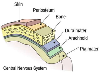
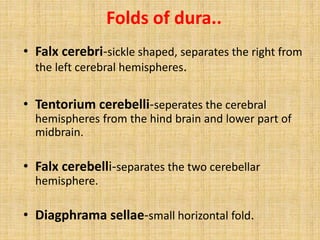
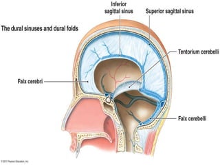
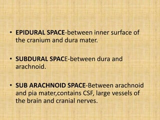
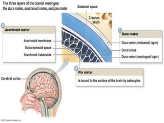
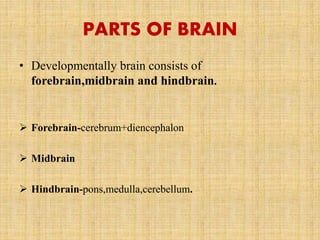
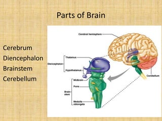
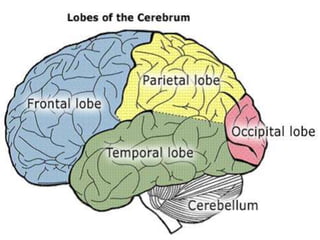
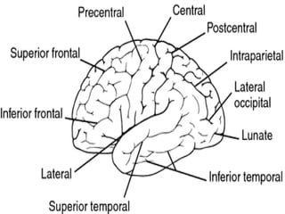
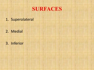
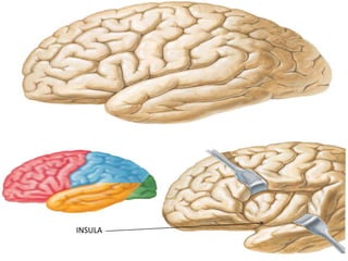
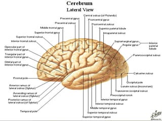
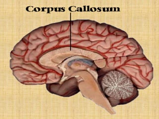
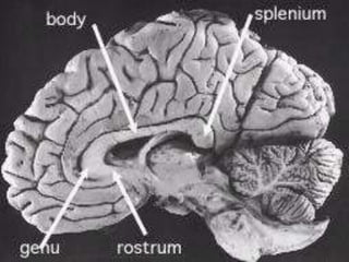
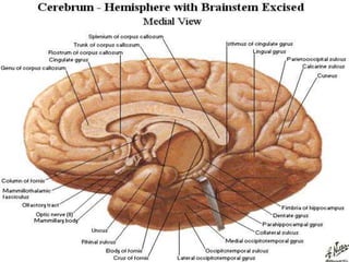
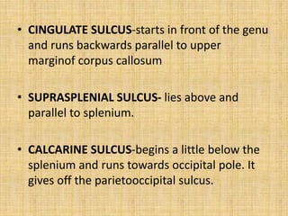
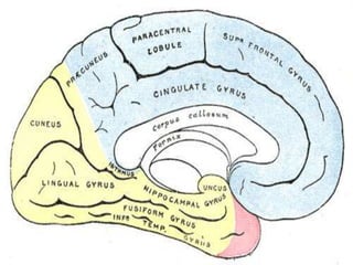
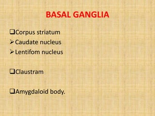
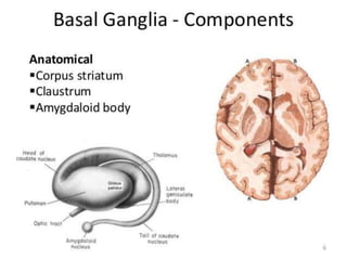
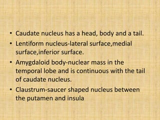
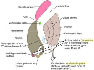
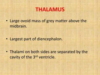
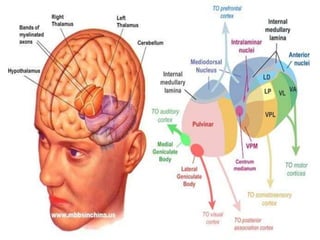
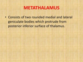
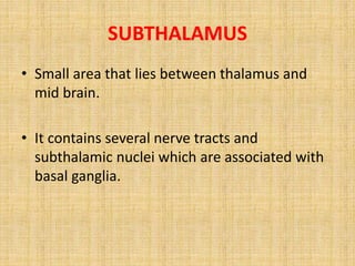
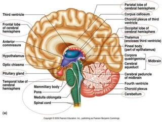
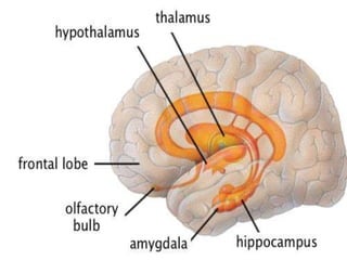
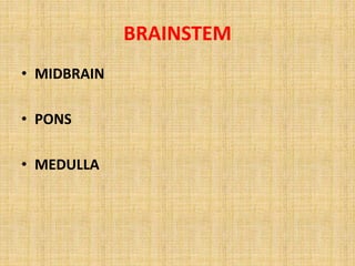
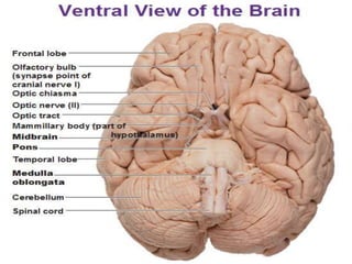
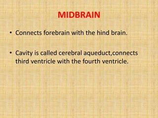
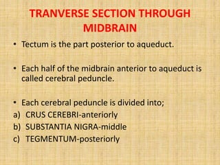
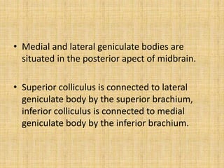
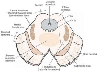
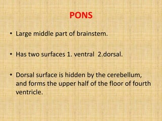
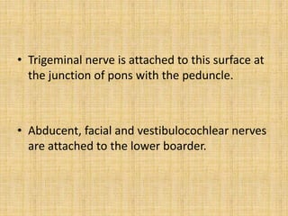
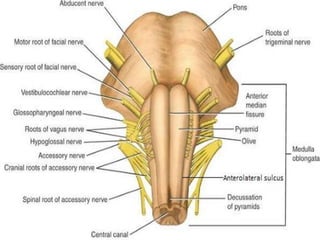
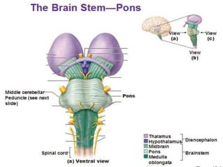
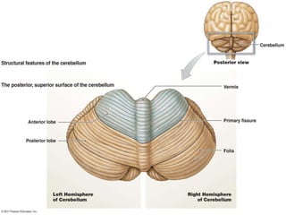
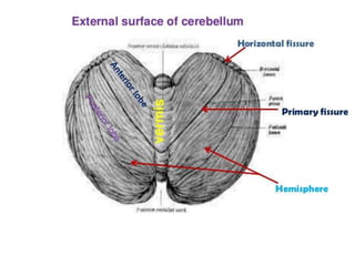
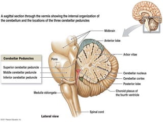
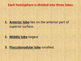
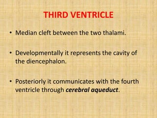
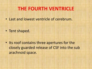
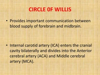
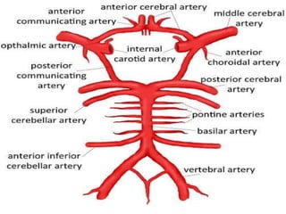
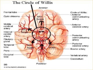
ANATOMY OF BRAIN
PRESENTER: DR THOMAS
JOHN
CENTRAL NERVOUS SYSTEM
• CNS consists of brain and spinal cord. Brain is
situated within the cranial cavity and the spinal cord
lies within the vertebral column.
• The adult brain constitutes about one fiftieth of total
body weight and weighs about 1400g in males and
1200g in females.
• BRAIN- gray matter is present around the periphery
and white matter in the centre.
BRAIN IS PROTECTED BY.....
1. Bony covering-THE CRANIUM
2. The meninges
3. CSF-in the sub arachnoid space
MENINGES
1) DURA MATER-outer endosteal layer and
inner meningeal layer enclosing the cranial
venous sinuses between the two.
2) ARACHNOID-thin,transparent and do not dip
into the sulci.
3) PIA MATER-thin, vascular and closely invests
the brain
• Dura mater is called as pachymeninges.
• Arachnoid and pia are togather known as
leptomeninges.
• Meningeal layer of dura mater forms four
folds which divide the cavity into
intercommunicating compartments.
Folds of dura..
• Falx cerebri-sickle shaped, separates the right from
the left cerebral hemispheres.
• Tentorium cerebelli-seperates the cerebral
hemispheres from the hind brain and lower part of
midbrain.
• Falx cerebelli-separates the two cerebellar
hemisphere.
• Diagphrama sellae-small horizontal fold.
• Superior sagittal sinus lies along the superior
margin of falx cerebri-recieves in its course the
superior cerebral vein.
• Inferior sagittal sinus lies along the inferior
margin of falx cerebri-joins great cerebral vein.
• Straight sinus-lies at the confluence of falx
cerebri and tentorium cerebelli.
ARACHNOID MATER
• It provides sheath for the cranial nerves as far
as their exit from the skull.
• Arachnoid villi are small finger like processes
of arachnoid tissue projecting into the venous
sinuses. They absorb CSF. With age these villi
enlarge to form pedunculated tufts called
arachnoid granulations.
• EPIDURAL SPACE-between inner surface of
the cranium and dura mater.
• SUBDURAL SPACE-between dura and
arachnoid.
• SUB ARACHNOID SPACE-Between arachnoid
and pia mater,contains CSF, large vessels of
the brain and cranial nerves.
• The function of CSF is primarily as a shock
absorber and a cushion for the brain so that it
doesn't move around so much in the head.
• CSF is secreted by specialized cells within the
walls of the ventricles known as choroid
plexus.
• Re-absorption occurs in the subarachnoid
space by venous structures known as
"arachnoid villi"
PARTS OF BRAIN
• Developmentally brain consists of
forebrain,midbrain and hindbrain.
 Forebrain-cerebrum+diencephalon
 Midbrain
 Hindbrain-pons,medulla,cerebellum.
Parts of Brain
Cerebrum
Diencephalon
Brainstem
Cerebellum
CEREBRUM
• Longitudinal cerebral fissure-divides it into two
hemispheres.
• Corpus callosum- joins the two hemispheres across the
median plane
• Each cerbral hemisphere consists of
 Outer grey matter
 Inner white matter
 Basal ganglia/basal nuclei
 Lateral ventricle.
Each cerebral hemisphere has 3 poles
1. FRONTAL POLE
2. TEMPORAL POLE
3. OCCIPITAL POLE.
LOBES OF CEREBRAL CORTEX
• FRONTAL
• PARIETAL
• TEMPORAL
• OCCIPITAL
SULCI AND GYRI
• In order to increase the surface area of brain it
is folded at places creating sulci and gyri.
• The grooves that make these wrinkles are
called sulci and the ridges between them are
called gyri.
MEDICOLEGAL IMPORTANCE
• In generalised edema of the brain flattening
of the cerebral convulsions with obliteration
of sulci is seen.
• In such cases it is difficult to appreciate the
sulci and gyri correctly.
SULCI AND GYRI
• Central sulcus
• Lateral sulcus
• Parieto occipital sulcus
.
• CENTRAL SULCUS- begins on the medial
surface and runs on the superolateral sulcus
obliquely downwards and forwardsand ends a
little abive the posterior ramus of the lateral
sulcus
• LATERAL SULCUS- inferiorly separates the
orbital and tentorial surfaces,then reaches
superolaterally and divides into anterior,
ascending and posterior rami
• PARIETO OCCIPITAL SULCUS-present on the
medial aspect.
• PREOCCIPITAL NOTCH-indentation on the
inferolateral boarder about 5cm in front of
occipital pole.
SURFACES
1. Superolateral
2. Medial
3. Inferior
INSULA
MEDIAL SURFACE OF BRAIN
• Central part of medial suface is occupied by
corpus callosum.
• Below this lies the septum pellucidum, the
fornix and the thalamus.
.
CORPUS CALLOSUM
Parts-
1. GENU-Anterior end
2. BODY
3. SPLENIUM-Posterior end
• CINGULATE SULCUS-starts in front of the genu
and runs backwards parallel to upper
marginof corpus callosum
• SUPRASPLENIAL SULCUS- lies above and
parallel to splenium.
• CALCARINE SULCUS-begins a little below the
splenium and runs towards occipital pole. It
gives off the parietooccipital sulcus.
INFERIOR SURFACE
• Divided into orbital surface and tentorial
surface.
• ORBITAL surface-parallel to medial orbital
boarder there is olfatory sulcus. The rest of
the orbital surface is divided by an ‘H’ shaped
sulcus.
TENTORIAL SURFACE-medially collateral
sulcus and laterally occipitotemporal sulcus.
On the medial side of temporal pole there is
rhinal sulcus.
• Part medial to collateral sulcus is
parahippocampal gyrus.
• Part lateral to collateral sulcus is divided into
medial and lateral occipitotemporal gyri by
the occipitotemporal sulcus.
BOARDERS
1. SUPEROMEDIAL
2. INFEROLATERAL
3. MEDIAL ORBITAL
4. MEDIAL OCCIPITAL
BASAL GANGLIA
• Large subcortical masses of gray matter
located within the white matter in the basal
part of cerebral hemisphere.
• Functionally it includes substantia nigra, red
nucleus and subthalamus.
BASAL GANGLIA
Corpus striatum
Caudate nucleus
Lentifom nucleus
Claustram
Amygdaloid body.
• Caudate nucleus has a head, body and a tail.
• Lentiform nucleus-lateral surface,medial
surface,inferior surface.
• Amygdaloid body-nuclear mass in the
temporal lobe and is continuous with the tail
of caudate nucleus.
• Claustrum-saucer shaped nucleus between
the putamen and insula
• Corpus stritum is divided by internal capsule into
caudate nucleus and lentiform nucleus.
• Lentiform nucleus is divided into a lateral part
putamen and a medial part called globus
pallidus.
• Caudate nucleus and putamen are togather
known as striatum whereas globus pallidus is the
pallidum.
FUNCTIONS
• Corpus striatum regulates muscle tone and
help in smoothening voluntary movements.
• It controls automatic associated movements
and the coordinated movements of different
parts of the body for emotional expression.
INTERNAL CAPSULE
• Large band of fibres situated in the
inferomedial part of cerebral hemisphere.
• In horizontal sections of brain it appears V
shaped with its concavity directed laterally.
• It consists of fibres going from and to the
cerebral cortex.
PARTS
1. Anterior limb
2. Posterior limb
3. Genu
4. Retrolentiform part
5. Sublentiform part.
• Anterior limb-between head of caudate
nucleus and lentiform nucleus.
• Posterior limb-between thalamus and
lentiform nucleus.
• Genu-bend between anterior and posterior
limbs.
• Retrolentiform part-lies behind the lentiform
nucleus.
• Sublentiform part-below the lentiform
nucleus.
DIENCEPHALON
• Is a middle structure which is embedded in the
cerebrum.
1. Thalamus
2. Hypothalamus
3. Metathalamus
4. Epithalamus
5. Subthalamus.
THALAMUS
• Large ovoid mass of grey matter above the
midbrain.
• Largest part of diencephalon.
• Thalami on both sides are separated by the
cavity of the 3rd ventricle.
HYPOTHALAMUS
• Most inferior part of diencephalon and
contains several small nuclei and tracts.
• Mamillary bodies appear as rounded
elevations on the base of the brain in the
region of interpeduncular fossa.
METATHALAMUS
• Consists of two rounded medial and lateral
geniculate bodies which protrude from
posterior inferior surface of thalamus.
EPITHALAMUS
• Lies posterosuperior to thalamus.
• Consists of pineal gland and habenular nuclei.
SUBTHALAMUS
• Small area that lies between thalamus and
mid brain.
• It contains several nerve tracts and
subthalamic nuclei which are associated with
basal ganglia.
LIMBIC SYSTEM
• The limbic system is a complex set of
structures that lies on both sides of the
thalamus, just under the cerebrum.
• It includes the hypothalamus, the
hippocampus, the amygdala, and several
other nearby areas.
• It appears to be primarily responsible for our
emotional life, and has a lot to do with the
formation of memories.
• HIPPOCAMPUS- is concerned with long term
memories.
BRAINSTEM
• MIDBRAIN
• PONS
• MEDULLA
MIDBRAIN
• Connects forebrain with the hind brain.
• Cavity is called cerebral aqueduct,connects
third ventricle with the fourth ventricle.
TRANVERSE SECTION THROUGH
MIDBRAIN
• Tectum is the part posterior to aqueduct.
• Each half of the midbrain anterior to aqueduct is
called cerebral peduncle.
• Each cerebral peduncle is divided into;
a) CRUS CEREBRI-anteriorly
b) SUBSTANTIA NIGRA-middle
c) TEGMENTUM-posteriorly
• Medial and lateral geniculate bodies are
situated in the posterior apect of midbrain.
• Superior colliculus is connected to lateral
geniculate body by the superior brachium,
inferior colliculus is connected to medial
geniculate body by the inferior brachium.
PONS
• Large middle part of brainstem.
• Has two surfaces 1. ventral 2.dorsal.
• Dorsal surface is hidden by the cerebellum,
and forms the upper half of the floor of fourth
ventricle.
VENTRAL SURFACE
• Ventral surface is convex towards both sides
and is transversely striated.
• In the median plane it is shows a vertical
basilar sulcus which lodges the basilar artery.
• Laterally the surface is continuous with the
middle cerebellar peduncle.
• Trigeminal nerve is attached to this surface at
the junction of pons with the peduncle.
• Abducent, facial and vestibulocochlear nerves
are attached to the lower boarder.
MEDULLA
• Lies in the anterior part of posterior cranial fossa
extending down to the foramen magnum.
• Medulla is divided into right and left halves by
the anterior and posterior median fissures.
• Each half is divided into anterior,lateral and
posterior regions by anterolateral and
posterolateral sulci.
• Anterior region is in the form of an elevation
called the pyramid.
• Upper part of the lateral region shows an oval
elevation- the olive.
• The rootlets of hypoglossal nerve emerge
from the anterolateral sulcus between the
pyramid and the olive.
• Rootlets of 9th 10th and of the cranial part of
accessory nerve emerge through the
posterolateral fissure.
• Upper part of the posterolateral region shows
a V shaped depression which is the lower part
of floor of fourth ventricle.
• Below the floor three elevations are seen:
fasciculus gracillis, fasciculuc cuneatus and
inferior cerebellar peduncle.
• Upper ends of fasciculus gracillis and
fasciculus cuneatus expands to form the
gracile and cuneate tubercles.
• In the lower part of medulla there is another
elevation called tubercinerium produced by
spinal nucleus of trigeminal nerve.
• Medulla is divided into two parts, the lower
closed part with a central canal; upper open
part where central canal opens out to form
the fourth ventricle.
CEREBELLUM
• Largest part of hind brain.
• Grey matter is highly folded and the
arrangement is called “arbor vitae”(vital tree
of life)
• Consists of two cerebellar hemispheres united
to each ither through a median vermis.
• Superior surface is convex, the two
hemispheres are continuous.
• Inferior surface has a notch called vallecula
that separates the two hemispheres.
Each hemisphere is divided into three lobes
1. Anterior lobe-lies on the anterior part of
superior surface.
2. Middle lobe-largest
3. Flocculonodular lobe-smallest.
• Middle lobe is limited in front by the fissura
prima and by the posterolateral fissure on the
inferior surface.
• Cerebellum is divided into numerous small
parts by fissures.
• Horizontal fissure-separates superior surface
from the inferior.
• Fissura prima-separates anterior lobe from the
middle lobe on the superior surface.
• Posterolateral fissure- separates middle lobe
from the flocculonodular lobe on the inferior
surface.
VENTRICLES OF BRAIN
• LATERAL VENTRICLES- irregular cavities
situated on each cerebral hemisphere.
• Each communicate with the third through an
interventricular foramen(foramen of Monroe).
• Ech lateral ventricle has;
 Central part
 Three horns-anterior, posterior, inferior.
THIRD VENTRICLE
• Median cleft between the two thalami.
• Developmentally it represents the cavity of
the diencephalon.
• Posteriorly it communicates with the fourth
ventricle through cerebral aqueduct.
THE FOURTH VENTRICLE
• Last and lowest ventricle of cerebrum.
• Tent shaped.
• Its roof contains three apertures for the
closely guarded release of CSF into the sub
arachnoid space.
• Dorsally on the lower part of the roof there is
a median aperture-foramen of Magendie.
• On either sides it communicates with the
subarachnoid space through lateral apertures-
foramina of Luchka.
CIRCLE 0F WILLIS
• Provides important communication between
blood supply of forebrain and midbrain.
• Internal carotid artery (ICA) enters the cranial
cavity bilaterally and divides into the Anterior
cerebral artery (ACA) and Middle cerebral
artery (MCA).
• The anterior cerebral arteries are then united
by an anterior communicating (ACOM) artery.
• Posteriorly, the basilar artery, formed by the
left and right vertebral arteries, branches into
a left and right Posterior cerebral artery(PCA),
forming the posterior circulation.
• The PCAs complete the circle of Willis by
joining the internal carotid system anteriorly
via the posterior communicating (PCOM)
arteries.
ARTERIAL CIRCLE OF WILLIS
THANK YOU

anatomyofbrain-180502045850.pdf

  • 1.
  • 2.
    CENTRAL NERVOUS SYSTEM •CNS consists of brain and spinal cord. Brain is situated within the cranial cavity and the spinal cord lies within the vertebral column. • The adult brain constitutes about one fiftieth of total body weight and weighs about 1400g in males and 1200g in females. • BRAIN- gray matter is present around the periphery and white matter in the centre.
  • 3.
    BRAIN IS PROTECTEDBY..... 1. Bony covering-THE CRANIUM 2. The meninges 3. CSF-in the sub arachnoid space
  • 4.
    MENINGES 1) DURA MATER-outerendosteal layer and inner meningeal layer enclosing the cranial venous sinuses between the two. 2) ARACHNOID-thin,transparent and do not dip into the sulci. 3) PIA MATER-thin, vascular and closely invests the brain
  • 6.
    • Dura materis called as pachymeninges. • Arachnoid and pia are togather known as leptomeninges. • Meningeal layer of dura mater forms four folds which divide the cavity into intercommunicating compartments.
  • 7.
    Folds of dura.. •Falx cerebri-sickle shaped, separates the right from the left cerebral hemispheres. • Tentorium cerebelli-seperates the cerebral hemispheres from the hind brain and lower part of midbrain. • Falx cerebelli-separates the two cerebellar hemisphere. • Diagphrama sellae-small horizontal fold.
  • 8.
    • Superior sagittalsinus lies along the superior margin of falx cerebri-recieves in its course the superior cerebral vein. • Inferior sagittal sinus lies along the inferior margin of falx cerebri-joins great cerebral vein. • Straight sinus-lies at the confluence of falx cerebri and tentorium cerebelli.
  • 10.
    ARACHNOID MATER • Itprovides sheath for the cranial nerves as far as their exit from the skull. • Arachnoid villi are small finger like processes of arachnoid tissue projecting into the venous sinuses. They absorb CSF. With age these villi enlarge to form pedunculated tufts called arachnoid granulations.
  • 11.
    • EPIDURAL SPACE-betweeninner surface of the cranium and dura mater. • SUBDURAL SPACE-between dura and arachnoid. • SUB ARACHNOID SPACE-Between arachnoid and pia mater,contains CSF, large vessels of the brain and cranial nerves.
  • 12.
    • The functionof CSF is primarily as a shock absorber and a cushion for the brain so that it doesn't move around so much in the head. • CSF is secreted by specialized cells within the walls of the ventricles known as choroid plexus. • Re-absorption occurs in the subarachnoid space by venous structures known as "arachnoid villi"
  • 14.
    PARTS OF BRAIN •Developmentally brain consists of forebrain,midbrain and hindbrain.  Forebrain-cerebrum+diencephalon  Midbrain  Hindbrain-pons,medulla,cerebellum.
  • 15.
  • 16.
    CEREBRUM • Longitudinal cerebralfissure-divides it into two hemispheres. • Corpus callosum- joins the two hemispheres across the median plane • Each cerbral hemisphere consists of  Outer grey matter  Inner white matter  Basal ganglia/basal nuclei  Lateral ventricle.
  • 18.
    Each cerebral hemispherehas 3 poles 1. FRONTAL POLE 2. TEMPORAL POLE 3. OCCIPITAL POLE.
  • 20.
    LOBES OF CEREBRALCORTEX • FRONTAL • PARIETAL • TEMPORAL • OCCIPITAL
  • 22.
    SULCI AND GYRI •In order to increase the surface area of brain it is folded at places creating sulci and gyri. • The grooves that make these wrinkles are called sulci and the ridges between them are called gyri.
  • 23.
    MEDICOLEGAL IMPORTANCE • Ingeneralised edema of the brain flattening of the cerebral convulsions with obliteration of sulci is seen. • In such cases it is difficult to appreciate the sulci and gyri correctly.
  • 24.
    SULCI AND GYRI •Central sulcus • Lateral sulcus • Parieto occipital sulcus .
  • 25.
    • CENTRAL SULCUS-begins on the medial surface and runs on the superolateral sulcus obliquely downwards and forwardsand ends a little abive the posterior ramus of the lateral sulcus
  • 28.
    • LATERAL SULCUS-inferiorly separates the orbital and tentorial surfaces,then reaches superolaterally and divides into anterior, ascending and posterior rami
  • 29.
    • PARIETO OCCIPITALSULCUS-present on the medial aspect. • PREOCCIPITAL NOTCH-indentation on the inferolateral boarder about 5cm in front of occipital pole.
  • 30.
  • 31.
  • 33.
    MEDIAL SURFACE OFBRAIN • Central part of medial suface is occupied by corpus callosum. • Below this lies the septum pellucidum, the fornix and the thalamus. .
  • 34.
    CORPUS CALLOSUM Parts- 1. GENU-Anteriorend 2. BODY 3. SPLENIUM-Posterior end
  • 39.
    • CINGULATE SULCUS-startsin front of the genu and runs backwards parallel to upper marginof corpus callosum • SUPRASPLENIAL SULCUS- lies above and parallel to splenium. • CALCARINE SULCUS-begins a little below the splenium and runs towards occipital pole. It gives off the parietooccipital sulcus.
  • 40.
    INFERIOR SURFACE • Dividedinto orbital surface and tentorial surface. • ORBITAL surface-parallel to medial orbital boarder there is olfatory sulcus. The rest of the orbital surface is divided by an ‘H’ shaped sulcus.
  • 41.
    TENTORIAL SURFACE-medially collateral sulcusand laterally occipitotemporal sulcus. On the medial side of temporal pole there is rhinal sulcus.
  • 42.
    • Part medialto collateral sulcus is parahippocampal gyrus. • Part lateral to collateral sulcus is divided into medial and lateral occipitotemporal gyri by the occipitotemporal sulcus.
  • 44.
    BOARDERS 1. SUPEROMEDIAL 2. INFEROLATERAL 3.MEDIAL ORBITAL 4. MEDIAL OCCIPITAL
  • 46.
    BASAL GANGLIA • Largesubcortical masses of gray matter located within the white matter in the basal part of cerebral hemisphere. • Functionally it includes substantia nigra, red nucleus and subthalamus.
  • 47.
    BASAL GANGLIA Corpus striatum Caudatenucleus Lentifom nucleus Claustram Amygdaloid body.
  • 49.
    • Caudate nucleushas a head, body and a tail. • Lentiform nucleus-lateral surface,medial surface,inferior surface. • Amygdaloid body-nuclear mass in the temporal lobe and is continuous with the tail of caudate nucleus. • Claustrum-saucer shaped nucleus between the putamen and insula
  • 50.
    • Corpus stritumis divided by internal capsule into caudate nucleus and lentiform nucleus. • Lentiform nucleus is divided into a lateral part putamen and a medial part called globus pallidus. • Caudate nucleus and putamen are togather known as striatum whereas globus pallidus is the pallidum.
  • 51.
    FUNCTIONS • Corpus striatumregulates muscle tone and help in smoothening voluntary movements. • It controls automatic associated movements and the coordinated movements of different parts of the body for emotional expression.
  • 52.
    INTERNAL CAPSULE • Largeband of fibres situated in the inferomedial part of cerebral hemisphere. • In horizontal sections of brain it appears V shaped with its concavity directed laterally. • It consists of fibres going from and to the cerebral cortex.
  • 53.
    PARTS 1. Anterior limb 2.Posterior limb 3. Genu 4. Retrolentiform part 5. Sublentiform part.
  • 55.
    • Anterior limb-betweenhead of caudate nucleus and lentiform nucleus. • Posterior limb-between thalamus and lentiform nucleus. • Genu-bend between anterior and posterior limbs. • Retrolentiform part-lies behind the lentiform nucleus. • Sublentiform part-below the lentiform nucleus.
  • 56.
    DIENCEPHALON • Is amiddle structure which is embedded in the cerebrum. 1. Thalamus 2. Hypothalamus 3. Metathalamus 4. Epithalamus 5. Subthalamus.
  • 57.
    THALAMUS • Large ovoidmass of grey matter above the midbrain. • Largest part of diencephalon. • Thalami on both sides are separated by the cavity of the 3rd ventricle.
  • 59.
    HYPOTHALAMUS • Most inferiorpart of diencephalon and contains several small nuclei and tracts. • Mamillary bodies appear as rounded elevations on the base of the brain in the region of interpeduncular fossa.
  • 60.
    METATHALAMUS • Consists oftwo rounded medial and lateral geniculate bodies which protrude from posterior inferior surface of thalamus.
  • 61.
    EPITHALAMUS • Lies posterosuperiorto thalamus. • Consists of pineal gland and habenular nuclei.
  • 62.
    SUBTHALAMUS • Small areathat lies between thalamus and mid brain. • It contains several nerve tracts and subthalamic nuclei which are associated with basal ganglia.
  • 64.
    LIMBIC SYSTEM • Thelimbic system is a complex set of structures that lies on both sides of the thalamus, just under the cerebrum. • It includes the hypothalamus, the hippocampus, the amygdala, and several other nearby areas.
  • 66.
    • It appearsto be primarily responsible for our emotional life, and has a lot to do with the formation of memories. • HIPPOCAMPUS- is concerned with long term memories.
  • 67.
  • 69.
    MIDBRAIN • Connects forebrainwith the hind brain. • Cavity is called cerebral aqueduct,connects third ventricle with the fourth ventricle.
  • 70.
    TRANVERSE SECTION THROUGH MIDBRAIN •Tectum is the part posterior to aqueduct. • Each half of the midbrain anterior to aqueduct is called cerebral peduncle. • Each cerebral peduncle is divided into; a) CRUS CEREBRI-anteriorly b) SUBSTANTIA NIGRA-middle c) TEGMENTUM-posteriorly
  • 71.
    • Medial andlateral geniculate bodies are situated in the posterior apect of midbrain. • Superior colliculus is connected to lateral geniculate body by the superior brachium, inferior colliculus is connected to medial geniculate body by the inferior brachium.
  • 73.
    PONS • Large middlepart of brainstem. • Has two surfaces 1. ventral 2.dorsal. • Dorsal surface is hidden by the cerebellum, and forms the upper half of the floor of fourth ventricle.
  • 74.
    VENTRAL SURFACE • Ventralsurface is convex towards both sides and is transversely striated. • In the median plane it is shows a vertical basilar sulcus which lodges the basilar artery. • Laterally the surface is continuous with the middle cerebellar peduncle.
  • 75.
    • Trigeminal nerveis attached to this surface at the junction of pons with the peduncle. • Abducent, facial and vestibulocochlear nerves are attached to the lower boarder.
  • 77.
    MEDULLA • Lies inthe anterior part of posterior cranial fossa extending down to the foramen magnum. • Medulla is divided into right and left halves by the anterior and posterior median fissures. • Each half is divided into anterior,lateral and posterior regions by anterolateral and posterolateral sulci.
  • 78.
    • Anterior regionis in the form of an elevation called the pyramid. • Upper part of the lateral region shows an oval elevation- the olive. • The rootlets of hypoglossal nerve emerge from the anterolateral sulcus between the pyramid and the olive.
  • 79.
    • Rootlets of9th 10th and of the cranial part of accessory nerve emerge through the posterolateral fissure. • Upper part of the posterolateral region shows a V shaped depression which is the lower part of floor of fourth ventricle.
  • 80.
    • Below thefloor three elevations are seen: fasciculus gracillis, fasciculuc cuneatus and inferior cerebellar peduncle. • Upper ends of fasciculus gracillis and fasciculus cuneatus expands to form the gracile and cuneate tubercles.
  • 81.
    • In thelower part of medulla there is another elevation called tubercinerium produced by spinal nucleus of trigeminal nerve. • Medulla is divided into two parts, the lower closed part with a central canal; upper open part where central canal opens out to form the fourth ventricle.
  • 83.
    CEREBELLUM • Largest partof hind brain. • Grey matter is highly folded and the arrangement is called “arbor vitae”(vital tree of life)
  • 84.
    • Consists oftwo cerebellar hemispheres united to each ither through a median vermis. • Superior surface is convex, the two hemispheres are continuous. • Inferior surface has a notch called vallecula that separates the two hemispheres.
  • 88.
    Each hemisphere isdivided into three lobes 1. Anterior lobe-lies on the anterior part of superior surface. 2. Middle lobe-largest 3. Flocculonodular lobe-smallest.
  • 89.
    • Middle lobeis limited in front by the fissura prima and by the posterolateral fissure on the inferior surface. • Cerebellum is divided into numerous small parts by fissures.
  • 90.
    • Horizontal fissure-separatessuperior surface from the inferior. • Fissura prima-separates anterior lobe from the middle lobe on the superior surface. • Posterolateral fissure- separates middle lobe from the flocculonodular lobe on the inferior surface.
  • 91.
    VENTRICLES OF BRAIN •LATERAL VENTRICLES- irregular cavities situated on each cerebral hemisphere. • Each communicate with the third through an interventricular foramen(foramen of Monroe). • Ech lateral ventricle has;  Central part  Three horns-anterior, posterior, inferior.
  • 93.
    THIRD VENTRICLE • Mediancleft between the two thalami. • Developmentally it represents the cavity of the diencephalon. • Posteriorly it communicates with the fourth ventricle through cerebral aqueduct.
  • 94.
    THE FOURTH VENTRICLE •Last and lowest ventricle of cerebrum. • Tent shaped. • Its roof contains three apertures for the closely guarded release of CSF into the sub arachnoid space.
  • 95.
    • Dorsally onthe lower part of the roof there is a median aperture-foramen of Magendie. • On either sides it communicates with the subarachnoid space through lateral apertures- foramina of Luchka.
  • 97.
    CIRCLE 0F WILLIS •Provides important communication between blood supply of forebrain and midbrain. • Internal carotid artery (ICA) enters the cranial cavity bilaterally and divides into the Anterior cerebral artery (ACA) and Middle cerebral artery (MCA).
  • 98.
    • The anteriorcerebral arteries are then united by an anterior communicating (ACOM) artery. • Posteriorly, the basilar artery, formed by the left and right vertebral arteries, branches into a left and right Posterior cerebral artery(PCA), forming the posterior circulation. • The PCAs complete the circle of Willis by joining the internal carotid system anteriorly via the posterior communicating (PCOM) arteries.
  • 99.
  • 101.