Download to read offline


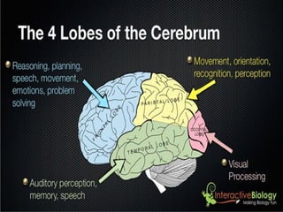


The document summarizes the main components and structures of the central and peripheral nervous systems. The central nervous system consists of the brain and spinal cord. The brain is divided into the forebrain, midbrain, and hindbrain. The forebrain includes the cerebrum, diencephalon, and limbic system. The cerebrum is made up of four lobes - frontal, parietal, temporal, and occipital - each with different functions. The peripheral nervous system includes spinal nerves, cranial nerves, and the autonomic nervous system. The document provides details on the structures and functions of these various components of the nervous system.


