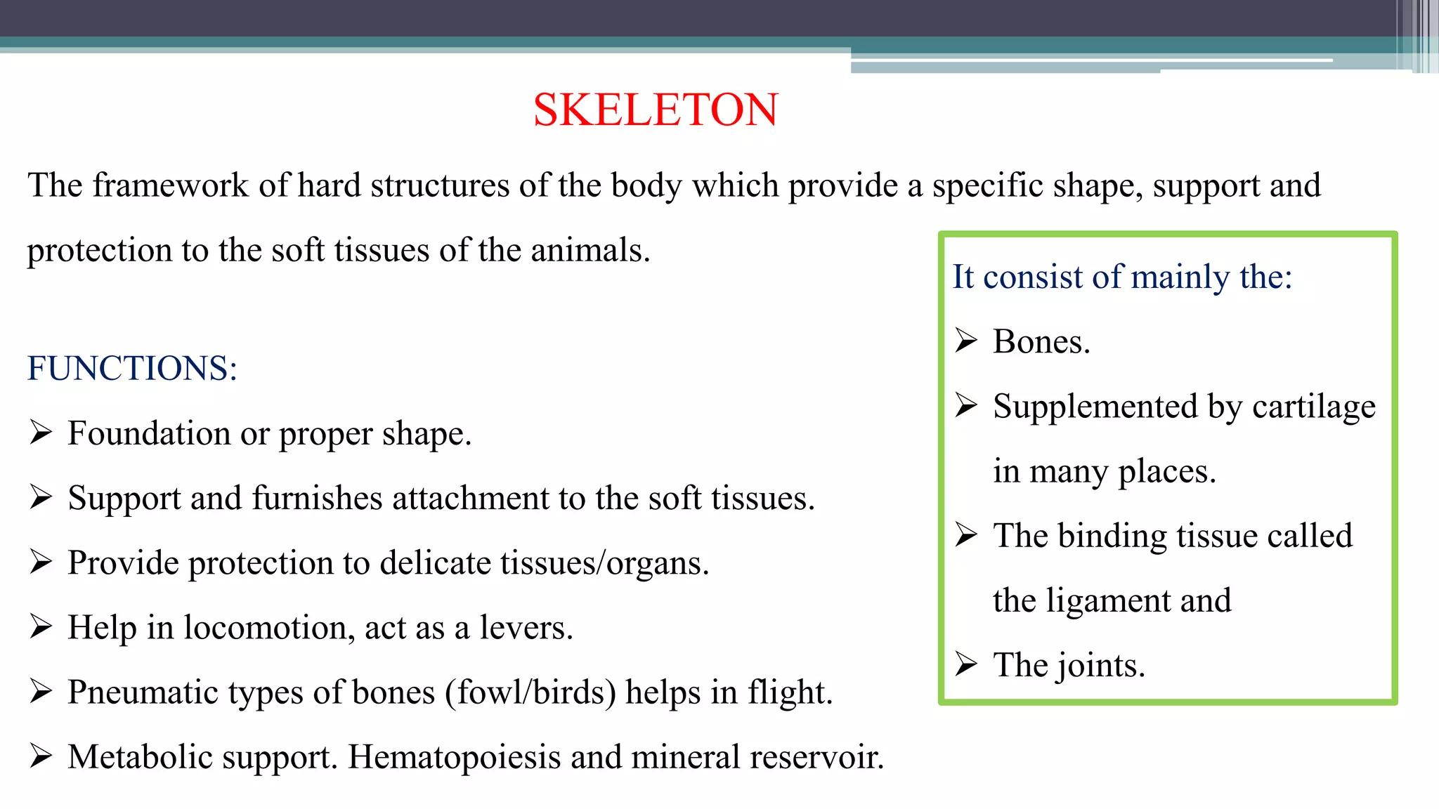
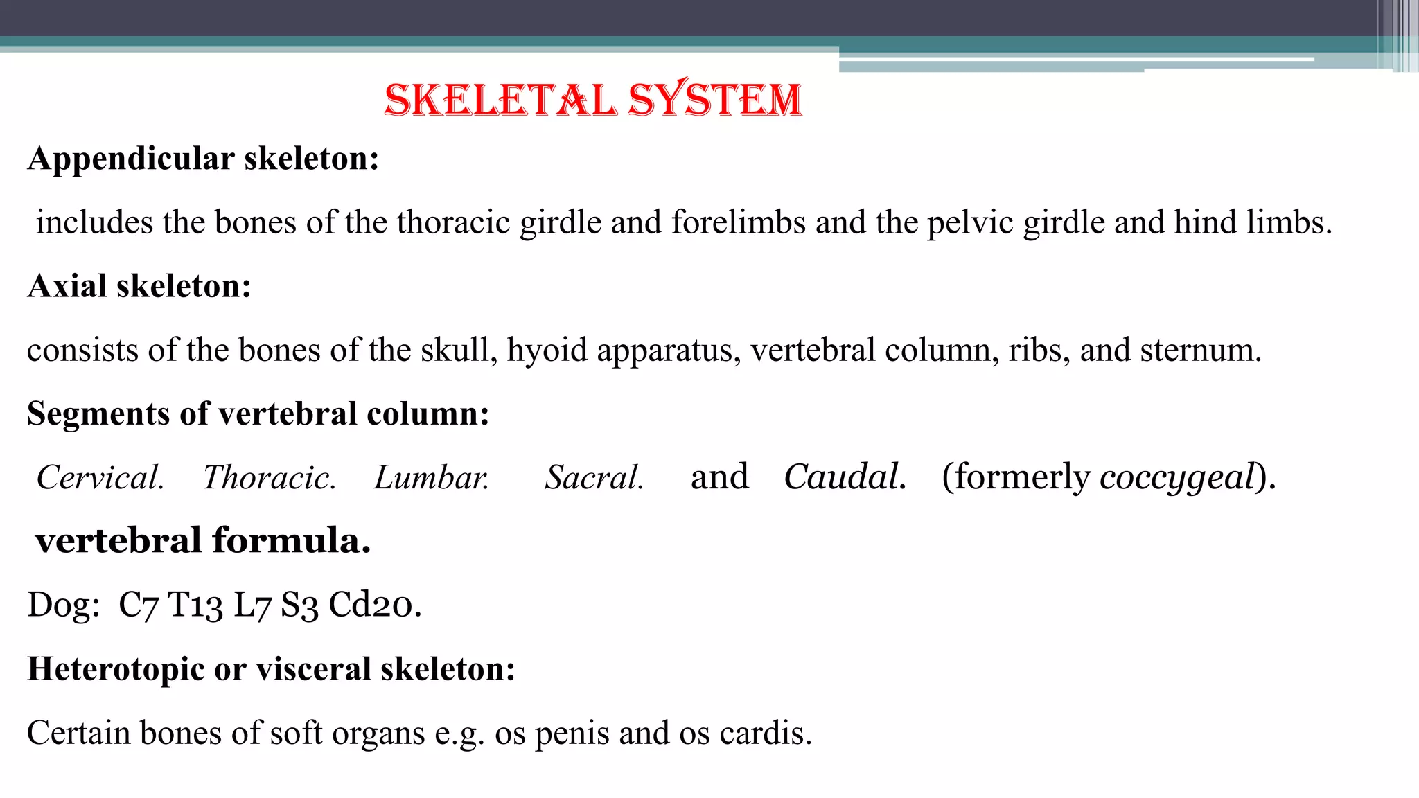
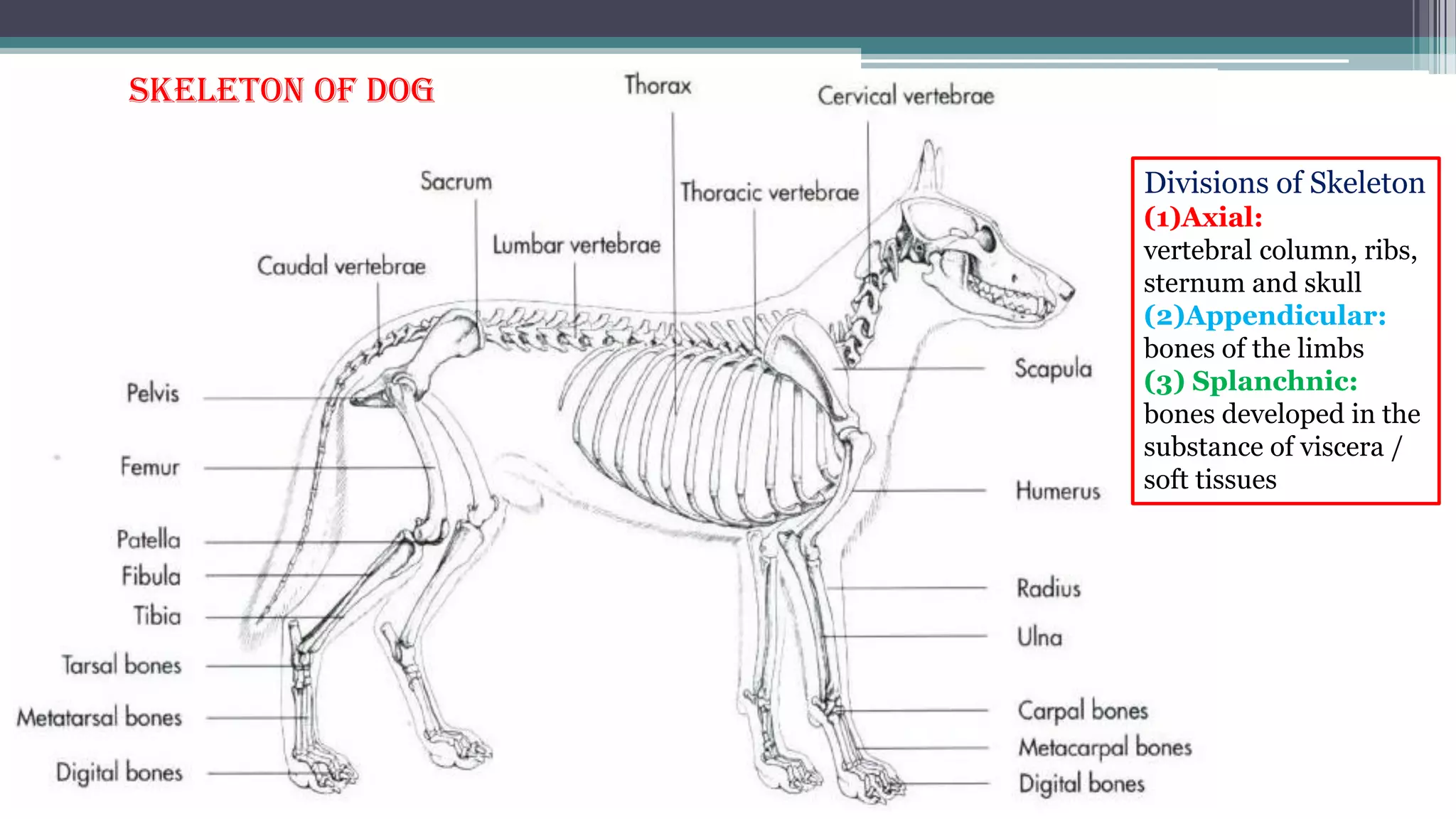
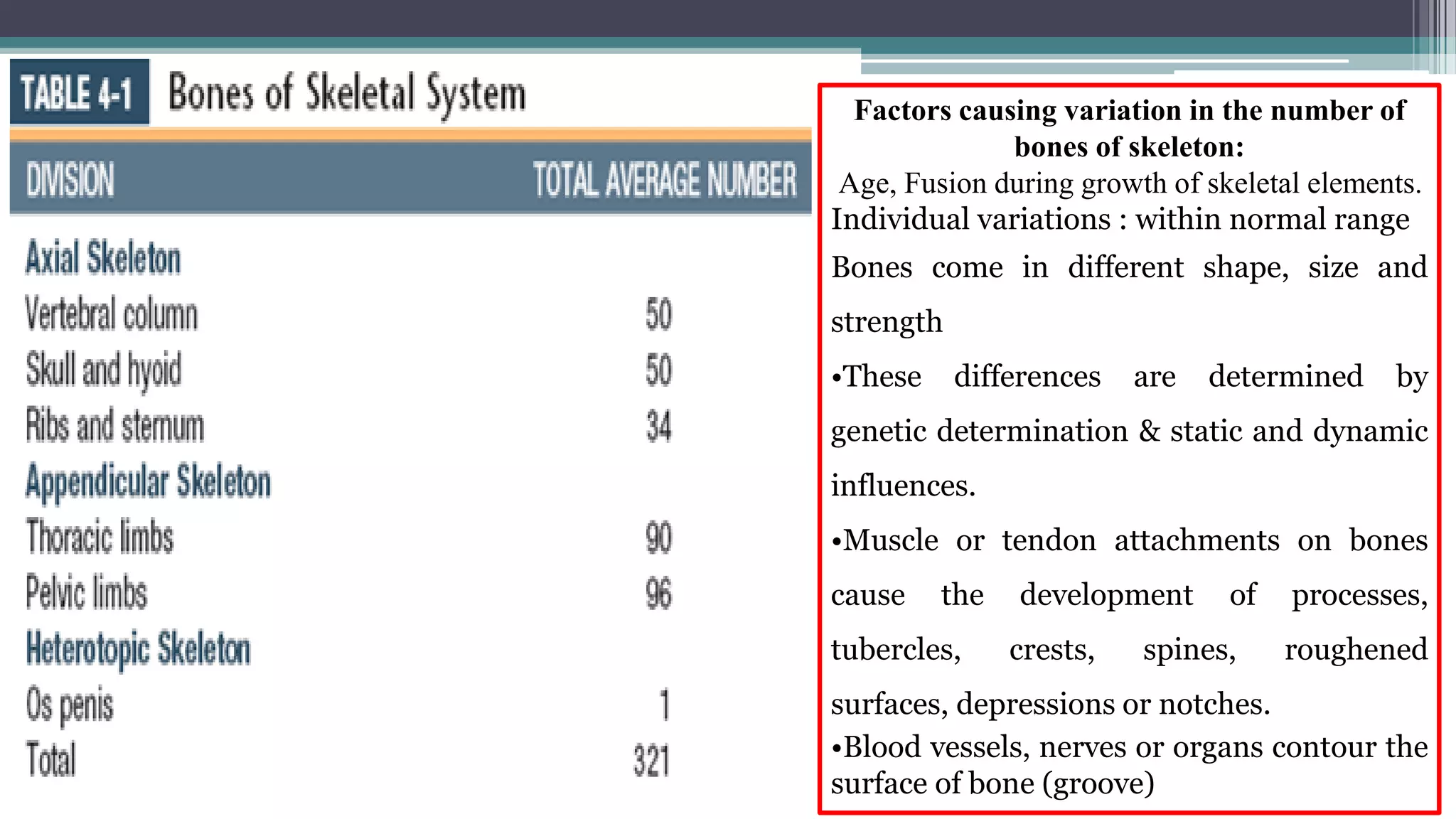
Locomotor System – I
Skeleton (BONES AND Cartilage)
ANATOMY-I (ANAT-02101) Crd Hr 3(1-2)
LECTURE 2
BY
SULTAN ALI
DVM (SAU, TANDOJAM)
MPHIL (UVAS, LAHORE)
Learning outcomes
At the end of this lecture students will be expected to know about:
Components of locomotor system .
The skeleton, its divisions, Major bones of the body and regional locations.
Classification, properties, structure and functions of bones.
The structure of a typical long bone (Epiphysis, Metaphysis, Diaphysis-Medullary
cavity, Periosteum, Endosteum)
(Complex organ system)
SKELETON
The framework of hard structures of the body which provide a specific shape, support and
protection to the soft tissues of the animals.
FUNCTIONS:
 Foundation or proper shape.
 Support and furnishes attachment to the soft tissues.
 Provide protection to delicate tissues/organs.
 Help in locomotion, act as a levers.
 Pneumatic types of bones (fowl/birds) helps in flight.
 Metabolic support. Hematopoiesis and mineral reservoir.
It consist of mainly the:
 Bones.
 Supplemented by cartilage
in many places.
 The binding tissue called
the ligament and
 The joints.
SKELETAL SYSTEM
Appendicular skeleton:
includes the bones of the thoracic girdle and forelimbs and the pelvic girdle and hind limbs.
Axial skeleton:
consists of the bones of the skull, hyoid apparatus, vertebral column, ribs, and sternum.
Segments of vertebral column:
Cervical. Thoracic. Lumbar. Sacral. and Caudal. (formerly coccygeal).
vertebral formula.
Dog: C7 T13 L7 S3 Cd20.
Heterotopic or visceral skeleton:
Certain bones of soft organs e.g. os penis and os cardis.
Skeleton of dog
Divisions of Skeleton
(1)Axial:
vertebral column, ribs,
sternum and skull
(2)Appendicular:
bones of the limbs
(3) Splanchnic:
bones developed in the
substance of viscera /
soft tissues
Factors causing variation in the number of
bones of skeleton:
Age, Fusion during growth of skeletal elements.
Individual variations : within normal range
Bones come in different shape, size and
strength
•These differences are determined by
genetic determination & static and dynamic
influences.
•Muscle or tendon attachments on bones
cause the development of processes,
tubercles, crests, spines, roughened
surfaces, depressions or notches.
•Blood vessels, nerves or organs contour the
surface of bone (groove)
Regional location of bones and joints:
Coracoid
*Dorsal as well as palmar sesamoid bones.
JOINTS
SHOULDER
ELBOW
KNEE
FETLOCK
PASTERN
COFFIN
JOINTS
HIP
STIFYL
HOCK
FETLOCK
PASTERN
COFFIN
P1
P2
P3
Classification of bones on the basis of shape / appearance:
Bone: The hard semi rigid calcified connective tissue
forming the skeleton.
Five general divisions:
 Long Bones.
 Short Bones and,
 Sesamoid bones.
(are characteristic of the limbs)
 Flat Bones.
 Irregular bones.
(are characteristic of the skull and vertebral column)
CHEMICAL COMPOSITION
Dried bone consists of organic and inorganic
matter in the ratio of 1 : 2
Organic Ground Substance: Collagen fibers.
Contributing flexibility and tensile strength
Bone mineral: Calcium phosphate (85-90 %)
Calcium carbonate (8-10 %), Magnesium
phosphate (1.5 %) and calcium fluoride (0.3 %)
Contributing stiffness and compressive
strength
PHYSICAL PROPERTIES OF BONE
Fresh dead bone is yellowish-white in color
Macerated or boiled and bleached is bright white
Tensile strength nearly twice that of oak
Characterized by:
a body/shaft -Diaphysis
a proximal and distal ends/extremities -Epiphyses
and the growth plate -Metaphysis
•Diaphysis encloses the central marrow
or medullary cavity.
•Epiphysis consist of core of spongy
bone covered by compact bone and
Articular cartilage (hyaline cartilage)
•Shaft is lined by periosteum and
endosteum on the outside and inside
respectively.
Long bones:
Typical in limbs
Main weight bearing
Ends are enlarged
Structure of a typical long bone:
Medullary cavity: the space in shafts
of long bones in the adult are filled
largely with yellow bone marrow chiefly
fat, in fetus and the newborn, red bone
marrow functions in forming red blood
cells.
Periosteum: the fibrous layer of
connective tissue covers the non
articular surfaces of all bones in the
fresh state.
aid in growth, repair, nutrition &
attachment for ligaments and tendons.
Endosteum: is lines the large
medullary cavities similar to periosteum
but thinner.
Perichondrium: The connective tissue
covering of cartilage does not differ
histologically from periosteum.
Vessels and Nerves of Bone
• Nutrient foramen and canals
• Long bones and many flat and irregular bones have a conspicuous
• Nutrient (medullary) artery and vein
• Lymph vessels are present in the periosteum as
perivascular sheaths
• and probably also as unaccompanied vessels within the bone
marrow.
• Nerves in bone are principally sensory
Short bones - vary greatly in shape : cylindrical, cuboid
or spheroid (six surfaces) to irregularly compressed rods
(one flat) articular surface.
Bears weight, helps in joints movement
They are occupied by a three-dimensional lattice of
spongy bone with intervening haemoreticular tissue.
Include the bones of the carpus, tarsus and spine
Sesamoid bones
Seasame (seed in Greek), found in Freely moving joints.
develop in the capsules of some joints or in tendons Chief
function is to protect tendons, where greatest friction is
developed.
Largest bone is patella
• Flat bones protect the soft organs , serve for muscle
attachment.
• consists of two layers of solid bone (tabulae) with spongy bone
(diploe) or air-filled cavities (sinus) between them.
• Include the scapula and skull bones
• Some of the flat bones of the skull are pneumatized.
Irregular bones
• Jutting processes are the characteristic features
• Muscular and ligamentous attachments; some articulation.
• Vertebral column, some bones of the skull, and the os coxae.
Pneumatic bones:
contain air-spaces or sinuses within the compact substance
instead of spongy bone and marrow.
COMPACT BONE/Lamellar/Cortical bone:
Collagen fibres arranged in parallel and concentric
Haversian Lamellae Outer and Inner circumferential
lamellae covered by periosteum and endosteum,
respectively Composed of cylindrical units Osteons
(Haversian system)
OSTEON
•A central vascular channel (Haversian canal)
surrounded by Haversian Lamellae
•Bone cells are located between the lamellae
•The Haversian or nutrient canals communicate with the
marrow cavity and the external surface via transverse
channels, Volkmann canals
•Forms the outer layer of the whole of long, short,
irregular and flat bones
•Forms the shaft of the long bones
SPONGY BONE/Cancellous / Trabecular bone :
•Consists of delicate intercrossing bony plates and
spicules
•The intervals (marrow spaces) between the plates are
occupied by marrow and lined by endosteum
•Forms the bulk of short/irregular bones and of the
extremities and inner aspect of shafts of long bones,
diploe of flat bones
PNEUMATIC BONES contain air-spaces or sinuses
within the compact substance instead of spongy bone and
marrow.
MARKING ON THE BONES/ BONY CONTOURS OF A BONE.
Tuberosity:
A large usually rounded roughened projection which is non articular.
Tubercle:
A small rounded projection.
Trochanter:
A large blunt projection found only on the femur
Trochlea:
Pulley shaped articular projection/structure
Marking on the bones
Condyles:
A large articular prominence, usually smooth and convex at the end of the
long bones e.g. condyles of humerus and femur.
Epicondyle:
A prominence just above the condyles usually roughened.
 Spine:
A sharp thin elongated process e.g. scapula
Crest:
Prominent or well developed linear sharp ridge on the bone e.g. humerus,
tibia.
Line: Less prominent or faint ridge
Marking on the bones
Foramen:
An opening in the bone (for blood vessel and nerve).
Canal:
Long foramen which has a second opening.
Fissure:
A narrow cleft like opening between bones
Groove:
Small narrow furrow accommodating vessels, nerve or tendon.
Sinus:
A large air cavity with in a bone
• Head. Neck, Fovea
• Notch: Depression on the edge of bone
• Facet. A smooth flat surface.
 Mostly covered by hyaline cartilage

LEC # 02 Skeleton.pdf

  • 1.
    Locomotor System –I Skeleton (BONES AND Cartilage) ANATOMY-I (ANAT-02101) Crd Hr 3(1-2) LECTURE 2 BY SULTAN ALI DVM (SAU, TANDOJAM) MPHIL (UVAS, LAHORE)
  • 2.
    Learning outcomes At theend of this lecture students will be expected to know about: Components of locomotor system . The skeleton, its divisions, Major bones of the body and regional locations. Classification, properties, structure and functions of bones. The structure of a typical long bone (Epiphysis, Metaphysis, Diaphysis-Medullary cavity, Periosteum, Endosteum)
  • 3.
  • 4.
    SKELETON The framework ofhard structures of the body which provide a specific shape, support and protection to the soft tissues of the animals. FUNCTIONS:  Foundation or proper shape.  Support and furnishes attachment to the soft tissues.  Provide protection to delicate tissues/organs.  Help in locomotion, act as a levers.  Pneumatic types of bones (fowl/birds) helps in flight.  Metabolic support. Hematopoiesis and mineral reservoir. It consist of mainly the:  Bones.  Supplemented by cartilage in many places.  The binding tissue called the ligament and  The joints.
  • 5.
    SKELETAL SYSTEM Appendicular skeleton: includesthe bones of the thoracic girdle and forelimbs and the pelvic girdle and hind limbs. Axial skeleton: consists of the bones of the skull, hyoid apparatus, vertebral column, ribs, and sternum. Segments of vertebral column: Cervical. Thoracic. Lumbar. Sacral. and Caudal. (formerly coccygeal). vertebral formula. Dog: C7 T13 L7 S3 Cd20. Heterotopic or visceral skeleton: Certain bones of soft organs e.g. os penis and os cardis.
  • 6.
    Skeleton of dog Divisionsof Skeleton (1)Axial: vertebral column, ribs, sternum and skull (2)Appendicular: bones of the limbs (3) Splanchnic: bones developed in the substance of viscera / soft tissues
  • 7.
    Factors causing variationin the number of bones of skeleton: Age, Fusion during growth of skeletal elements. Individual variations : within normal range Bones come in different shape, size and strength •These differences are determined by genetic determination & static and dynamic influences. •Muscle or tendon attachments on bones cause the development of processes, tubercles, crests, spines, roughened surfaces, depressions or notches. •Blood vessels, nerves or organs contour the surface of bone (groove)
  • 10.
    Regional location ofbones and joints: Coracoid *Dorsal as well as palmar sesamoid bones. JOINTS SHOULDER ELBOW KNEE FETLOCK PASTERN COFFIN JOINTS HIP STIFYL HOCK FETLOCK PASTERN COFFIN P1 P2 P3
  • 11.
    Classification of boneson the basis of shape / appearance: Bone: The hard semi rigid calcified connective tissue forming the skeleton. Five general divisions:  Long Bones.  Short Bones and,  Sesamoid bones. (are characteristic of the limbs)  Flat Bones.  Irregular bones. (are characteristic of the skull and vertebral column) CHEMICAL COMPOSITION Dried bone consists of organic and inorganic matter in the ratio of 1 : 2 Organic Ground Substance: Collagen fibers. Contributing flexibility and tensile strength Bone mineral: Calcium phosphate (85-90 %) Calcium carbonate (8-10 %), Magnesium phosphate (1.5 %) and calcium fluoride (0.3 %) Contributing stiffness and compressive strength PHYSICAL PROPERTIES OF BONE Fresh dead bone is yellowish-white in color Macerated or boiled and bleached is bright white Tensile strength nearly twice that of oak
  • 13.
    Characterized by: a body/shaft-Diaphysis a proximal and distal ends/extremities -Epiphyses and the growth plate -Metaphysis •Diaphysis encloses the central marrow or medullary cavity. •Epiphysis consist of core of spongy bone covered by compact bone and Articular cartilage (hyaline cartilage) •Shaft is lined by periosteum and endosteum on the outside and inside respectively. Long bones: Typical in limbs Main weight bearing Ends are enlarged
  • 14.
    Structure of atypical long bone: Medullary cavity: the space in shafts of long bones in the adult are filled largely with yellow bone marrow chiefly fat, in fetus and the newborn, red bone marrow functions in forming red blood cells. Periosteum: the fibrous layer of connective tissue covers the non articular surfaces of all bones in the fresh state. aid in growth, repair, nutrition & attachment for ligaments and tendons. Endosteum: is lines the large medullary cavities similar to periosteum but thinner. Perichondrium: The connective tissue covering of cartilage does not differ histologically from periosteum.
  • 16.
    Vessels and Nervesof Bone • Nutrient foramen and canals • Long bones and many flat and irregular bones have a conspicuous • Nutrient (medullary) artery and vein • Lymph vessels are present in the periosteum as perivascular sheaths • and probably also as unaccompanied vessels within the bone marrow. • Nerves in bone are principally sensory
  • 17.
    Short bones -vary greatly in shape : cylindrical, cuboid or spheroid (six surfaces) to irregularly compressed rods (one flat) articular surface. Bears weight, helps in joints movement They are occupied by a three-dimensional lattice of spongy bone with intervening haemoreticular tissue. Include the bones of the carpus, tarsus and spine Sesamoid bones Seasame (seed in Greek), found in Freely moving joints. develop in the capsules of some joints or in tendons Chief function is to protect tendons, where greatest friction is developed. Largest bone is patella
  • 18.
    • Flat bonesprotect the soft organs , serve for muscle attachment. • consists of two layers of solid bone (tabulae) with spongy bone (diploe) or air-filled cavities (sinus) between them. • Include the scapula and skull bones • Some of the flat bones of the skull are pneumatized. Irregular bones • Jutting processes are the characteristic features • Muscular and ligamentous attachments; some articulation. • Vertebral column, some bones of the skull, and the os coxae. Pneumatic bones: contain air-spaces or sinuses within the compact substance instead of spongy bone and marrow.
  • 19.
    COMPACT BONE/Lamellar/Cortical bone: Collagenfibres arranged in parallel and concentric Haversian Lamellae Outer and Inner circumferential lamellae covered by periosteum and endosteum, respectively Composed of cylindrical units Osteons (Haversian system) OSTEON •A central vascular channel (Haversian canal) surrounded by Haversian Lamellae •Bone cells are located between the lamellae •The Haversian or nutrient canals communicate with the marrow cavity and the external surface via transverse channels, Volkmann canals •Forms the outer layer of the whole of long, short, irregular and flat bones •Forms the shaft of the long bones
  • 21.
    SPONGY BONE/Cancellous /Trabecular bone : •Consists of delicate intercrossing bony plates and spicules •The intervals (marrow spaces) between the plates are occupied by marrow and lined by endosteum •Forms the bulk of short/irregular bones and of the extremities and inner aspect of shafts of long bones, diploe of flat bones PNEUMATIC BONES contain air-spaces or sinuses within the compact substance instead of spongy bone and marrow.
  • 25.
    MARKING ON THEBONES/ BONY CONTOURS OF A BONE. Tuberosity: A large usually rounded roughened projection which is non articular. Tubercle: A small rounded projection. Trochanter: A large blunt projection found only on the femur Trochlea: Pulley shaped articular projection/structure
  • 26.
    Marking on thebones Condyles: A large articular prominence, usually smooth and convex at the end of the long bones e.g. condyles of humerus and femur. Epicondyle: A prominence just above the condyles usually roughened.  Spine: A sharp thin elongated process e.g. scapula Crest: Prominent or well developed linear sharp ridge on the bone e.g. humerus, tibia. Line: Less prominent or faint ridge
  • 28.
    Marking on thebones Foramen: An opening in the bone (for blood vessel and nerve). Canal: Long foramen which has a second opening. Fissure: A narrow cleft like opening between bones Groove: Small narrow furrow accommodating vessels, nerve or tendon. Sinus: A large air cavity with in a bone • Head. Neck, Fovea • Notch: Depression on the edge of bone • Facet. A smooth flat surface.  Mostly covered by hyaline cartilage