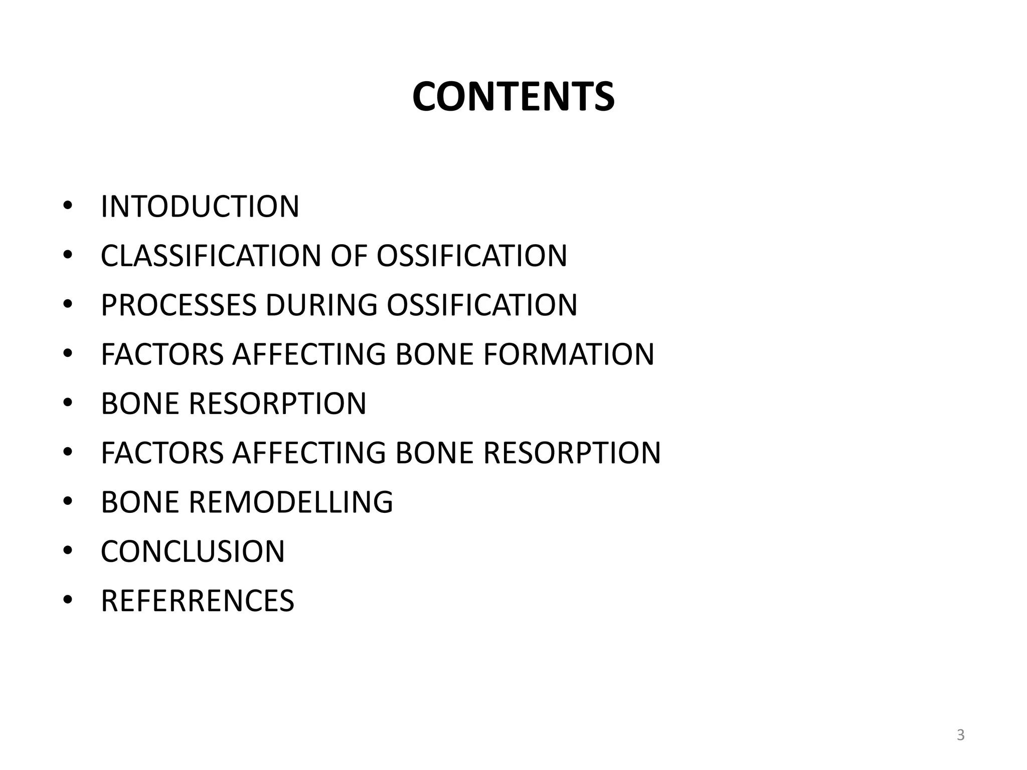
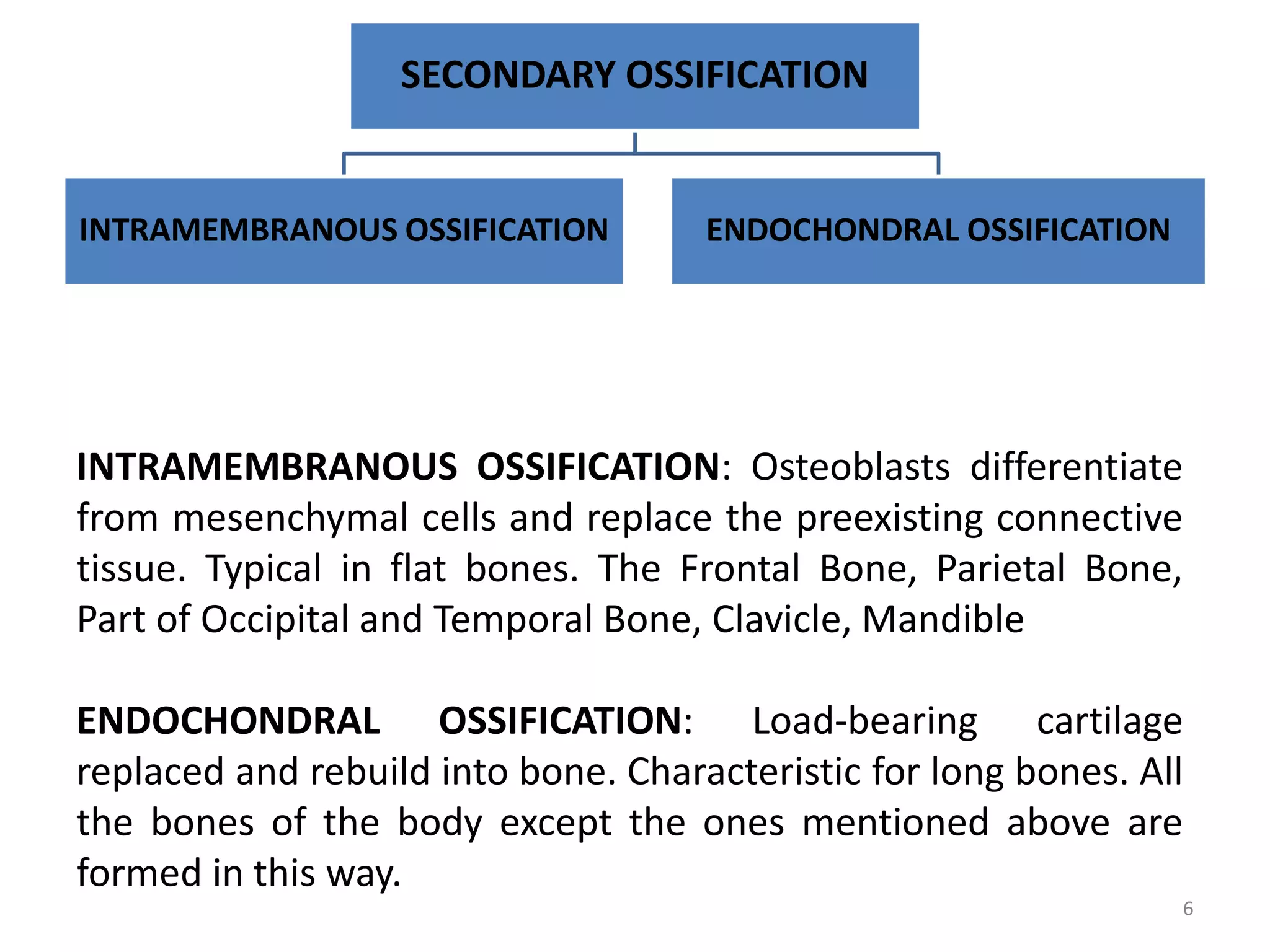
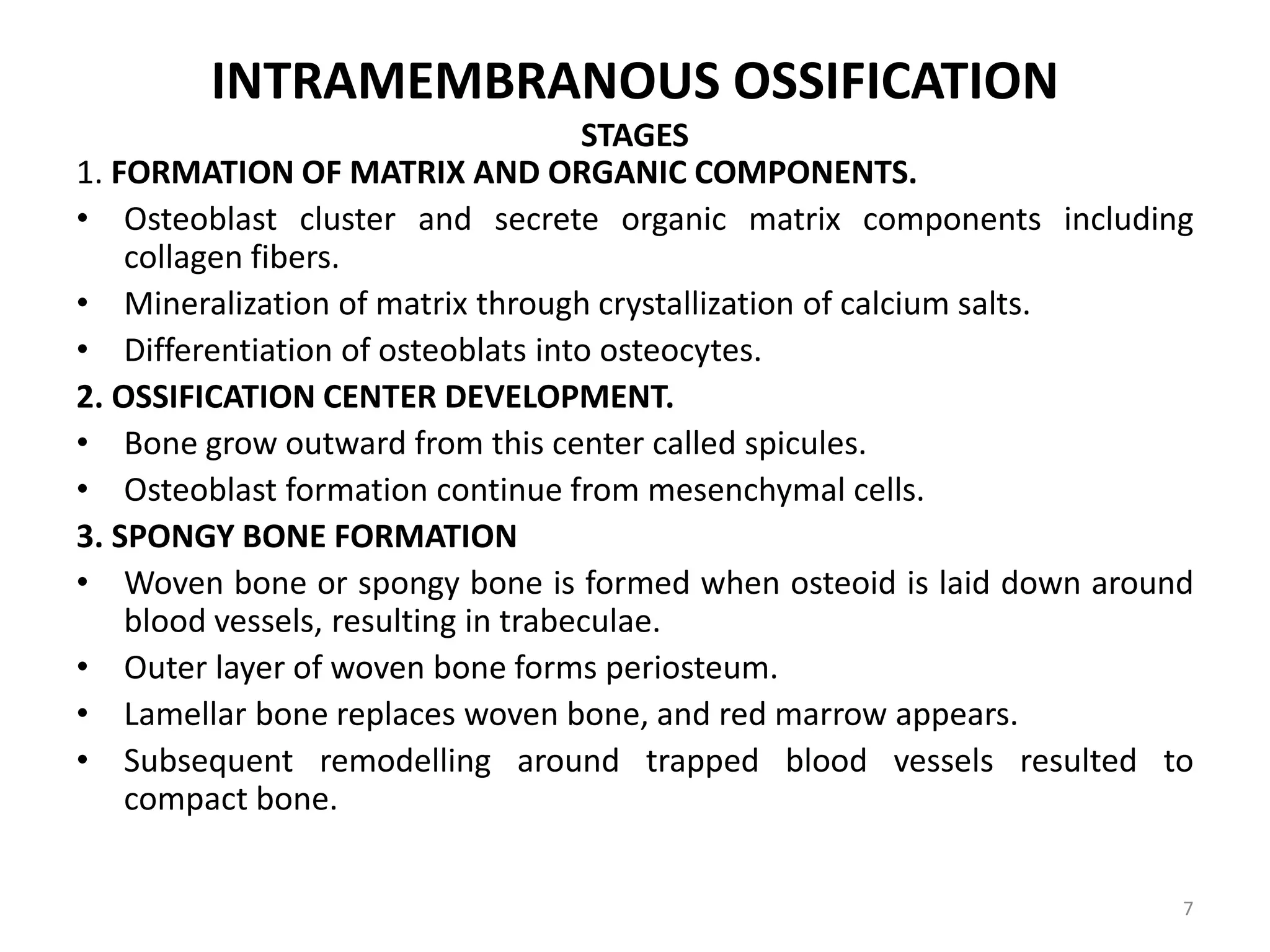
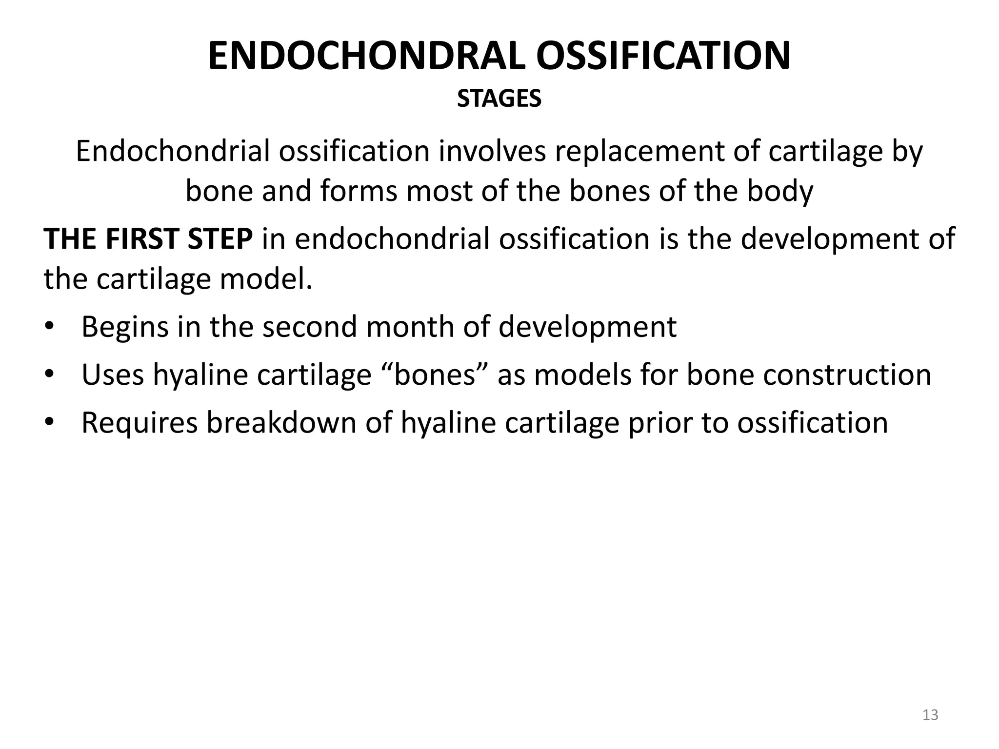
This document provides an overview of ossification, the process of bone formation. It discusses the classification of ossification into primary and secondary ossification, with the latter including intramembranous and endochondral ossification. The stages and factors involved in intramembranous and endochondral ossification are described in detail. Bone resorption, remodeling and the factors that influence these processes are also summarized.
Ossification (Intracartilaginous and Intramembranous)Mohiuddin Masum
Ossification is the process of bone formation. There are two types of ossification: intramembranous ossification and intracartilaginous (endochondral) ossification. In intramembranous ossification, bone is laid down directly in fibrous membranes by osteoblasts differentiating from mesenchymal cells. Bones formed this way include the skull and clavicle. In intracartilaginous ossification, a cartilaginous model is first formed and then replaced by bone. Long bones develop through this process, with osteoblasts eroding and replacing the cartilage matrix.
There are two main methods of ossification: intramembranous ossification and endochondral ossification. Intramembranous ossification forms flat bones directly from mesenchymal tissue through condensation, vascularization, osteoblast differentiation, and osteoid formation and calcification. Endochondral ossification forms long bones through a cartilage model, with primary ossification centers forming within the cartilage followed by invasion of blood vessels and osteoprogenitor cells leading to replacement of cartilage by bone except at joint surfaces. Growth in long bone length occurs through the epiphyseal plate where columns of chondrocytes undergo proliferation, hypertrophy, and calcification before being replaced by bone.
The document discusses the anatomy and physiology of bone. It covers the functions, classification, microscopic structure, development, growth and common disorders of bone. Key points include that bone provides structure, protection and movement; is made of collagen fibers and hydroxyapatite; develops through intramembranous or endochondral ossification; and can be affected by conditions like fractures, osteoporosis or cancer.
1. Enchondral bone formation begins with the development of a cartilage model surrounded by perichondrium.
2. Chondrocytes in the center of the model hypertrophy and the perichondrium is converted to periosteum which secretes bone around the model to form a bone collar.
3. Osteoclasts invade the cartilage model and erode the cartilage, forming cavities that are filled with bone and marrow in a process that replaces cartilage with bone until maturation is complete.
This document discusses cartilage and bone. It defines cartilage as a specialized connective tissue that functions as a supporting or weight-bearing tissue. There are three main types of cartilage: hyaline, elastic, and fibrocartilage. Bone is a highly vascularized living tissue with a calcified matrix. It provides structure and support for the body. The document outlines the basic components, cells, and structures of both cartilage and bone in detail.
Bone grows through the processes of intramembranous and endochondral ossification. Intramembranous ossification develops bone directly from connective tissue, while endochondral ossification replaces cartilaginous skeletal parts with bone. During endochondral ossification, hyaline cartilage is present near the epiphyses of long bones and breaks down as cartilage cell columns are replaced by osteoblasts depositing new bone tissue. Bone age can be determined by examining an x-ray of the epiphyseal plates and timing of their fusion with the diaphyses, which occurs at different ages for each bone.
CONTENTS
FORMATION OF BONE
CLASSIFICATION OF BONES
STRUCTURE OF BONE
BLOOD SUPPLY
COMPOSITION OF BONE
FRACTURE HEALING
CARTILAGE
TYPES OF CARTILAGE
BONE (syn – Os; Osteon)
Osseous tissue, a specialised form of dense connective
tissue consisting of bone cells (osteocytes)
Embedded in a matrix of calcified intercelluar
substance
Bone matrix contains collagen fibres and the minerals
calcium phosphate and calcium carbonate
There are two types of bone ossification: intramembranous and endochondral. Intramembranous ossification forms bones like the skull and clavicles directly in connective tissue. Endochondral ossification replaces cartilage with bone to form long bones. This process begins with mesenchymal cells forming cartilage, which then undergoes interstitial and appositional growth. Osteoblasts eventually deposit bone matrix around the calcified cartilage, forming trabeculae and replacing the cartilage with bone from the primary ossification center outward.
Bone is a composite material formed mostly of calcium phosphate. There are two types of bone tissue: compact bone and spongy bone. Cortical bone accounts for 80% of the total bone mass in the adult skeleton. There are two processes of bone formation: intramembranous ossification which forms flat bones of the skull, and endochondral ossification which forms most other bones through a cartilage model. Bone is constantly remodeled through the actions of osteoblasts which build bone and osteoclasts which break it down.
The document discusses the structure and function of bone. It defines bone as a mineralized connective tissue composed of osteocytes, osteoblasts, and osteoclasts within a matrix of collagen fibers and hydroxyapatite. Bone has two layers: a dense compact bone and a porous spongy bone. The microscopic structure of compact bone consists of cylindrical osteons containing central canals and concentric lamellae. Osteoblasts build new bone matrix while osteoclasts resorb old bone. The periosteum and endosteum provide nutrients and new osteoblasts to bones.
a brief ppt description about cartilage which may be usefull for teaching for first year mbbs, bds and paramedical students, hope it is helpfull to everyone
Bone develops through two main processes: endochondral and intramembranous ossification. Endochondral ossification involves the development of a cartilage model that is later replaced by bone tissue. It occurs in long bones and involves chondroblasts forming cartilage, blood vessels infiltrating and bringing osteoprogenitor cells, and the formation of primary and secondary ossification centers. Intramembranous ossification occurs in flat bones where mesenchymal cells directly develop into bone, forming woven bone that matures into lamellar bone. Bone is made up of an organic collagen matrix and inorganic hydroxyapatite, with three main cell types involved in formation and maintenance.
Bone is a specialized connective tissue composed of cells, fibers, and minerals. It forms through two processes: endochondral ossification and intramembranous ossification. Endochondral ossification involves cartilage models that are replaced by bone, forming long bones and portions of flat bones. It begins in the second month of development. Intramembranous ossification involves mesenchymal cells directly forming bone, without a cartilage intermediate, forming bones like the skull and clavicle. Bone growth is regulated by hormones like growth hormone and sex hormones. Common bone diseases include osteogenesis imperfecta and achondroplasia.
Cartilage is a connective tissue that provides support and flexibility to various regions of the body. There are three main types of cartilage - hyaline, elastic, and fibrocartilage. Hyaline cartilage is the most abundant and is characterized by chondrocytes embedded in a matrix with collagen fibers and proteoglycans. It is found in locations like the fetal skeleton, nose, and joints. Elastic cartilage contains elastic fibers that allow for flexibility and is present in the ear and epiglottis. Fibrocartilage consists of thick collagen fibers and is found in joints like the pubic symphysis where it can withstand compressive forces.
Bone tissue is a specialized form of connective tissue composed of cells and a mineralized extracellular matrix. The matrix is made up of collagen fibers and hydroxyapatite crystals that give bone its rigidity. There are two types of bone tissue: compact bone which forms the dense outer layer, and spongy or cancellous bone which is found at the ends of long bones and has a spongy, mesh-like structure. Bones develop through two processes - intramembranous ossification which forms flat bones, and endochondral ossification where cartilage is replaced by bone to form most other bones including long bones.
general anatomy and development of bonesTaimurKhan87
Bones can be classified based on location in the skeletal system or shape. There are two types of bone tissue: compact bone forming the outer walls and spongy bone in the interior. Bones form through two processes - intramembranous ossification where connective tissue is replaced with bone, and endochondral ossification where cartilage is replaced with bone. Blood supply to long bones comes from nutrient arteries in the diaphysis and metaphyses and periosteal vessels on the surface.
The document discusses the process of ossification where cartilage is changed to bone during human development. It explains that ossification is impacted by three key cells: osteoblasts, which build bone; osteocytes, which are trapped osteoblasts that give bone its star-shaped appearance; and osteoclasts, which break down old or damaged bone. The document also outlines factors like nutrition, vitamins, hormones, and growth that influence bone growth and remodeling throughout life.
The document discusses the skeletal system and connective tissues. It covers the definitions of osteology and arthrology, the study of bones and joints. The skeletal system is composed of bones, cartilage, ligaments and other connective tissues. Cartilage is weaker but more flexible than bone. There are three types of cartilage - hyaline, fibrocartilage, and elastic cartilage. Bones provide structure, protection, movement, mineral storage and blood cell formation. The two types of ossification that form bones are intramembranous and endochondral ossification.
This document provides an overview of long bone anatomy and classification of joints. It describes the main parts of long bones including the diaphysis, epiphyses, metaphysis and their structures. It also classifies bones based on shape and classifies joints based on both structure and function, describing the characteristics of synovial, cartilaginous and fibrous joints. Key joint types include ball-and-socket, hinge, pivot, gliding and saddle joints.
There are three main types of joints in the body - fibrous, cartilaginous, and synovial joints. Fibrous joints are immovable, cartilaginous joints allow slight movement, and synovial joints can move freely. Synovial joints are further classified by their shape and include hinge, ball-and-socket, and saddle joints. Each joint has a fibrous capsule, ligaments, synovial membrane, articular cartilage, and in some cases articular discs or bursae to facilitate movement and reduce friction between bones.
A joint is the site where two or more bones meet. There are three main types of joints: fibrous, cartilaginous, and synovial. Synovial joints are the most common and provide the most movement. They contain synovial fluid and have articular cartilage covering the bone ends. Common synovial joints include the ball and socket shoulder joint, hinge elbow joint, pivot radioulnar joint, condyloid wrist joint, saddle thumb joint, and gliding vertebral joints.
Cartilage is a connective tissue composed of cells called chondrocytes embedded in an extracellular matrix. There are three main types of cartilage - hyaline, elastic, and fibrocartilage. Hyaline cartilage is found in joints, respiratory airways, and growing bones. It contains type II collagen and proteoglycans that allow it to bear mechanical stress and provide cushioning. Chondrocytes maintain the extracellular matrix by synthesizing its components. Cartilage grows through both interstitial and appositional growth and has limited ability for repair due to its avascular nature.
BONE – AN INTRODUCTION
A bone is a rigid organ that constitutes part of the vertebrate skeleton.
There are around 270 to 300+ bones in Infants which gets reduced to 206 bones in adults.
Bones are dynamic structures that are undergoing constant change and remodelling in
response to the ever-changing environment.
Bones support and protect the various organs of the body, produce red and white blood cells,
store minerals, provide structure and support for the body, and enable mobility.
It has a honeycomb-like matrix internally, which helps to give the bone rigidity.
The largest bone in the body is the femur or thigh-bone, and the smallest is the stapes in
the middle ear.
classification of joints and characteristics of each type jointmuti ullah
This document defines and classifies different types of joints in the body. It begins by defining a joint as the area where two or more bones meet. Joints can be classified based on their structure, development, and function. The three main types of joints are fibrous joints, cartilaginous joints, and synovial joints. Fibrous joints are fixed or slightly movable joints connected by dense fibrous tissue. Cartilaginous joints are also slightly movable and connected by cartilage, found between vertebrae. Synovial joints are the most movable joints, containing a synovial fluid-filled cavity between bones. Common synovial joints include ball-and-socket and hinge joints in the limbs.
Bone tissue is the major structural and supportive connective tissue of the body. Osseous tissue forms the rigid part of the bones that make up the skeletal system.
This document provides an overview of alveolar bone structure and function. It begins with introductions to bone composition, development, and cell types. Key bone cells include osteoblasts, which form bone, and osteoclasts, which resorb bone. The document then discusses alveolar bone morphology, blood supply, and functions. Importantly, alveolar bone is in a constant state of flux, undergoing remodeling as bone is broken down and rebuilt through the coupled actions of osteoblasts and osteoclasts. Healing of alveolar bone after tooth extraction and age-related changes are also covered.
PHYSIOLOGY OF BONE AND ITS PROSTHODONTIC IMPLICATIONSGauri Patil
This document summarizes the physiology of bone. It begins by describing the macroscopic and microscopic structures of bone, including compact and spongy bone. It then discusses the cellular components of bone including osteoblasts, osteoclasts, osteocytes, and bone lining cells. The document outlines the process of bone formation, remodeling, and resorption. It discusses disorders like osteoporosis, rickets, and Paget's disease. It concludes by covering prosthodontic implications such as residual ridge resorption and bone substitutes.
The skeleton is composed of bone and cartilage.
Importance of skeletal system
Mechanical support
Role in mineral homeostasis
Hematopoietic elements
Protection
Size and shape of body
Bone is a type of connective tissue.
Composed of
Inorganic and
Organic components
This document provides information on bone structure and cells. It discusses the composition of bone as mineralized connective tissue composed of organic matrix and inorganic salts. It classifies bones based on shape and development. It describes the microscopic structure of compact and spongy bone. It details the types of bone cells - osteoprogenitor cells, osteoblasts, osteocytes, and osteoclasts - and their roles and functions in bone formation and remodeling. It also addresses factors that regulate the activity of bone cells.
Bone & Its Importance to ProsthodontistSelf employed
Bone is a hard connective tissue that forms the skeleton. It has an inorganic mineral component made up of calcium salts and an organic collagen matrix. Bone comes in two types - dense cortical bone forming the hard outer shell and spongy cancellous bone on the inside. Bone is constantly remodeled through the actions of bone-forming osteoblasts and bone-resorbing osteoclasts. Diseases like osteoporosis weaken the bones through decreased bone density while Paget's disease involves abnormal bone deposition and resorption.
This document discusses metabolic bone diseases. It begins by describing the basic structure and function of bone, including its cellular components like osteoblasts and osteoclasts. It then discusses the constituents of bone matrix, including collagen, proteoglycans, and minerals. Bone development and homeostasis are explained, involving processes like remodeling. Specific metabolic bone diseases are then outlined in more detail, including osteogenesis imperfecta (brittle bone disease), osteomalacia/rickets due to abnormal mineralization, and hypophosphatasia due to a genetic disturbance in alkaline phosphatase synthesis. Throughout, the document provides microscopic and radiographic characteristics of these conditions.
This document summarizes bone formation and resorption. It classifies bones based on shape, development, and microscopic structure. It describes the composition of bone including bone cells like osteoblasts, osteoclasts, and osteocytes. Bone formation is influenced by growth factors while resorption involves acid secretion and enzyme activity by osteoclasts. Bone remodeling maintains bone mass through coupled formation and resorption, regulated by hormones and cytokines. Markers like TRAP indicate the rate of resorption.
This document provides an overview of bone formation, resorption, and remodeling. It discusses the classification of bones based on shape and development. It describes the composition of bone including cells like osteoblasts, osteoclasts, and osteocytes. Bone formation is mediated by growth factors while resorption involves acid secretion and enzyme activity by osteoclasts. Remodeling is a continuous process where old bone is replaced, maintaining bone strength through the coupled activities of formation and resorption. Markers of bone turnover provide information about these dynamic processes.
This document summarizes bone formation and resorption. It classifies bones based on shape, development, and microscopic structure. It describes the composition of bone including bone cells like osteoblasts, osteocytes, and osteoclasts. Bone formation is influenced by growth factors and hormones. Bone resorption involves osteoclasts acidifying the bone surface and releasing enzymes to degrade the organic matrix. Bone remodeling maintains bone strength through coupled bone resorption and formation mediated by hormones and growth factors.
CHONDROBLAST:Progenitor of chondrocytes
Lines border between perichondrium and matrix
Secretes type II collagen and other ECM components
CHONDROCYTE: Mature cartilage cell
Reside in a space called the lacuna
Clear areas = Golgi and lipid droplets,RER
PERICHONDRIUM:Dense irregularly arranged connective tissue
Ensheaths the cartilage
Houses the blood vessels that nourish chondrocytes
CARTILAGE GROWTH:Appositional
Increasing in WIDTH; chondroblasts deposit matrix on surface of pre-existing cartilage
Interstitial
Increasing in LENGTH; chondrocytes divide and secrete matrix from w/in lacunae
Alveolar bone houses the roots of teeth and helps distribute occlusal forces. It has both cortical and spongy components and undergoes remodeling through the coupled processes of bone resorption by osteoclasts and bone formation by osteoblasts. This maintains bone mass and replaces bone during tooth movement or repair. Osteoclasts resorb bone while osteoblasts form new bone matrix that becomes mineralized. This balanced process is regulated by local functional factors and systemic factors like PTH, calcitonin, and vitamins.
Alveolar bone forms tooth sockets and provides attachment for the periodontal ligament. It is composed of outer cortical and inner cancellous bone. Osteoblasts form bone matrix containing collagen fibers and hydroxyapatite crystals. Osteoclasts resorb bone. Bone is remodeled through the balanced actions of osteoblasts and osteoclasts, regulated by hormones and growth factors.
bone formation _Osteogenesis Flowchart.pptssuser7ec6af
The document discusses the two types of ossification (bone formation): intramembranous and endochondral. Intramembranous ossification involves the direct development of bone from connective tissue membranes, forming flat bones like the skull. Endochondral ossification involves the replacement of cartilage by bone, which is how most long bones form via ossification centers with cartilage templates that are later replaced by bone. Both processes involve osteoblasts secreting bone matrix that eventually hardens into mature bone through remodeling.
This document discusses bone formation, types of bone, bone repair and healing, and bone grafts. It covers the following key points:
1. There are two types of bone formation - intramembranous and endochondral ossification. There are also two types of bone tissue - cortical/compact bone and cancellous/trabecular bone.
2. Bone repair occurs in three stages - inflammatory, repair, and remodeling. Fractures heal via primary or secondary bone repair depending on the severity.
3. Bone grafts can aid bone healing via osteoinduction, osteoconduction, and osteogenesis. Autografts from the patient are generally most effective but allografts
Bone provides structural support and protects vital organs. It is a specialized connective tissue composed of cells embedded in a mineralized matrix. There are three main bone cell types: osteocytes found in bone matrix, osteoblasts which build bone matrix, and osteoclasts which break down bone matrix. Bone is either compact cortical bone or cancellous trabecular bone. Bone develops through intramembranous or endochondral ossification and is remodeled throughout life by coordinated bone resorption and formation. The skeleton acts as a calcium reserve and its mineral content is maintained through dynamic exchange with body fluids. Joints connect bones and allow various ranges of movement.
6. alveolar bone in health part b dr-ibrahim_shaikhDrIbrahim Shaikh
This document discusses the cells and components that make up healthy alveolar bone. It describes the main cell types, including osteoprogenitor cells that develop into osteoblasts or osteoclasts. Osteoblasts secrete osteoid and regulate mineralization, while osteoclasts are responsible for bone resorption. The bone matrix contains collagen fibers and hydroxyapatite crystals, along with noncollagenous proteins. Alveolar bone undergoes physiological remodeling through the coordinated actions of osteoblasts and osteoclasts, allowing adaptation to tooth movement and replacement over time.
The document discusses calcium homeostasis and bone remodeling. It describes how calcium is stored in bones and regulated by the small intestine, kidneys and parathyroid hormone. Osteoblasts, osteocytes and osteoclasts maintain bone structure and calcium levels. The hormonal regulation of calcium by parathyroid hormone, vitamin D and calcitonin controls calcium absorption, resorption and excretion to keep blood calcium levels stable.
10 Benefits an EPCR Software should Bring to EMS Organizations Traumasoft LLC
The benefits of an ePCR solution should extend to the whole EMS organization, not just certain groups of people or certain departments. It should provide more than just a form for entering and a database for storing information. It should also include a workflow of how information is communicated, used and stored across the entire organization.
Histololgy of Female Reproductive System.pptxAyeshaZaid1
Dive into an in-depth exploration of the histological structure of female reproductive system with this comprehensive lecture. Presented by Dr. Ayesha Irfan, Assistant Professor of Anatomy, this presentation covers the Gross anatomy and functional histology of the female reproductive organs. Ideal for students, educators, and anyone interested in medical science, this lecture provides clear explanations, detailed diagrams, and valuable insights into female reproductive system. Enhance your knowledge and understanding of this essential aspect of human biology.
Mercurius is named after the roman god mercurius, the god of trade and science. The planet mercurius is named after the same god. Mercurius is sometimes called hydrargyrum, means ‘watery silver’. Its shine and colour are very similar to silver, but mercury is a fluid at room temperatures. The name quick silver is a translation of hydrargyrum, where the word quick describes its tendency to scatter away in all directions.
The droplets have a tendency to conglomerate to one big mass, but on being shaken they fall apart into countless little droplets again. It is used to ignite explosives, like mercury fulminate, the explosive character is one of its general themes.
Osteoporosis - Definition , Evaluation and Management .pdfJim Jacob Roy
Osteoporosis is an increasing cause of morbidity among the elderly.
In this document , a brief outline of osteoporosis is given , including the risk factors of osteoporosis fractures , the indications for testing bone mineral density and the management of osteoporosis
5-hydroxytryptamine or 5-HT or Serotonin is a neurotransmitter that serves a range of roles in the human body. It is sometimes referred to as the happy chemical since it promotes overall well-being and happiness.
It is mostly found in the brain, intestines, and blood platelets.
5-HT is utilised to transport messages between nerve cells, is known to be involved in smooth muscle contraction, and adds to overall well-being and pleasure, among other benefits. 5-HT regulates the body's sleep-wake cycles and internal clock by acting as a precursor to melatonin.
It is hypothesised to regulate hunger, emotions, motor, cognitive, and autonomic processes.
Adhd Medication Shortage Uk - trinexpharmacy.comreignlana06
The UK is currently facing a Adhd Medication Shortage Uk, which has left many patients and their families grappling with uncertainty and frustration. ADHD, or Attention Deficit Hyperactivity Disorder, is a chronic condition that requires consistent medication to manage effectively. This shortage has highlighted the critical role these medications play in the daily lives of those affected by ADHD. Contact : +1 (747) 209 – 3649 E-mail : sales@trinexpharmacy.com
DECLARATION OF HELSINKI - History and principlesanaghabharat01
This SlideShare presentation provides a comprehensive overview of the Declaration of Helsinki, a foundational document outlining ethical guidelines for conducting medical research involving human subjects.
3. CONTENTS
• INTODUCTION
• CLASSIFICATION OF OSSIFICATION
• PROCESSES DURING OSSIFICATION
• FACTORS AFFECTING BONE FORMATION
• BONE RESORPTION
• FACTORS AFFECTING BONE RESORPTION
• BONE REMODELLING
• CONCLUSION
• REFERRENCES
3
4. INTRODUCTION
Human skeleton is made up of 206 bones
out of which 28 bones form the skull and
facial skeleton.
Formation of bony skeleton begins in
second month of development.
Postnatal bone growth occurs until early
adulthood. Bone remodeling and repair
are lifelong processes.
Up to about week 8, fibrous membranes
and hyaline cartilage of fetal skeleton are
replaced with bone tissue. Ossification is
the term used to describe process of bone
formation by deposition of calcium in the
fetal hyaline cartilage and mesenchymal
tissue.
4
5. CLASSIFICATION OF OSSIFICATION
OSSIFICATION
PRIMARY OSSIFICATION SECONDARY OSSIFICATION
5
Primary Ossification or Primary angiogenic ossification of Krompecher :
Bone is synthesized de novo by osteoblasts differentiated from
mesenchymal cells found in the adventitia of small vessels – in human it is
not seen.
Secondary ossification : Bone replaces a former tissue type, the former
tissue forms into a shape producing, load-bearing tissue
6. SECONDARY OSSIFICATION
INTRAMEMBRANOUS OSSIFICATION ENDOCHONDRAL OSSIFICATION
6
INTRAMEMBRANOUS OSSIFICATION: Osteoblasts differentiate
from mesenchymal cells and replace the preexisting connective
tissue. Typical in flat bones. The Frontal Bone, Parietal Bone,
Part of Occipital and Temporal Bone, Clavicle, Mandible
ENDOCHONDRAL OSSIFICATION: Load-bearing cartilage
replaced and rebuild into bone. Characteristic for long bones. All
the bones of the body except the ones mentioned above are
formed in this way.
7. INTRAMEMBRANOUS OSSIFICATION
STAGES
1. FORMATION OF MATRIX AND ORGANIC COMPONENTS.
• Osteoblast cluster and secrete organic matrix components including
collagen fibers.
• Mineralization of matrix through crystallization of calcium salts.
• Differentiation of osteoblats into osteocytes.
2. OSSIFICATION CENTER DEVELOPMENT.
• Bone grow outward from this center called spicules.
• Osteoblast formation continue from mesenchymal cells.
3. SPONGY BONE FORMATION
• Woven bone or spongy bone is formed when osteoid is laid down around
blood vessels, resulting in trabeculae.
• Outer layer of woven bone forms periosteum.
• Lamellar bone replaces woven bone, and red marrow appears.
• Subsequent remodelling around trapped blood vessels resulted to
compact bone.
7
13. ENDOCHONDRAL OSSIFICATION
STAGES
Endochondrial ossification involves replacement of cartilage by
bone and forms most of the bones of the body
THE FIRST STEP in endochondrial ossification is the development of
the cartilage model.
• Begins in the second month of development
• Uses hyaline cartilage “bones” as models for bone construction
• Requires breakdown of hyaline cartilage prior to ossification
13
14. Development of Cartilage
model
• Mesenchymal cells form a
cartilage model of the bone
during development
Growth of Cartilage model
• In length by chondrocyte cell
division and matrix formation (
interstitial growth)
• In width by formation of new
matrix on the periphery by
new chondroblasts from the
perichondrium (appositional
growth)
14
15. • Formation of bone collar
• Cavitation of the hyaline: cartilage cells in mid-
region burst and change pH triggering
calcification and chondrocyte death
• Invasion of internal cavities by the periosteal bud,
and spongy bone formation
• Formation of the medullary cavity; appearance of
secondary ossification centers in the epiphyses
• Ossification of the epiphyses, with hyaline
cartilage remaining only in the epiphyseal plates
15
16. Development of Primary
Ossification Center
• Perichondrium lays down
periosteal bone collar
• Nutrient artery penetrates
center of cartilage model
• Periosteal bud brings
osteoblasts and osteoclasts
to center of cartilage model
• Osteoblasts deposit bone
matrix over calcified
cartilage forming spongy
bone trabeculae
• Osteoclasts form medullary
cavity
16
17. Development of Secondary
Ossification Center
• Blood vessels enter the
epiphyses around time of
birth
• Spongy bone is formed
but no medullary cavity
Formation of articular
cartilage
• Cartilage on ends of bone
remains as articular
cartilage and epiphysial
plate.
17
19. FACTORS AFFECTING BONE FORMATION
• Platelet derived growth factor
Collagen synthesis and rate of bone apposition
• Acidic fibroblast growth factors and basic fibroblast growth factor
Increases collagen synthesis
• Insulin like growth factor
Increase preosteoblasts replication and stimulates collagen synthesis
• Transforming growth factor
TGF-α – resorption
TGF-β – formation
• Bone morphogenetic proteins (BMPs)
During repair they are released and are required for healing
• Nutrition
Adequate levels of minerals and vitamins like calcium and phosphorus for
bone growth; Vitamin C for collagen formation and Vitamins K and B12 for
protein synthesis.
19
20. BONE RESORPTION
INVOLVES 3 PHASES
First phase -
Formation of osteoclast
Second phase -
Activation and alteration of osteoclast
Third phase -
Resorption of bone
FORMATION OF OSTEOCLAST
The monocyte phagocytic system is the precursor of osteoclasts. Osteoclast
formation requires the presence of RANKL (receptor activator of nuclear
factor κβ ligand) and M-CSF (macrophage colony-stimulating factor). These
membrane-bound proteins are produced by neighbouring stromal cells and
osteoblasts, thus requiring direct contact between these cells and osteoclast
precursors.
20
21. ALTARATION OF OSTEOCLAST
Activated osteoclasts assume polarity of structure and function.
- The two distinct alterations are the
• Development of a ruffled border
• Sealing zone at the plasma membrane.
- The osteoclasts have a ruffled border. The cytoplasm adjacent
to ruffled border is devoid of cell organelles, contains actin
microfilaments surrounded by vinculin ( an actin binding
protein)rings known as clear zone.
- When osteoclasts arrive at resorption site, they use the sealing
zone to attach themselves to the bone surface.
21
22. REMOVAL OF HYDROXYAPATITE
The initial phase involves the
dissolution of the mineral by HCl
secreted by osteoclasts.
The protons for the acid arise from the
activity of cytoplasmic carbonic
anhydrase II, which is synthesized in
osteoclast.
The protons are then released across
the ruffled border into the resorption
zone by an ATP consuming proton
pump.
This leads to a fall in pH to 2.5 to 3.0 in
the osteoclast resorption space.
22
23. DEGRADATION OF ORGANIC MATRIX:
• Proteolytic enzymes are synthesized by osteoclasts- cathepsin k and MMP-
9, MMP-13.
• Cathepsin k is the most important enzyme in bone. It degrades major
amount of type I collagen & other non collagen proteins
• MMP-9(collagenase B) - osteoclast migration.
• MMP-13 - bone resorption and osteoclast differentiation.
REMOVAL OF DEGRADATION PRODUCTS FROM LACUNAE
- Once liberated from bone, the free organic and non organic particles of
bone matrix are taken in or endocytosed from resorption lacunae, across
the ruffled border, into the osteoclast.
- These are then packed into membrane bound vesicles within cytoplasm of
osteoclast.
- These vesicles and their contents pass across the cell and fuse with
functional secretory domain (FSD) a specialized region of the basement
membrane.
- Then the vesicles are released by exocytosis.
23
24. FACTORS ASSOCIATED WITH MECHANISM OF BONE
RESORPTION
• Interleukin 1 – IL-1α, IL-1β
It stimulates production and release of prostaglandin PGE2
• Interleukin-6 (IL-6)
• Tumor necrosis factor
• Lymphotoxin
• Gamma interferon – inhibits resorption
• Colony stimulating factors
• Prostaglandins and other arachidonic acid metabolites
24
25. BONE REMODELLING
The process by which overall size and shape of bone is established is called bone
modelling.
REASONS OF REMODELLING
• To prevent accumulation of damaged bone by regenerating new bone.
• Allowing to respond to the changes in mechanical forces.
• Mineral homeostasis.
MECHANISM
•First the osteoclasts tunnel into surface of bone resorb the haversian lamellae, and
form a resorption tunnel or cutting cone.
•After sometime resorption ceases and osteoclasts are replaced by osteoblasts. These
osteoblasts lay down a new set of haversian lamellae,encircling a vessel upon a
reversal line.
•This cement line is a thin layer of glycoproteins comprising bone sialoprotein and
osteopontin that acts as a cohesive mineralized layer between the old bone and new
bone to be secreted.
•The entire area of osteon, where active formation occurs is termed the filling cone.
•The osteoblasts get entrapped in new bone and are called osteocytes. Fragments of
lamellae from old bone haversian systems are left behind as interstitial lamellae
25