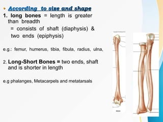
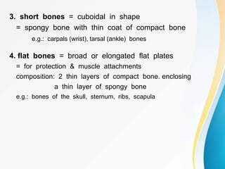
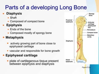
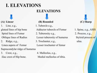
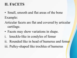
2. • osteology
• Bony & cartilaginous framework of the body
• Exoskeleton
• Endoskeleton = visceral and somatic
• Functions-
• Rigid framework of the body
• Protection to the viscera
• Provides leverage for body movements.
• stores ca++
• erytrhropoiesis
• electrolyte balance
• detoxification
• assist in respiration
3. Bone
Highly vascular mineralized connective tissue ,
consisting of cells & dense intercellular organic matrix
impregnated with organic salts.
5. •
1. Intramembranous = starts as fibrous membrane,calcium gradually
deposited until structure becomes ossified intramembranous
ossification
embryonic mesenchye directly forms bony tissue; usually occurs in embryo
e.g.: bones of the skull and face, clavicle
2. Cartilagenous = starts as cartilage, gradually ossified
enchondral or intracartilagenous ossification.
e.g.: long bones except clavicle, short bone and irregular bones
3. Membrano-cartiligenous
at the junction of vault ad base of skull bones are formed by this method
6. • According to structure
1. compact = solid mass; dense & hard
= forms the outer layer of bone structure
= functional unit --- Haversian system
2. cancellous or spongy = contain spaces
filled with bone marrow and lamellae are arranged flat
= incomplete Haversian system
8. •
A X I A L
skull
Cranium & fascial 22
hyoid 1
ossicles 6
Vertebrae 26
Ribs & sternum
24+1_
80
APPENDICULAR
Upper Extremities Lower Extremities
clavicle 2 hip bone 2
scapula 2 femur 2
humerus 2 patella 2
radius 2 tibia 2
ulna 2 fibula 2
carpals 16 tarsals 14
metacarpals 10 metatarsals 10
phalanges 28 phalanges 28
64 62
9.
10.
11.
12.
13. •
1. long bones = length is greater
than breadth
= consists of shaft (diaphysis) &
two ends (epiphysis)
e.g.: femur, humerus, tibia, fibula, radius, ulna,
2. Long-Short Bones = two ends, shaft
and is shorter in length
e.g phalanges, Metacarpels and metatarsals
14. 3. short bones = cuboidal in shape
= spongy bone with thin coat of compact bone
e.g.: carpals (wrist), tarsal (ankle) bones
4. flat bones = broad or elongated flat plates
= for protection & muscle attachments
composition: 2 thin layers of compact bone. enclosing
a thin layer of spongy bone
e.g.: bones of the skull, sternum, ribs, scapula
15.
16. 5. Irregular bones = all other bones not assigned to the
previous groups
e.g: vertebrae, pelvic bones ,bones of the base of the skull
5. MISCELLANEOUS CLASSIFICATION
a. Sesamoid bone = short bone that develops in a tendon e.g:patella ,
pisiform
b. Pneumatic bones- flat or irregular bones possessing a hollow space with in
their body, contains air.
e.g.: ethmoid, maxilla ,mastoid part of temporal bone.
c. Accessory bones- develop in relation to axial skeleton
e.g:cervical rib, lumbar rib , two frontal bones
d. Supernumerary bones- develop in relation to appendicular skeleton
e.g polydactyle
e. wormian bone- rarely found. some times a centre of ossification appears in
the middle primitive frontal lobe which forms wormian bone
17.
18.
19. Parts of a developing Long Bone
Diaphysis
Shaft
Composed of compact bone
Epiphysis
Ends of the bone
Composed mostly of spongy bone
Metaphysis
actively growing part of bone close to
epiphyseal cartilage
vascular and responsible for bone growth
Epiphyseal cartilage
plate of cartilegenous tissue present
between epiphysis and diaphysis
Figure 5.2a
20. Gross Structures of a Long Bone
Medullary cavity
Cavity of the shaft, walls are hard that
make the shaft strong
ends are articular, smoot and covered by
hyaline cartilage
Nutrient foramen
lined by endosteum
contains gellatinous material
Yellow marrow (fibro-fatty mass) in
adults
Red marrow (for blood cell formation,
erythropoiesis) in infants
found in ends of long bones bodies of
verebrae, sternum and falt bones of skull
no marrow cavity in clavicle and ribs
21. Periosteum
Outside covering of the diaphysis
Fibrous connective tissue
membrane
Sharpey’s fibers
peroiosteal collagenous fribers
Secure periosteum to underlying
bone
it is sensitive due to rich nerve
supply derived from the nerves
supplying the muscles attached to
the bone
22. Blood Supply of Bones
Numerous, supply spongy and compact bone
» enter through nutrient foramen
» reach medullary cavity
» gives ascending and desecnding branches
» anastomose with periosteal and end-blood vessels
» branchs also enters volkman’s and haversian canals
» branches are called “nutritiae
» arises from two sources
» a) periarticular anastomosis = these vessels are called
juxta epiphyseal vessels of lexer
» b) from the arteries passing over that region
23. here
additional epiphyseal and metaphyseal
arteries are present
periosteal arteries
supplied by
periosteal and nutrient arteries which
anastomose with eachother
periosteal arteries
periosteal and middle meningeal artery
24. I. ELEVATIONS
ELEVATIONS
(A) Linear (B) Rounded (C) Sharp
1. Line, e.g., 1. Tubercle e.g.,
gluteal lines of hip bone Adductor tubercle of Femur 1. Spine, e.g., ASIS
Spiral lines of Femur 2. Tuberosity e.g., 2. Process, e.g.,
Oblique lines of Radius Lesser tuberosity of humerus Styloid process of
2. Ridge, e.g., 3. Trochanter, e.g., ulna.
Linea aspera of Femur Lesser trochanter of femur
Supracondylar ridge of humerus
3. Crest, e.g., 4. Malleolus, e.g.,
iliac crest of Hip bone. Medial melleolus of tibia.
25. II. FACETS
• Small, smooth and flat areas of the bone
Example:
Articular facets are flat and covered by articular
cartilage.
• Facets may show variations in shape.
i. knuckle-like in condyles of femur
ii. Rounded like in head of humerus and femur
iii. Pulley-shaped like trochlea of humerus
26. III. DEPRESSIONS
• They are of following types
(A) Pit or Fovea
Very small and shallow depression
(B) Fossa
Slightly large and deep depression
(C) Notch or incisura
Depression bridged by a ligament
(D) Groove or sulcus
Depression with some length
(E) Foramen
Perforated depression or hole in the bone
(F) Canal or Meatus
Foramen with some length
27. CARTILAGE
The cartilage is a phylogenetically old type of connective
tissue, with wide spread distribution in vertebrates, which is
characterized by the presence of rigid but flexible matrix.
General Features
1. Covered by fibro-vasculo-cellular membrane called
pericondrium.
2. High tensile strength and resistence
3. Avascular
4. Metabolic rate is low
5. No lymphatics and nerves
6. Regeneration is not effective
28. CARTILAGE
• Types
It is composed of :
1. Cells (chondroblasts & chondrocytes)
2. Matrix (consists of ground substances and
connective tissue fibers)
29. • Depending on the matrix there are three types of
cartilage.