SlideShare a Scribd company logo
Blood
Done By
A Mahesh Kumar
1st
Year Pg student
Dept of Pedodontics
Drs S&N SIDS
1
CONTENTS
1.INTRODUCTION
2.PROPERTIES
3.FUNCTIONS
4.COMPOSITION
5.RBC
6.WBC
7.REFERENCES
2
REFERENCES :
1.Text book of Medical Physiology- 10th
edition- Guyton & Hall
2.Concise Medical Physiology- 4th
edition- Chaudhuri
3.Essentials of Medical physiology-4th
edition-Sembulingam
4.Per haavardsholm finne and Sverre halvorsen. Regulation of
Erythropoiesis in the Fetus and Newborn. Archives of Disease in
Childhood 1972; 47: 683-687
5.Narla Mohandas and Patrick G. Gallagher. Red cell membrane:
past, present, and future. Blood 2008; 112: 3939-3948
3
6.Michael Fo¨ller1, Stephan M. Huber2 and Florian Lang.
Erythrocyte Programmed Cell Death. Life 2008; 60(10): 661–8
7.Petra Kleinbongard et all. Red blood cells express a functional
endothelial nitric oxide synthase. Blood 2006; 107: 2943-51
8. Veronique Witko-Sarsat et al. Neutrophils: Molecules, Functions
and Pathophysiological Aspects. Laboratory investigations 2000;
80(5): 617-653
9. SM Rashmi et al. Neutrophils in health and disease: An overview.
J Oral Maxillofac Pathol 2006; 10: 3-8
4
INTRODUCTION
• Specialized connective tissue
• Contains cells suspended in a fluid matrix
• Fluid of life
• Fluid of growth
• Fluid of health
5
PROPERTIES
Colour : Red
Volume : In adults 5 ltrs
New born 450 ml
pH : slightly alkaline 7.4
Specific gravity : 1.052 – 1.061 total blood
1.092 – 1.101 blood cells
1.022 – 1.026 plasma
Viscosity : five times more viscous than water
6
FUNCTIONS:
1.Respiratory function
2.Excretory function
3.Nutrient function
4.Role of blood in various homeostatic processes
a)Body temperature and blood
b)pH and blood
7
5. Role of blood in various defence mechanism
6. Transport of hormones and enzymes
7. Storage function
8. Regulation of water balance
8
COMPOSITION OF BLOOD
1.Plasma - 55%
2.Formed elements - 45%
a) RBC or Erythrocytes
b) WBC or Leukocytes
c) Platelets or Thrombocytes
9
10
11
CONTENTS
1.MORPHOLOGY
2.PROPERTIES
3.ERYTHROPOIESIS
4.HEMOGLOBIN
5.IRON METABOLISM
6.LAB INVESTIGATIONS
12
MORPHOLOGY
• Circular, biconcave cell without a nucleus
• Diameter - 7.5µ
• Thickness at the periphery - 2µ
• Thickness at the centre - 1µ
13
Advantages of biconcave shape
 Thickness of an RBC in its central part is not great,
so oxygen doesnot have to travel a great distance for the
diffusion
 Biconcavity increases the surface area of the RBC,
so oxygen gets a bigger area for diffusion
 The erythrocyte can squeeze itself through a
capillary more easily.
14
Red cell membrane
It is a tri laminar structure having a bimolecular lipid layer
interposed between the two layers of proteins.
Lipids:
Glycolipids,
Phospholipids,
Cholesterol.
15
The proteins comprising the erythrocyte cytoskeleton
include
1.Ankyrin
2.spectrin
3.Band 4.1
4.Band 3
5.Actin filaments
16
Protein 4.1R-dependent multiprotein complex: New
insights into the structural organization of the red
blood cell membrane
17
PROPERTIES OF RED BLOOD CELLS
1.Rouleaux formation
2.Specific gravity : 1.092 to 1.101
3.Packed cell volume
4.Suspension stability
18
ERYTHROPOIESIS
Erythropoiesis is the process by which the origin,
development and maturation of erythrocytes occur.
Hemopoiesis is the process which includes origin,
development and maturation of all the blood cells.
19
 Blood forming tissues
Myeloid tissue
Lymphoid tissue
 Myleloid tissue
means RBM
produces RBCs, granulocytes, monocytes and platelets.
 Lymphoid
lymphnode, thymus, and spleen
Produce lymphocytes.
20
Site of Erythropoiesis
In fetal life
21
Age Site
Upto 5-6 years RBM of all bones
6-20 years RBM of long bones & all
membranous (flat) bones
After 20 years All membranous bones &
ends of long bones
In Post natal life and in adults
22
Bone marrow
2 types
• Red bone marrow
• Yellow bone marrow
RBM – found in flat bones (cranial, ribs, sternum ,
pelvic bones)
YBM – shafts of long bones in fully grown adults.
23
Extra medullary hemopoiesis
when there is necessity of increased erythropoiesis the
YBM is converted into RBM. If it is more intense liver
and spleen also start producing RBC.
(Medulla = bone marrow)
24
Histology of RBM
Consists of
1.Large no of sinusoids
2.Adventitious cells outside the sinusoids
3. Blood forming cells in between adventitious cells
25
• Sinusoids are basically capillaries with larger
diameter.
• Their walls contain big pores – big molecules,
blood cells pass.
• Adventitious cells – become fat cells
• The blood cells – Precursors of erythrocytes,
leucocytes, and platelets
• Normally – blood cells : fat cells – 1:1
26
RBM blood picture
Cells Percentage
Granulocytes & their precursors 60%
Erythrocytes & their precursors 20%
Lymphocytes, monocytes &
precursors
10%
Others ( non identifiable,
degenerated cells)
10%
27
Stages of development
28
29
30
Different stages of RBC development
Progenitor cells – BFU-E (Burst forming unit)
CFU-E
Blast cells – Pronormoblast (E1)
Early normoblast(E2)
Intermediate normoblast(E4)
Late normoblast (E5)
Reticulo cyte (E6)
Matured RBC
31
1. Pronormoblast : (proerythroblast)
 It is a large cell - deeply basophilic
cytoplasm with a large central
nucleus.
 The deep blue colour - high content
of RNA associated with protein
synthesis.
32
Basophilic Normoblast
•This is a round cell having a
diameter of 12-16 um with a
large nucleus.
•The nucleus is more condensed
than the pronormoblast and
contains basophilic cytoplasm.
•This cell undergoes rapid
proliferation.
33
Polychromatic normoblast
(Intermediate)
•The nucleus - coarse and deeply
basophilic.
•The cytoplasm is polychromatic with
mixture of basophilic RNA and
acidophilic hemoglobin.
34
Orthochromatic (late) normoblast
•This is the final stage in the
maturation of nucleated red cells.
•The cell is smaller - small pyknotic
nucleus with dark nuclear chromatin.
•The cytoplasm - acidophilic due to
large amounts of hemoglobin.
35
Reticulocytes
•Reticulocytes - devoid of nucleus but
contain RNA
•These cells in the peripheral blood
smear - slightly basophilic hue.
36
Precursors of RBC
37
Factors influencing erythropoiesis:
1. Hemopoietic growth factor
Erythropoietin
Stem Cell Factor (SCF)
Interleukins
(GM-CSF)
Colony Stimulating Factor (CSF)
Thrombopoietin
2. Vitamin B12, Folic Acid, Pyridoxine, Vitamin C
3. Iron and Copper
38
Erythropoietin (EPO)
39
Applied physiology
•COPD – high RBC count due to high production of
EPO
•Chronic damage of kidney – anemia due to lower
levels of EPO
40
Interleukins (IL)
•IL-1: Stimulates granulocytic cells. Induces
expression of GM-CSF
•IL-6 & IL-1: Stimulates proliferation of progenitor
cells.
•IL-3: Stimulates growth in all phagocytes.
41
INTERLEUKINS:
• Involved in erythropoiesis are
• IL-3
• IL-6
• IL-11
42
Stem Cell Factor (SCF):
Stimulates early stem cells to differentiate.
(TGF-beta)
Inhibits hematopoieses.
Directly inhibits proliferation of progenitor
cells.
43
Role of Vit B12
44
Folic acid
intestine
polyglutamate monoglutamate
methylmonoglutamate &
absorbed & enter blood
reduced to become
methyltetrahydrofolate
this form reaches tissues
they loose methyl group & become
tetrahydrofolate
accepted by Vit B12 methylcobalamin
45
THF (tetrahydrofolate)
formyl THF (folinic acid )
5,10 methylene THF (active form of folate )
dihydrofolate(FH2 )
46
Hemoglobin: (Hb )
• Present inside the RBC.
• Chromoprotein specialized for transport of oxygen
and carbon dioxide
• Conjugated protein with mol wt 68,000 daltons
• Forms 95% dry weight and 35 – 40% of volume of
RBC
47
NORMAL VALUES
AGE NORMAL VALUE
At birth 25gm%
After 3rd
month 20gm%
After 1 year 17gm%
In adult males 15gm%
In adult females 14.5gm%
48
DERIVATIVES OF HEMOGLOBIN
1.OXYHEMOGLOBIN
2.REDUCED HEMOGLOBIN OR FERROHEMOGLOBIN
3.CARBHEMOGLOBIN
4.CARBOXYHEMOGLOBIN
5.SULFHEMOGLOBIN
6.NITROUS OXIDE HEMOGLOBIN
7.METHEMOGLOBIN OR FERRIHEMOGLOBIN
49
FUNCTIONS OF HEMOGLOBIN
1.Transport of O2
2.Transport of CO2
3.Acts as blood buffer
50
Hb Chemistry &
synthesis:
Hb molecule contains
two ingredients:
•Haem ( iron &
protoporphyrin )
•Globin
51
Heme formation
• Heme formation takes place in the mitochondria then
the cytoplasm of erythroid precursors through the
reticulocyte stage.
• It begins with production of a protoporphyrin ring.
• Iron then incorporates with protoporphyrin to form
heme.
• Heme synthesis is stimulated by erythropoietin
52
Heme structure:
4 pyrrole structures
They are linked up with one another by methine ( = CH - )
53
Globin formation
•The polypeptide chains of globin are produced on the
ribosomes.
•The four most common chain types are alpha, beta,
gamma and delta chains.
•Each of these chains differs from the others in their
amino acid sequence.
•Each globin molecule is made of 2 pairs of chains and
each chain is made of 141-146 amino acids.
54
Globin structure:
•contains 4 polypeptide chains
2 alpha - containing 141 amino acids
2 beta - containing 146 amino acids
•with each polypeptide chain 1 molecule of haem is
attached.
55
56
57
Synthesis of Hb
•Hb is synthesized in the erythroid series in the red bone
marrow
•Hb first appears at the intermediate normoblast
•Protoporphyrin can be synthesized by normoblasts from
products of metabolism and aminoacids like succinyl Co
A , glycine etc
•Iron has to be obtained from food or iron contained in
the hemoglobin of dead RBC’s
58
Basic Chemical Steps : (synthesis of Hb )
1.2 succinyl-CoA + 2 glycine ---------------> pyrrole
molecule
2.4 pyrrole -----------------> protoporphyrin IX
3.Protoporphyrin + ferrous iron -------> heme
4.Heme + polypeptide (globin) ---------> hemoglobin
chain
5.2 alpha +2 beta ------------- > hemoglobin
59
Factors necessary for Hb maturation
1.Proteins and aminoacids
2.Iron
3.Copper – necessary for absorption of iron from gut
4.Cobalt and nickel for utilization of iron during Hb
formation
60
Iron metabolism
•Imp for oxygen transport
•Imp for formation of Hb and myoglobin
•Also necessary for formation of other substances
like cytochrome, cytochrome oxidase, peroxidase,
and catalase
61
Normal values
Total quantity of iron in the body is 4gms
Hb 65-68%
Muscle as myoglobin 4%
Intracellular oxidative
heme compound
1%
In the plasma as
transferrin
0.1%
Reticuloendothelial
system
25-30%
62
•Dietary iron available 2 forms
•Heme iron
•Non heme iron
•Heme iron- RBC , absorbs easily
•Non heme – vegetables, grains, cereals. not absorbed
easily
63
Absorption of iron
Iron is absorbed mainly from the small intestine.
Bile is essential for absorption
Immediately after absorption into the blood, iron
combines with a β globulin called – apotransferrin.
Which results in formation of transferrin
64
65
Fate Of RBC
•Life span of RBC is about 120 days.
• As age of the RBC increases, the enzymes which
protect the erythrocyte from the damaging effects of
oxygen begin to lose their efficiency, therefore oxidative
damages begin to appear & the RBC becomes fragile.
•Now the cell ruptures , self-destruct in spleen
66
LAB INVESTIGATIONS
67
RED CELL COUNT:
Equipment
• Red cell pipette with a red bead in the bulb & markings of
0.5, 1 & 101
• Improved Neubauer chamber
68
Diluting Fluid
1.Hayem’s fluid
Mercuric chloride 0.5 g
Sodium chloride 1.0 g
Sodium sulfate 5.0 g
Distilled water 200 ml
69
RBC count = N
1/5 x 1/10 x 1/200
= N x 10000
Normal Range :
Adult male 4.5-5.5 million/ cumm
Adult female 3.8-5.2 million/cumm
At birth 4.0-6.0 million/cumm
70
VARIATIONS IN NUMBER OF RED BLOOD CELLS
Physiological variations
1.Increase in RBC
a)Age
b)Sex
c)High altitudes
d)Muscular exercise
e)Emotional conditions
f)Increased environmental temperature
g)After meals
71
2. Decrease in RBC
a)High barometric pressures
b)After sleep
c)Pregnancy
72
Pathological Variations
1.Primary Polycythemia – Polycythemia Vera
Myeloproliferative disorders like malignancy of red
bone marrow
2.Secondary Polycythemia
a)Respiratory disorders like emphysema
b)Congenital heart disease
c)Ayerza’s disease
d)Chronic carbon monoxide poisoning
e)Poisoning by chemicals like P & As
f)Repeated mild hemorrhages
73
VARIATIONS IN SIZE OF RBC
1.Microcytes
a)Iron deficiency anemia
b)Prolonged forced breathing
c)Increased osmotic pressure in blood
2.Macrocytes
a)Megaloblastic anemia
b)Muscular exercise
c)Decreased osmotic pressure in blood
3.Anisocytes
a)Pernicious anemia
74
VARIATIONS IN SHAPE OF RED BLOOD CELLS
1.Crenation
2.Spherocytosis
3.Elliptocytosis
4.Sickle cell
5.Poikilocytosis
75
VARIATIONS IN STRUCTURE OF RED BLOOD CELLS
1.Punctate Basophilism:
•Dots of basophilic materials (porphyrin) appear in RBC
•Lead poisoning
2. Ring:
•Twisted strands of basophilic materials appear in the
periphery
•Goblet ring
3.Howell – Jolly bodies:
•Nuclear fragments in the RBC
76
Hb estimation methods
1.Calorimetric Methods
a)Visual Methods
b)Methods using photoelectric colorimeter
2. Gasometric Method
3.Chemical Method
4.Specific Gravity Method
77
Visual
1.Tall Quist paper method - inaccurate
2.Sahlis Acid hematin
3.WHO Hb colour scale – simple, reliable,
inexpensive, suitable for where calorimeter is not
available.
78
Sahli’s Method
Tube with marking
Principle :
Blood is added to N/10 Hcl – Acid
Hematin.
Acid hematin is matched against the
brown colour.
79
• Add N/10 Hcl – into Hb meter tube
• Fill the Hb pipette with 0.02ml of blood
• Acid act on the RBC for 10min to lyse the cells and
convert Hb to acid hematin.
• Match colour of the solution with comparator in
natural light.
• Darker – add distilled water
• Continue till the colour of solution matches
80
Disadvantages:
• Visual error
• After 10min acid hematin starts fading
81
Tall Quist Paper Method
• Includes a color chart
and 150 test papers
• Allow blood to absorb
into one of the test
papers and compare the
color scale.
82
WHO Hb colour scale
• Place a drop of blood on the
test strip provided
• Wait about 30 seconds
• Match immediately the colour
• simple, reliable, inexpensive,
suitable for where colorimeter
is not available.
83
Methods using photoelectric colorimeter
1.Cyanmet haemoglobin method
2.Oxyhemoglobin method
3.Alkaline haematin Method
84
Cyanmethemoglobin Method
Principle :
Hb is oxidized to - Met Hb by potassium ferricyanide
Met Hb – Cyanmet Hb
Technique
Take 5ml of Drabkin’s solution
( Drabkin’s reagent contains sodium bicarbonate, potassium
ferricyanide, and potassium cyanide)
Add 0.02ml of blood
Wait for 10min
Hb = OD of test sample x Conc. Standardx250
OD of standard x1000
Oxyhemoglobin Method
• 0.04% of ammonia is used
• Which lyses the RBC
• Colour is compared to standard
Alkaline Hb method
• N/10 NaOH is used.
• Colour of alkaline hematin is compared with standard
86
Gasometric Method – oxygen carrying capacity of blood
is measured in van slyke like apparatus.
Not suitable for routine use
Specific Gravity Method – simple , rapid & inexpensive.
Rough estimation of Hb is obtained from specific gravity
of blood, used for mass screening
87
PCV
•Hematocrit is the % volume of red cells in a given sample
of blood.
•It gives an estimate of relative volume of cells and plasma.
•It is useful for evaluating absolute values like MCV and
MCHC
88
Wintrobe’s method :
•Collect 2ml of blood
•Take wintrobe’s tube
• 0-100 – ESR
• 100-0 – PCV
•Take the blood in pasteur’s pippete and withdraw the
blood into the tube, till it reaches 100 marking.
89
normal range
Males – 40- 55%
Females – 35- 48 %
90
MCV: ( normal value 80 – 100 fl )
It is the volume of an average RBC (single RBC)
expressed in femotoliters or fl
PCV X 1000
MCV = -----------------------------------------
RBC in millions / cmm.
Normal : 82-98 fl
91
MCH - Amount of Hb in the red cell
Hb x10
MCH = ----------------------- pg
RBC count
Normal = 28-32pg
92
MCHC: ( normal value 31 – 36% )
represents the average concentration of Hb in a given volume
of packed red cells.
Hb
MCHC = -------------------------------------------
PCV ( % )
93
Erythrocyte Sedementation Rate : (ESR)
It is a measure of the settling of red blood cells in a tube
of blood during one hour.
normal value - 0-20mm in female in 1st
hr
- 0-10mm in male in 1st
hr
Stages of sedimentation :
•Rouleaux formation – first 15 min
•Formation of fine threads – next 15 mins
•Rapid fall – next 15min
•Packing phase – packing of RBC next 15min
94
• Two methods to determine ESR.
Westergrens Method:-Westergrens tube - 300mm long(30cm),
2.5mm internal diameter & opened on both ends, calibrated
from 0-200.
Procedure
•1.6 -2ml of blood mixed with 0.4-0.5ml of 3.8% sodium
citrate - (anticoagulant) and mix well, loaded in westergrens
tube up to zero mark.
95
• The tube fitted to the stand vertically and left
undisturbed at room temperature.
• Reading taken at the end of 1 hour (Distance from 0
mark to top of RBC column is recorded as ESR)
96
Anticoagulated whole
blood has just been added.
(Time: 0)
Red blood cells have settled, leaving
plasma at the top of the tube. Reading:
18 mm/hour
(Time: one hour)
WESTERGREN’S TUBES
97
Modified Westergrens method
•EDTA instead of citrate as anticoagualant
•2ml of EDTA diluted with 0.5ml of 3.8% sodium
citrate or 0.5ml of 0.85% sodium chloride
•Undiluted EDTA blood gives poor precision
98
Procedure
•Collect 2ml of blood
• Blood loaded in the tube up to’0’mark,
tube is placed vertically on the Wintrobe’s
stand.
•The reading taken after one hour
Wintrobes Method
•Wintrobe’s tube short tube (110mm long,
diameter 2.5mm) opened on only one end.
99
NORMAL VALUES OF ESR
Westergren’s method
•Males – 0-15mm 1st
hr
•Females - 0-20mm 1st
hr
Wintrobe’s method
•Males – 0-7mm 1st
hr
•Females - 0-14mm 1st
hr
100
Factors That May Influence ESR
Factors that increase
ESR
Old age
Female
Pregnancy
Anemia
Macrocytosis
Factors that decrease ESR
Polycythemia
Red blood cell abnormalities
Spherocytosis
Microcytosis
101
LEUCOCYTES
102
Introduction :
• Leukos = white, cytes = cells
• Mobile units of the body’s protective system.
• White blood cells are the colorless and nucleated formed
elements of blood.
• These cells are larger in size and their number is less
compared to that of RBC’s.
• They play a very important role in defense mechanism
103
104
Type of leucocyte Normal range Lifespan
Neutrophil 50-75 % 2-5 days
Eosinophil 1-6 % 7-12 days
Basophil 0-1% 12-15 days
Lymphocyte 25-45% ½-1 day
Monocyte 3-8 % 2-5 days
105
NEUTROPHILS
•Diameter : 10-14µ
•Nucleus :
Young neutrophil : horse-shoe shaped nucleus
Mature neutrophil : multilobed (2-6 lobes)
lobes connected by chromatin filaments
•Cytoplasm : pale bluish in colour & full of fine granules
Granules take both acidic & basic stain & look violet-
pink in colour
106
•Condensed chromatin along the inner surface of nuclear
envelope
•More central region of each lobe appears paler
•Cytoplasm contains few mitochondria & a small Golgi
complex
•Azurophilic granules are round or oval & more electron
dense than specific granules.
107
NEUTROPHILS - DEVELOPMENT
MYELOBLAST :
• Earliest recognizable cell in the granulocytic maturation
process.
• 15-20µm in diameter
• Large round to oval nucleus
• Small amount of basophilic cytoplasm
• Nucleus contains 2 to 5 nucleoli
• Nuclear chromatin is fine and reticular
108
PROMYELOCYTE :
• Slightly larger in size than myeloblast.
• Primary or azurophilic granules appear at the
promyelocyte stage.
• Nucleus contains nucleoli as in myeloblast stage
• But nuclear chromatin shows slight condensation.
109
MYELOCYTE :
• Characterized by the appearance of secondary or specific
granules.
• Smaller cell with round to oval eccentrically placed
nucleus.
• More condensation of chromatin than in promyelocyte
stage and absence of nucleoli.
110
• Cytoplasm is relatively greater in amount than in
promyelocyte stage.
• Contains both primary and secondary granules.
• Last cell capable of mitotic division
111
METAMYELOCYTE :
• Nucleus becomes indented and kidney shaped.
• Nuclear chromatin becomes moderately coarse
• Cytoplasm contains both primary and secondary
granules
112
BAND STAGE ( STAB FORM ) :
• Characterized by band-like shape of the nucleus
with constant diameter throughout
• Condensed nuclear chromatin
113
SEGMENTED NEUTROPHIL:
• With Leishman’s stain,
nucleus appears deep purple
with 2-5 lobes joined by thin
filamentous strands.
• Nuclear chromatin pattern is
coarse.
• Cytoplasm stains light pink
and has small, specific granules
114
115
Granules
•Number of granules : 500-1500/granulocyte
•Large amount of protein, traces of lipids & nucleic acids
1.Primary or azurophilic granules
2.Secondary or specific granules
3.Tertiary or gelatinase granules
4.Secretory vesicles
116
Azurophilic granules
•Myeloperoxidase, defensins, lysozyme, azurocidin etc
•These granules fuse with phagocytes vesicles resulting in
the delivery of their contents to the ingested organism
•Greenish coloration to pus is imparted by
myeloperoxidase
117
Secondary or specific granules :
• 3times more common in cytoplasm
• Lysozyme, Lactoferrin, collagenase, histaminase may
modify the inflammatory process
Tertiary granules :
• Gelatinases
118
Life history of neutrophils :
•Released from the bone marrow
•Exist in two populations
•Circulating pool
•Marginal pool
•Rapid exchange between the two pools
• Activated by numerous stimuli
119
 Disposed of internally by cells of reticuloendothelial
system
 External loss : emigration through gingiva into the
saliva & excretion in urine ( common)
 Neutrophils occur in the secretions of uterus during
second half of menstrual cycle
 Dead neutrophils – granulocyte-inducing factor
120
Old senile neutrophils are characterized by:
•Loss of motility
•Poorly stained granules
•Increased nuclear lobulation
•Easy breakability while making blood smear
121
Nitroblue tetrazolium reduction test :
•Ability of neutrophils to destroy micoorganisms with
intracellular enzymes can be evaluated by NBT
•Normal neutrophils contain enzymes that convert colorless
NBT to dark blue granules within the cell
•When dark blue granules are not seen, neutrophils will not
destroy bacteria
122
FUNCTIONS :
First line of defence
1.PHAGOCYTOSIS
2.REACTION OF INFLAMMATION
3.FEBRILE RESPONSE
123
VARIATION IN COUNTS
Neutrophilia - >1000 cumm
Physiological causes
•New born babies
•After exercise
•After meals
•Pregnancy
•Menstruation
•Parturition
•Lactation
•Mental & emotional stress
124
Pathological causes
•Acute bacterial infections
Lobar pneumonia
Bronchopneumonia
Pyogenic meningitis
Cellulitis
Infected burns
Diphtheria
125
•Acute inflammatory diseases like
Acute rheumatic fever
Acute appendicitis
•Acute stress states like
Post surgery
Post haemorrhage
Myocardial infarction
•Chronic myeloproliferative disorders like
CML
Polycythemia vera
126
NEUTROPENIA:
CAUSES
•Typhoid and paratyphoid fever
•Physical agents like radiation
•Chemicals : Benzene
•Antimetabolite drugs : Cyclophosphamide, Methotrexate
•Hematologic disorders like megaloblastic anemia, aplastic
anemia, subleukemic leukemia, cyclic neutropenia,
127
COOKE’S ARNETH COUNT
Stage Nuclear lobes Normal count
Stage I (N1) 1 lobe 5-10 %
Stage II (N2) 2 lobes 20-30 %
Stage III (N3) 3 lobes 40-50 %
Stage IV (N4) 4 lobes 10-15 %
Stage V (N5) 5 lobes or more 3-5 %
128
Clinical Significance :
1.Left shift
•N1+N2+N3 > 80%
•More younger cells
•Indicates hyperactive bone marrow (high rate of
formation)
2.Right shift
•N4+N5 > 20%
•More mature cells
•Hypoactive bone marrow (slow rate of formation)
129
Variation in neutrophil morphology :
1.Variation in granules
2.Formation of vacuoles in cytoplasm
3.Formation of Dohle bodies in cytoplasm
4.Presence of sex chromatin with nuclear lobes
5.Hypersegmented neutrophils
6.Band and straw form neutrophil
7.Pelger – Huet anamoly
130
DOHLE BODIES
131
SEX CHROMATIN
132
HYPERSEGMENTED NEUTROPHILS
133
PELGER HUET ANAMOLY
134
If most of the neutrophils appear bilobed, this is
indicative of an uncommon condition known as Pelger-
Huet anomaly, an inherited condition.
This is the heterozygous form.
The homozygous form is fatal. Just be aware of this
condition when you get back a manual differential count
with mostly bands, but the WBC count is normal or the
patient shows no signs of infection or inflammation.
135
EOSINOPHILS :
• Forms via same stages as the neutrophil.
• Diameter = 12µ - 17µ
• Nucleus: bilobed, purple colored, spectacle shaped, nucleoli
– absent
Condensed chromatin stains less intensely than that of
neutrophil
136
Cytoplasm: acidophilic and appears bright pink
Contains deep red staining granules which do not cover the
nucleus.
•Condensed chromatin is peripherally distributed along the
inner surface of nuclear envelope.
•Specific granules : ovoid with electron dense crystalloid
cores that consist chiefly of basic proteins.
•Only other prominent organelles : Golgi apparatus,
mitochondria
137
In RBM ( 5-6 days)
Circulatory pool (8-12 hours)
Emigrates into tissue
Death and removed
138
Cytoplasm contains two types of granules
1. Specific granules
Large numerous elongated granules
Contain Crystalloid bodies
- Contain four major proteins
1. Major basic protein
2. Eosinophil cationic protein
3. Eosinophil peroxidase
4. Eosinophil derived neurotoxin
Also contains histaminase, arylsulfatase, collagenase,
cathepsins
139
Azurophilic granules
•Lysosomes, contains acid hydrolases
•Function in the destruction of parasites
•Hydrolysis of antigen – antibody complexes.
140
FUNCTIONS OF EOSINOPHILS :
1.ROLE IN PARASITIC INFESTATIONS:
•Major basic protein (MBP) : damage the parasites by
causing distension and detachment of the tegumental
sheath of these organisms.
•Eosinophil cationic protein : major destroyer of
helminths.
10 times more toxic than MBP
Destroys parasites by complete disintegration
141
• Eosinophil peroxidase : capable of destroying helminths,
bacteria and tumor cells
• Eosinophilic derived neurotoxin : destroys the nerve
fibers particularly myelinated nerve fibers.
2. ROLE IN ALLERGIC REACTION
• Capable of destroying inflammation inducing substances
like histamines and bradykinin.
142
• Destroy antigen-antibody complexes & thus prevent
spread of local inflammatory process.
• Eosinophil arylsulfatase can degrade SRS-A liberated
by mast cells and basophils
• Produces histaminase that can inactivate histamine.
143
Eosinophilia :
•Allergic conditions like hay fever, bronchial asthma
•Parasitic infestations
•Skin diseases like urticaria
•Scarlet fever
Eosinopenia :
•ACTH & steroid therapy
•Stressful conditions
•Acute pyogenic infections
144
BASOPHILS :
•Diameter = 10-12µ
•Nucleus – bilobed , stains less deeply than that of
neutrophil
•Cytoplasm : basophilic, full of granules
granules : deep purple or blue
145
• Membrane bound granules – 0.5µ
• metachromatic & similar to mast cell granules.
• Chemical mediators liberated when basophils
degranulate include histamine, SRS-A, ECF-A
• The cell surface receptors on basophils closely
resemble those on mast cells.
146
FUNCTIONS
1.ROLE IN ALLERGIC REACTION :
release histamine, bradykinin, SRSA, seratonin.
2. ROLE IN PREVENTING SPREAD OF ALLERGIC
INFLAMMATORY PROCESS :
•releases eosinophil chemotactic factor
•eosinophils then phagocytose & destroy antigen – antibody
complexes & prevent spread of local inflammatory process
147
3. RELEASE OF HEPARIN
• Prevents clotting of the blood.
• Activates the enzyme lipoprotein lipase which
removes fat particles from the blood after a fatty meal
148
BASOPHILIA : >100 cumm
•Viral infections
•Allergic diseases
•Chronic myeloid leukemia
BASOPENIA :
•Corticosteroid therapy
•Drug-induced reactions
•Acute pyogenic infections
149
MONOCYTES
•12-20µ in diameter, Largest leukocytes
CFU-M
Monoblast
Promonocyte
Monocyte
150
• Nucleus : variable in appearance
ranging from indented ovoid or
roughly kidney shaped to wide horse
shoe
Chromatin less condensed
• Cytoplasm : pale grayish-blue color
Fine pinkish-purple granules can
also sometimes be seen
Specific granules lacking
151
• 2 or more nucleoli
• Fairly prominent Golgi apparatus, moderate amount of
ribosomes, small amount of rER, no. of small
mitochondria
• Small dense granules – 0.3-0.6µ
• Surface of monocytes is irregular
152
153
Functions
1.Phagocytosis :
second line of defence
2.Immediate precursors of macrophages
3.Releases IL-1, TNF-α, transferrin, lysozyme,
proteases, acid hydrolase
154
MONOCYTOSIS
•Parasitic conditions like malaria and kalaazar
•Infective conditions like subacute bacterial endocarditis,
tuberculosis
•Neoplastic states like acute monocytic leukemia, acute
myelomonocytic leukemia, chronic myelomonocytic
leukemia
155
LYMPHOCYTES
•20-50% of blood leukocytes
•Lack prominent cytoplasmic granules when seen in LM. 10%
- may contain reddish purple staining granules
•Based on size
1.Small lymphocyte : 6-9µ
2.Large lymphocyte : 9-15µ
156
Committed stem cells
Pre -T cell Pre B cell
T lymphocytes Blymphocytes
157
Small lymphocyte :
•Small spherical nucleus with a small indentation on one side.
•Condensed chromatin
•Remarkably little cytoplasm
•Cytoplasm is hardly seen in LM
158
•Cytoplamic organelles are
scant
•Few mitochondria, sparse
cisternae of rER, small Golgi
apparatus
159
Large lymphocyte :
•Nucleus slightly larger than small lymphocyte
•More cytoplasm
EM :
•Large numbers of mitochondria, free ribosomes,
more cisternae of rER, slightly larger Golgi apparatus
160
161
On the basis of functional properties
162
LYMPHOCYTOSIS : >45%
•Chronic infective conditions like tuberculosis,
syphilis & brucellosis
•Viral diseases like infectious mononucleosis,
measles, chicken-pox & viral fever
•Neoplastic conditions like CLL
163
LAB INVESTIGATIONS
TOTAL LEUCOCYTE COUNT
Principle :
Whole blood is diluted with WBC fluid that hemolyses the
red cells.
Nucleated cells – WBC s stained by Gentian Violet are
counted in a Neubauer chamber
164
Equipment:
Hemocytometer set consisting of WBC pipette and
improved Neubauer chamber with a cover slip
WBC pipette has a white bead in the bulb and markings
of 0.5, 1, 11
165
WBC Fluid ( Turk’s fluid )
Glacial Acetic Acid 2ml
1 % Gentian violet 5 drops
Add water to 100 ml
166
TLC = N x 20
4 x 0.1
= N x 50
Depth of the chamber = 0.1 mm
Dilution = 1 in 20
4 corner squares are counted
N = Number of WBCs in 4 large squares
167
Adults 4000 – 11000 cells / cumm
At birth 8000 – 28000 cells / cumm
Childhood 6000 – 15000 cells / cumm
Normal Value
168
Leucocytosis Leucopenia
> 11000 / cumm < 4000 / cumm
•Pregnancy
•Exercise
•Leukemoid reaction
•Diabetic and uremic coma
•Infectious mononucleosis
•Bacterial and viral infections
•Leukemias
•Aplastic anemia
•Hypersplenism
•Typhoid, paratyphoid fever
•Drug induced leucopenia
•Radiation & cytotoxic therapy
•Megaloblastic anemia
•Subleukemic leukemia
169
ABSOLUTE EOSINOPHIL COUNT
•Accurate assessment of eosinophils in blood & is useful
in diagnosis, treatment & follow-up of cases of
eosinophilia
•EQUIPMENT :
Improved Neubauer chamber, WBC pipette
170
Eosin 200mg dissolved in
10ml of distilled
water
Stains the granules
of eosinophils
orange red
Water 80ml Lyses the red cells
& white cells
except eosinophils
Acetone 10ml Fix the eosinophils
Dunger’s fluid
171
AEC = N X10
4 X 0.1
= N X 25
Normal Range = 40-450 cells /cumm
172
PERIPHERAL SMEAR AND DIFFERENTIAL
LEUCOCYTE COUNT
1.PREPARATION OF SMEAR
2.FIXATION OF THE SMEAR
•Fixed within 4hours to get good staining
•Methanol
3. STAINING OF THE SMEAR
173
Commonly used stains :
•Leishman stain
•Giemsa stain
•Wright’s stain
•Jenner’s stain
•Jenner – Giemsa stain
174
BLOOD GROUPS
•Discovered by Karl Landsteiner in 1901
•Nobel prize in 1930
•30 blood group systems given by International Society of
blood transfusion
175
176
177
International Society of Blood Transfusion. October 2006
178
LANDSTEINER’S LAW
1.If a particular antigen is present in the red blood cells,
corresponding antibody must be absent in the serum.
2.If a particular antigen is absent in the red blood cells,
the corresponding antibody must be present in the serum
Second part of law – not applicable to Rh factor
179
ABO Blood groups
Group Antigen in RBC Antibody in
serum
A A Anti B
B B Anti A
AB A and B No antibody
O No antigen Anti A and Anti
B
180
Population A B AB O
Europeans 42 9 3 46
Asians 25 25 5 45
181
DETERMINATION OF THE ABO GROUP
Blood grouping, blood typing or blood matching.
Principle
•Basis of agglutination
•Occurs antigen + antibody
Requisites
•Suspension of patients red blood cell
•Anti serum A & B
182
Procedure & Results
Agglutination with antiserum Blood group
Anti serumA A
Anti serum B B
Both A & B AB
No agglutination O
183
Universal donors O
Universal recipients AB
Cross matching
Mixing the serum of the recipient and the red blood
cells of donor
184
Gene receive from
parents
Group of the
offspring
Genotype
A+A A AA
A+O AO
B+B B B
B+O BO
A+B AB AB
O+O O OO
Inheritance
185
Transfusion reactions due to ABO incompatibility
•Jaundice
•Cardiac Shock
•Renal Shut down
186
Rh factor
• Antigen present in RBC
• Discovered by Landsteiner and Wiener in Rhesus
monkey
• Many Rh antigens only D is highly antigenic
• Among Asians 85% - Rh +ve
• 15% - Rh –ve
• Rh +ve more among blacks
187
Dd Dd Dd
Father
DD
Rh+
Mothe
r
dd
Rh -
Dd
All offsprings are Rh +
188
dd dd dd
Father
dd
Rh -
Mothe
r
dd
Rh -
dd
All offsprings are Rh -
189
dd Dd dd
Father
Dd
Rh+
Mothe
r
dd
Rh -
Dd
50% offsprings are Rh +
190
Transfusion reactions due to incompatibility
•Erythroblastosis Fetalis
•Hydrops Fetalis
•Kernicterus
191
REFERENCES :
1.Text book of Medical Physiology- 10th
edition- Guyton & Hall
2.Concise Medical Physiology- 4th
edition- Chaudhuri
3.Essentials of Medical physiology-4th
edition-Sembulingam
4.Per haavardsholm finne and Sverre halvorsen. Regulation of
Erythropoiesis in the Fetus and Newborn. Archives of Disease in
Childhood 1972; 47: 683-687
5.Narla Mohandas and Patrick G. Gallagher. Red cell membrane:
past, present, and future. Blood 2008; 112: 3939-3948
192
6.Michael Fo¨ller1, Stephan M. Huber2 and Florian Lang.
Erythrocyte Programmed Cell Death. Life 2008; 60(10): 661–
668
7.Petra Kleinbongard et all. Red blood cells express a functional
endothelial nitric oxide synthase. Blood 2006; 107: 2943-2951
8. Veronique Witko-Sarsat et al. Neutrophils: Molecules, Functions
and Pathophysiological Aspects. Laboratory investigations 2000;
80(5): 617-653
9. SM Rashmi et al. Neutrophils in health and disease: An overview.
J Oral Maxillofac Pathol 2006; 10: 3-8
193
194

More Related Content

What's hot

Hematopoiesis
HematopoiesisHematopoiesis
Hematopoiesis
Fayyaz Ahmad
 
White blood cells [wbc]
White blood cells [wbc]White blood cells [wbc]
White blood cells [wbc]
Beeula A
 
Platelets
PlateletsPlatelets
Platelets
Dr Kumar
 
Hematopoiesis: Formation of Blood Cells - An Overview
Hematopoiesis: Formation of Blood Cells - An OverviewHematopoiesis: Formation of Blood Cells - An Overview
Hematopoiesis: Formation of Blood Cells - An Overview
StudyFriend
 
Blood physiology
Blood physiologyBlood physiology
Blood physiology
Raniagaye Mansibang
 
Blood (RBC, WBC, PLATELET)
Blood (RBC, WBC, PLATELET)Blood (RBC, WBC, PLATELET)
Blood (RBC, WBC, PLATELET)
Avinandan Jana
 
RBC Indices- MCV, MCH, MCHC II Blood Physiology
RBC Indices- MCV, MCH, MCHC II Blood PhysiologyRBC Indices- MCV, MCH, MCHC II Blood Physiology
RBC Indices- MCV, MCH, MCHC II Blood Physiology
HM Learnings
 
Morphology of white blood cells
Morphology of white blood cellsMorphology of white blood cells
Morphology of white blood cells
Prince Lokwani
 
Hematopoiesis
HematopoiesisHematopoiesis
Hematopoiesis
NITISH SHAH
 
Erythropoiesis
ErythropoiesisErythropoiesis
Erythropoiesis
Sai Sailesh Kumar Goothy
 
ERYTHROPOIESIS
ERYTHROPOIESISERYTHROPOIESIS
ERYTHROPOIESIS
Dr Nilesh Kate
 
Physiology of Blood
Physiology of BloodPhysiology of Blood
Physiology of Blood
Eneutron
 
blood physiology
blood physiologyblood physiology
blood physiology
NITISH SHAH
 
Erythropoiesis
ErythropoiesisErythropoiesis
Erythropoiesis
Dr. Jayamala
 
Hemostasis
HemostasisHemostasis
Hemostasis
Rawan Abu Arra
 
Blood groups,blood transfusion,hazards,blood bank
Blood groups,blood transfusion,hazards,blood bankBlood groups,blood transfusion,hazards,blood bank
Blood groups,blood transfusion,hazards,blood bank
Ranadhi Das
 
PLATELETS
PLATELETSPLATELETS
PLATELETS
Dr Nilesh Kate
 

What's hot (20)

Hematopoiesis
HematopoiesisHematopoiesis
Hematopoiesis
 
White blood cells [wbc]
White blood cells [wbc]White blood cells [wbc]
White blood cells [wbc]
 
Physiology of platelets
Physiology of plateletsPhysiology of platelets
Physiology of platelets
 
Platelets
PlateletsPlatelets
Platelets
 
Hematopoiesis: Formation of Blood Cells - An Overview
Hematopoiesis: Formation of Blood Cells - An OverviewHematopoiesis: Formation of Blood Cells - An Overview
Hematopoiesis: Formation of Blood Cells - An Overview
 
Blood physiology
Blood physiologyBlood physiology
Blood physiology
 
Blood (RBC, WBC, PLATELET)
Blood (RBC, WBC, PLATELET)Blood (RBC, WBC, PLATELET)
Blood (RBC, WBC, PLATELET)
 
RBC Indices- MCV, MCH, MCHC II Blood Physiology
RBC Indices- MCV, MCH, MCHC II Blood PhysiologyRBC Indices- MCV, MCH, MCHC II Blood Physiology
RBC Indices- MCV, MCH, MCHC II Blood Physiology
 
Morphology of white blood cells
Morphology of white blood cellsMorphology of white blood cells
Morphology of white blood cells
 
Hematopoiesis
HematopoiesisHematopoiesis
Hematopoiesis
 
Erythropoiesis
ErythropoiesisErythropoiesis
Erythropoiesis
 
ERYTHROPOIESIS
ERYTHROPOIESISERYTHROPOIESIS
ERYTHROPOIESIS
 
Physiology of Blood
Physiology of BloodPhysiology of Blood
Physiology of Blood
 
blood physiology
blood physiologyblood physiology
blood physiology
 
Erythropoiesis
ErythropoiesisErythropoiesis
Erythropoiesis
 
Hemostasis
HemostasisHemostasis
Hemostasis
 
White blood cells
White blood cells White blood cells
White blood cells
 
Blood groups,blood transfusion,hazards,blood bank
Blood groups,blood transfusion,hazards,blood bankBlood groups,blood transfusion,hazards,blood bank
Blood groups,blood transfusion,hazards,blood bank
 
Blood Physiology - Ppt
Blood Physiology - PptBlood Physiology - Ppt
Blood Physiology - Ppt
 
PLATELETS
PLATELETSPLATELETS
PLATELETS
 

Similar to Blood physiology seminar

erythropioesis
erythropioesiserythropioesis
1. Body fluid and blood.pptx
1. Body fluid and blood.pptx1. Body fluid and blood.pptx
1. Body fluid and blood.pptx
AbhiDabra
 
Blood-and-Immunity.pptx
Blood-and-Immunity.pptxBlood-and-Immunity.pptx
Blood-and-Immunity.pptx
VynceDarwinSamelo
 
The cardiovascular system
The cardiovascular systemThe cardiovascular system
The cardiovascular system
Digitus tertius
 
Hematology_Comprehensive Blood Physiology Review
 Hematology_Comprehensive Blood Physiology Review Hematology_Comprehensive Blood Physiology Review
Hematology_Comprehensive Blood Physiology Review
Imhotep Virtual Medical School
 
Haemopoesis Erythropoesis
Haemopoesis ErythropoesisHaemopoesis Erythropoesis
Haemopoesis Erythropoesis
Dr .Priyanka Verma
 
Blood 1
Blood 1Blood 1
Blood 1
ROMAN BAJRANG
 
Rbcs & its clinical implications. Dr. Amit Suryawanshi .Oral & Maxillofacial ...
Rbcs & its clinical implications. Dr. Amit Suryawanshi .Oral & Maxillofacial ...Rbcs & its clinical implications. Dr. Amit Suryawanshi .Oral & Maxillofacial ...
Rbcs & its clinical implications. Dr. Amit Suryawanshi .Oral & Maxillofacial ...
All Good Things
 
Rbcs & its clinical implications by Dr. Amit T. Suryawanshi, Oral Surgeon, P...
Rbcs & its clinical implications by Dr. Amit T. Suryawanshi,  Oral Surgeon, P...Rbcs & its clinical implications by Dr. Amit T. Suryawanshi,  Oral Surgeon, P...
Rbcs & its clinical implications by Dr. Amit T. Suryawanshi, Oral Surgeon, P...
All Good Things
 
White Blood Cells
White Blood CellsWhite Blood Cells
White Blood Cells
Saran A K
 
Blood 4
Blood 4Blood 4
Blood 4
abhay joshi
 
Blood Physiology 2022.pptx
Blood Physiology 2022.pptxBlood Physiology 2022.pptx
Blood Physiology 2022.pptx
agent4731
 
HUMAN BLOOD - composition and functions of blood, hemopoeisis, blood grouping...
HUMAN BLOOD - composition and functions of blood, hemopoeisis, blood grouping...HUMAN BLOOD - composition and functions of blood, hemopoeisis, blood grouping...
HUMAN BLOOD - composition and functions of blood, hemopoeisis, blood grouping...
Kameshwaran Sugavanam
 
BLOOD / All About Blood/ Blood Group/ ABO Blood Group
BLOOD / All About Blood/ Blood Group/ ABO Blood Group BLOOD / All About Blood/ Blood Group/ ABO Blood Group
BLOOD / All About Blood/ Blood Group/ ABO Blood Group
MdMehbubAlam
 
hemopoiesis-I.ppt
hemopoiesis-I.ppthemopoiesis-I.ppt
hemopoiesis-I.ppt
UCMS
 
Hematology Part-I
Hematology Part-I    Hematology Part-I
Hematology Part-I
Jegan Nadar
 
RBCs (The Guyton and Hall Physiology)
RBCs (The Guyton and Hall Physiology)RBCs (The Guyton and Hall Physiology)
RBCs (The Guyton and Hall Physiology)
Maryam Fida
 
erythropoieses
erythropoieseserythropoieses
erythropoieses
annepaka eliya raju
 
BLOOD.pptx
BLOOD.pptxBLOOD.pptx
BLOOD.pptx
Simran Shivhare
 

Similar to Blood physiology seminar (20)

erythropioesis
erythropioesiserythropioesis
erythropioesis
 
1. Body fluid and blood.pptx
1. Body fluid and blood.pptx1. Body fluid and blood.pptx
1. Body fluid and blood.pptx
 
Blood-and-Immunity.pptx
Blood-and-Immunity.pptxBlood-and-Immunity.pptx
Blood-and-Immunity.pptx
 
The cardiovascular system
The cardiovascular systemThe cardiovascular system
The cardiovascular system
 
Hematology_Comprehensive Blood Physiology Review
 Hematology_Comprehensive Blood Physiology Review Hematology_Comprehensive Blood Physiology Review
Hematology_Comprehensive Blood Physiology Review
 
Haemopoesis Erythropoesis
Haemopoesis ErythropoesisHaemopoesis Erythropoesis
Haemopoesis Erythropoesis
 
Blood 1
Blood 1Blood 1
Blood 1
 
Rbcs & its clinical implications. Dr. Amit Suryawanshi .Oral & Maxillofacial ...
Rbcs & its clinical implications. Dr. Amit Suryawanshi .Oral & Maxillofacial ...Rbcs & its clinical implications. Dr. Amit Suryawanshi .Oral & Maxillofacial ...
Rbcs & its clinical implications. Dr. Amit Suryawanshi .Oral & Maxillofacial ...
 
Rbcs & its clinical implications by Dr. Amit T. Suryawanshi, Oral Surgeon, P...
Rbcs & its clinical implications by Dr. Amit T. Suryawanshi,  Oral Surgeon, P...Rbcs & its clinical implications by Dr. Amit T. Suryawanshi,  Oral Surgeon, P...
Rbcs & its clinical implications by Dr. Amit T. Suryawanshi, Oral Surgeon, P...
 
White Blood Cells
White Blood CellsWhite Blood Cells
White Blood Cells
 
Blood 4
Blood 4Blood 4
Blood 4
 
Blood Physiology 2022.pptx
Blood Physiology 2022.pptxBlood Physiology 2022.pptx
Blood Physiology 2022.pptx
 
HUMAN BLOOD - composition and functions of blood, hemopoeisis, blood grouping...
HUMAN BLOOD - composition and functions of blood, hemopoeisis, blood grouping...HUMAN BLOOD - composition and functions of blood, hemopoeisis, blood grouping...
HUMAN BLOOD - composition and functions of blood, hemopoeisis, blood grouping...
 
BLOOD / All About Blood/ Blood Group/ ABO Blood Group
BLOOD / All About Blood/ Blood Group/ ABO Blood Group BLOOD / All About Blood/ Blood Group/ ABO Blood Group
BLOOD / All About Blood/ Blood Group/ ABO Blood Group
 
hemopoiesis-I.ppt
hemopoiesis-I.ppthemopoiesis-I.ppt
hemopoiesis-I.ppt
 
Hematology Part-I
Hematology Part-I    Hematology Part-I
Hematology Part-I
 
RBCs (The Guyton and Hall Physiology)
RBCs (The Guyton and Hall Physiology)RBCs (The Guyton and Hall Physiology)
RBCs (The Guyton and Hall Physiology)
 
erythropoieses
erythropoieseserythropoieses
erythropoieses
 
BLOOD.pptx
BLOOD.pptxBLOOD.pptx
BLOOD.pptx
 
2 blood rev12
2 blood rev122 blood rev12
2 blood rev12
 

More from mahesh kumar

Radiology
RadiologyRadiology
Radiology
mahesh kumar
 
Nutrion and diet mah
Nutrion and diet mahNutrion and diet mah
Nutrion and diet mah
mahesh kumar
 
Clotting mechanism
Clotting mechanismClotting mechanism
Clotting mechanism
mahesh kumar
 
child abuse and neglect
child abuse and neglectchild abuse and neglect
child abuse and neglect
mahesh kumar
 
Stainless steel crowns
Stainless steel crownsStainless steel crowns
Stainless steel crowns
mahesh kumar
 
Apexification &amp; apexogenesis
Apexification &amp; apexogenesisApexification &amp; apexogenesis
Apexification &amp; apexogenesis
mahesh kumar
 
Pulpectomy
PulpectomyPulpectomy
Pulpectomy
mahesh kumar
 
Interceptive orthodontics
Interceptive orthodonticsInterceptive orthodontics
Interceptive orthodontics
mahesh kumar
 
prenatal and post natal growth of mandible
prenatal and post natal growth of mandibleprenatal and post natal growth of mandible
prenatal and post natal growth of mandible
mahesh kumar
 
pre natal &; post-natal growth of maxilla & palate
 pre natal &; post-natal growth of maxilla & palate  pre natal &; post-natal growth of maxilla & palate
pre natal &; post-natal growth of maxilla & palate
mahesh kumar
 
Sterilisation and disinfection
Sterilisation and disinfectionSterilisation and disinfection
Sterilisation and disinfection
mahesh kumar
 

More from mahesh kumar (11)

Radiology
RadiologyRadiology
Radiology
 
Nutrion and diet mah
Nutrion and diet mahNutrion and diet mah
Nutrion and diet mah
 
Clotting mechanism
Clotting mechanismClotting mechanism
Clotting mechanism
 
child abuse and neglect
child abuse and neglectchild abuse and neglect
child abuse and neglect
 
Stainless steel crowns
Stainless steel crownsStainless steel crowns
Stainless steel crowns
 
Apexification &amp; apexogenesis
Apexification &amp; apexogenesisApexification &amp; apexogenesis
Apexification &amp; apexogenesis
 
Pulpectomy
PulpectomyPulpectomy
Pulpectomy
 
Interceptive orthodontics
Interceptive orthodonticsInterceptive orthodontics
Interceptive orthodontics
 
prenatal and post natal growth of mandible
prenatal and post natal growth of mandibleprenatal and post natal growth of mandible
prenatal and post natal growth of mandible
 
pre natal &; post-natal growth of maxilla & palate
 pre natal &; post-natal growth of maxilla & palate  pre natal &; post-natal growth of maxilla & palate
pre natal &; post-natal growth of maxilla & palate
 
Sterilisation and disinfection
Sterilisation and disinfectionSterilisation and disinfection
Sterilisation and disinfection
 

Recently uploaded

Surat @ℂall @Girls ꧁❤8527049040❤꧂@ℂall @Girls Service Vip Top Model Safe
Surat @ℂall @Girls ꧁❤8527049040❤꧂@ℂall @Girls Service Vip Top Model SafeSurat @ℂall @Girls ꧁❤8527049040❤꧂@ℂall @Girls Service Vip Top Model Safe
Surat @ℂall @Girls ꧁❤8527049040❤꧂@ℂall @Girls Service Vip Top Model Safe
Savita Shen $i11
 
micro teaching on communication m.sc nursing.pdf
micro teaching on communication m.sc nursing.pdfmicro teaching on communication m.sc nursing.pdf
micro teaching on communication m.sc nursing.pdf
Anurag Sharma
 
Triangles of Neck and Clinical Correlation by Dr. RIG.pptx
Triangles of Neck and Clinical Correlation by Dr. RIG.pptxTriangles of Neck and Clinical Correlation by Dr. RIG.pptx
Triangles of Neck and Clinical Correlation by Dr. RIG.pptx
Dr. Rabia Inam Gandapore
 
HOT NEW PRODUCT! BIG SALES FAST SHIPPING NOW FROM CHINA!! EU KU DB BK substit...
HOT NEW PRODUCT! BIG SALES FAST SHIPPING NOW FROM CHINA!! EU KU DB BK substit...HOT NEW PRODUCT! BIG SALES FAST SHIPPING NOW FROM CHINA!! EU KU DB BK substit...
HOT NEW PRODUCT! BIG SALES FAST SHIPPING NOW FROM CHINA!! EU KU DB BK substit...
GL Anaacs
 
Knee anatomy and clinical tests 2024.pdf
Knee anatomy and clinical tests 2024.pdfKnee anatomy and clinical tests 2024.pdf
Knee anatomy and clinical tests 2024.pdf
vimalpl1234
 
ARTHROLOGY PPT NCISM SYLLABUS AYURVEDA STUDENTS
ARTHROLOGY PPT NCISM SYLLABUS AYURVEDA STUDENTSARTHROLOGY PPT NCISM SYLLABUS AYURVEDA STUDENTS
ARTHROLOGY PPT NCISM SYLLABUS AYURVEDA STUDENTS
Dr. Vinay Pareek
 
Prix Galien International 2024 Forum Program
Prix Galien International 2024 Forum ProgramPrix Galien International 2024 Forum Program
Prix Galien International 2024 Forum Program
Levi Shapiro
 
Superficial & Deep Fascia of the NECK.pptx
Superficial & Deep Fascia of the NECK.pptxSuperficial & Deep Fascia of the NECK.pptx
Superficial & Deep Fascia of the NECK.pptx
Dr. Rabia Inam Gandapore
 
Physiology of Chemical Sensation of smell.pdf
Physiology of Chemical Sensation of smell.pdfPhysiology of Chemical Sensation of smell.pdf
Physiology of Chemical Sensation of smell.pdf
MedicoseAcademics
 
Are There Any Natural Remedies To Treat Syphilis.pdf
Are There Any Natural Remedies To Treat Syphilis.pdfAre There Any Natural Remedies To Treat Syphilis.pdf
Are There Any Natural Remedies To Treat Syphilis.pdf
Little Cross Family Clinic
 
Cervical & Brachial Plexus By Dr. RIG.pptx
Cervical & Brachial Plexus By Dr. RIG.pptxCervical & Brachial Plexus By Dr. RIG.pptx
Cervical & Brachial Plexus By Dr. RIG.pptx
Dr. Rabia Inam Gandapore
 
The Normal Electrocardiogram - Part I of II
The Normal Electrocardiogram - Part I of IIThe Normal Electrocardiogram - Part I of II
The Normal Electrocardiogram - Part I of II
MedicoseAcademics
 
New Directions in Targeted Therapeutic Approaches for Older Adults With Mantl...
New Directions in Targeted Therapeutic Approaches for Older Adults With Mantl...New Directions in Targeted Therapeutic Approaches for Older Adults With Mantl...
New Directions in Targeted Therapeutic Approaches for Older Adults With Mantl...
i3 Health
 
BENIGN PROSTATIC HYPERPLASIA.BPH. BPHpdf
BENIGN PROSTATIC HYPERPLASIA.BPH. BPHpdfBENIGN PROSTATIC HYPERPLASIA.BPH. BPHpdf
BENIGN PROSTATIC HYPERPLASIA.BPH. BPHpdf
DR SETH JOTHAM
 
Ophthalmology Clinical Tests for OSCE exam
Ophthalmology Clinical Tests for OSCE examOphthalmology Clinical Tests for OSCE exam
Ophthalmology Clinical Tests for OSCE exam
KafrELShiekh University
 
Lung Cancer: Artificial Intelligence, Synergetics, Complex System Analysis, S...
Lung Cancer: Artificial Intelligence, Synergetics, Complex System Analysis, S...Lung Cancer: Artificial Intelligence, Synergetics, Complex System Analysis, S...
Lung Cancer: Artificial Intelligence, Synergetics, Complex System Analysis, S...
Oleg Kshivets
 
Flu Vaccine Alert in Bangalore Karnataka
Flu Vaccine Alert in Bangalore KarnatakaFlu Vaccine Alert in Bangalore Karnataka
Flu Vaccine Alert in Bangalore Karnataka
addon Scans
 
For Better Surat #ℂall #Girl Service ❤85270-49040❤ Surat #ℂall #Girls
For Better Surat #ℂall #Girl Service ❤85270-49040❤ Surat #ℂall #GirlsFor Better Surat #ℂall #Girl Service ❤85270-49040❤ Surat #ℂall #Girls
For Better Surat #ℂall #Girl Service ❤85270-49040❤ Surat #ℂall #Girls
Savita Shen $i11
 
basicmodesofventilation2022-220313203758.pdf
basicmodesofventilation2022-220313203758.pdfbasicmodesofventilation2022-220313203758.pdf
basicmodesofventilation2022-220313203758.pdf
aljamhori teaching hospital
 
Novas diretrizes da OMS para os cuidados perinatais de mais qualidade
Novas diretrizes da OMS para os cuidados perinatais de mais qualidadeNovas diretrizes da OMS para os cuidados perinatais de mais qualidade
Novas diretrizes da OMS para os cuidados perinatais de mais qualidade
Prof. Marcus Renato de Carvalho
 

Recently uploaded (20)

Surat @ℂall @Girls ꧁❤8527049040❤꧂@ℂall @Girls Service Vip Top Model Safe
Surat @ℂall @Girls ꧁❤8527049040❤꧂@ℂall @Girls Service Vip Top Model SafeSurat @ℂall @Girls ꧁❤8527049040❤꧂@ℂall @Girls Service Vip Top Model Safe
Surat @ℂall @Girls ꧁❤8527049040❤꧂@ℂall @Girls Service Vip Top Model Safe
 
micro teaching on communication m.sc nursing.pdf
micro teaching on communication m.sc nursing.pdfmicro teaching on communication m.sc nursing.pdf
micro teaching on communication m.sc nursing.pdf
 
Triangles of Neck and Clinical Correlation by Dr. RIG.pptx
Triangles of Neck and Clinical Correlation by Dr. RIG.pptxTriangles of Neck and Clinical Correlation by Dr. RIG.pptx
Triangles of Neck and Clinical Correlation by Dr. RIG.pptx
 
HOT NEW PRODUCT! BIG SALES FAST SHIPPING NOW FROM CHINA!! EU KU DB BK substit...
HOT NEW PRODUCT! BIG SALES FAST SHIPPING NOW FROM CHINA!! EU KU DB BK substit...HOT NEW PRODUCT! BIG SALES FAST SHIPPING NOW FROM CHINA!! EU KU DB BK substit...
HOT NEW PRODUCT! BIG SALES FAST SHIPPING NOW FROM CHINA!! EU KU DB BK substit...
 
Knee anatomy and clinical tests 2024.pdf
Knee anatomy and clinical tests 2024.pdfKnee anatomy and clinical tests 2024.pdf
Knee anatomy and clinical tests 2024.pdf
 
ARTHROLOGY PPT NCISM SYLLABUS AYURVEDA STUDENTS
ARTHROLOGY PPT NCISM SYLLABUS AYURVEDA STUDENTSARTHROLOGY PPT NCISM SYLLABUS AYURVEDA STUDENTS
ARTHROLOGY PPT NCISM SYLLABUS AYURVEDA STUDENTS
 
Prix Galien International 2024 Forum Program
Prix Galien International 2024 Forum ProgramPrix Galien International 2024 Forum Program
Prix Galien International 2024 Forum Program
 
Superficial & Deep Fascia of the NECK.pptx
Superficial & Deep Fascia of the NECK.pptxSuperficial & Deep Fascia of the NECK.pptx
Superficial & Deep Fascia of the NECK.pptx
 
Physiology of Chemical Sensation of smell.pdf
Physiology of Chemical Sensation of smell.pdfPhysiology of Chemical Sensation of smell.pdf
Physiology of Chemical Sensation of smell.pdf
 
Are There Any Natural Remedies To Treat Syphilis.pdf
Are There Any Natural Remedies To Treat Syphilis.pdfAre There Any Natural Remedies To Treat Syphilis.pdf
Are There Any Natural Remedies To Treat Syphilis.pdf
 
Cervical & Brachial Plexus By Dr. RIG.pptx
Cervical & Brachial Plexus By Dr. RIG.pptxCervical & Brachial Plexus By Dr. RIG.pptx
Cervical & Brachial Plexus By Dr. RIG.pptx
 
The Normal Electrocardiogram - Part I of II
The Normal Electrocardiogram - Part I of IIThe Normal Electrocardiogram - Part I of II
The Normal Electrocardiogram - Part I of II
 
New Directions in Targeted Therapeutic Approaches for Older Adults With Mantl...
New Directions in Targeted Therapeutic Approaches for Older Adults With Mantl...New Directions in Targeted Therapeutic Approaches for Older Adults With Mantl...
New Directions in Targeted Therapeutic Approaches for Older Adults With Mantl...
 
BENIGN PROSTATIC HYPERPLASIA.BPH. BPHpdf
BENIGN PROSTATIC HYPERPLASIA.BPH. BPHpdfBENIGN PROSTATIC HYPERPLASIA.BPH. BPHpdf
BENIGN PROSTATIC HYPERPLASIA.BPH. BPHpdf
 
Ophthalmology Clinical Tests for OSCE exam
Ophthalmology Clinical Tests for OSCE examOphthalmology Clinical Tests for OSCE exam
Ophthalmology Clinical Tests for OSCE exam
 
Lung Cancer: Artificial Intelligence, Synergetics, Complex System Analysis, S...
Lung Cancer: Artificial Intelligence, Synergetics, Complex System Analysis, S...Lung Cancer: Artificial Intelligence, Synergetics, Complex System Analysis, S...
Lung Cancer: Artificial Intelligence, Synergetics, Complex System Analysis, S...
 
Flu Vaccine Alert in Bangalore Karnataka
Flu Vaccine Alert in Bangalore KarnatakaFlu Vaccine Alert in Bangalore Karnataka
Flu Vaccine Alert in Bangalore Karnataka
 
For Better Surat #ℂall #Girl Service ❤85270-49040❤ Surat #ℂall #Girls
For Better Surat #ℂall #Girl Service ❤85270-49040❤ Surat #ℂall #GirlsFor Better Surat #ℂall #Girl Service ❤85270-49040❤ Surat #ℂall #Girls
For Better Surat #ℂall #Girl Service ❤85270-49040❤ Surat #ℂall #Girls
 
basicmodesofventilation2022-220313203758.pdf
basicmodesofventilation2022-220313203758.pdfbasicmodesofventilation2022-220313203758.pdf
basicmodesofventilation2022-220313203758.pdf
 
Novas diretrizes da OMS para os cuidados perinatais de mais qualidade
Novas diretrizes da OMS para os cuidados perinatais de mais qualidadeNovas diretrizes da OMS para os cuidados perinatais de mais qualidade
Novas diretrizes da OMS para os cuidados perinatais de mais qualidade
 

Blood physiology seminar

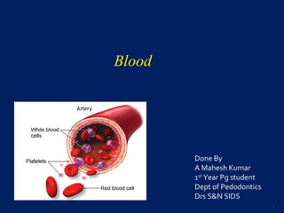
  • 1. Blood Done By A Mahesh Kumar 1st Year Pg student Dept of Pedodontics Drs S&N SIDS 1
  • 3. REFERENCES : 1.Text book of Medical Physiology- 10th edition- Guyton & Hall 2.Concise Medical Physiology- 4th edition- Chaudhuri 3.Essentials of Medical physiology-4th edition-Sembulingam 4.Per haavardsholm finne and Sverre halvorsen. Regulation of Erythropoiesis in the Fetus and Newborn. Archives of Disease in Childhood 1972; 47: 683-687 5.Narla Mohandas and Patrick G. Gallagher. Red cell membrane: past, present, and future. Blood 2008; 112: 3939-3948 3
  • 4. 6.Michael Fo¨ller1, Stephan M. Huber2 and Florian Lang. Erythrocyte Programmed Cell Death. Life 2008; 60(10): 661–8 7.Petra Kleinbongard et all. Red blood cells express a functional endothelial nitric oxide synthase. Blood 2006; 107: 2943-51 8. Veronique Witko-Sarsat et al. Neutrophils: Molecules, Functions and Pathophysiological Aspects. Laboratory investigations 2000; 80(5): 617-653 9. SM Rashmi et al. Neutrophils in health and disease: An overview. J Oral Maxillofac Pathol 2006; 10: 3-8 4
  • 5. INTRODUCTION • Specialized connective tissue • Contains cells suspended in a fluid matrix • Fluid of life • Fluid of growth • Fluid of health 5
  • 6. PROPERTIES Colour : Red Volume : In adults 5 ltrs New born 450 ml pH : slightly alkaline 7.4 Specific gravity : 1.052 – 1.061 total blood 1.092 – 1.101 blood cells 1.022 – 1.026 plasma Viscosity : five times more viscous than water 6
  • 7. FUNCTIONS: 1.Respiratory function 2.Excretory function 3.Nutrient function 4.Role of blood in various homeostatic processes a)Body temperature and blood b)pH and blood 7
  • 8. 5. Role of blood in various defence mechanism 6. Transport of hormones and enzymes 7. Storage function 8. Regulation of water balance 8
  • 9. COMPOSITION OF BLOOD 1.Plasma - 55% 2.Formed elements - 45% a) RBC or Erythrocytes b) WBC or Leukocytes c) Platelets or Thrombocytes 9
  • 10. 10
  • 11. 11
  • 13. MORPHOLOGY • Circular, biconcave cell without a nucleus • Diameter - 7.5µ • Thickness at the periphery - 2µ • Thickness at the centre - 1µ 13
  • 14. Advantages of biconcave shape  Thickness of an RBC in its central part is not great, so oxygen doesnot have to travel a great distance for the diffusion  Biconcavity increases the surface area of the RBC, so oxygen gets a bigger area for diffusion  The erythrocyte can squeeze itself through a capillary more easily. 14
  • 15. Red cell membrane It is a tri laminar structure having a bimolecular lipid layer interposed between the two layers of proteins. Lipids: Glycolipids, Phospholipids, Cholesterol. 15
  • 16. The proteins comprising the erythrocyte cytoskeleton include 1.Ankyrin 2.spectrin 3.Band 4.1 4.Band 3 5.Actin filaments 16
  • 17. Protein 4.1R-dependent multiprotein complex: New insights into the structural organization of the red blood cell membrane 17
  • 18. PROPERTIES OF RED BLOOD CELLS 1.Rouleaux formation 2.Specific gravity : 1.092 to 1.101 3.Packed cell volume 4.Suspension stability 18
  • 19. ERYTHROPOIESIS Erythropoiesis is the process by which the origin, development and maturation of erythrocytes occur. Hemopoiesis is the process which includes origin, development and maturation of all the blood cells. 19
  • 20.  Blood forming tissues Myeloid tissue Lymphoid tissue  Myleloid tissue means RBM produces RBCs, granulocytes, monocytes and platelets.  Lymphoid lymphnode, thymus, and spleen Produce lymphocytes. 20
  • 21. Site of Erythropoiesis In fetal life 21
  • 22. Age Site Upto 5-6 years RBM of all bones 6-20 years RBM of long bones & all membranous (flat) bones After 20 years All membranous bones & ends of long bones In Post natal life and in adults 22
  • 23. Bone marrow 2 types • Red bone marrow • Yellow bone marrow RBM – found in flat bones (cranial, ribs, sternum , pelvic bones) YBM – shafts of long bones in fully grown adults. 23
  • 24. Extra medullary hemopoiesis when there is necessity of increased erythropoiesis the YBM is converted into RBM. If it is more intense liver and spleen also start producing RBC. (Medulla = bone marrow) 24
  • 25. Histology of RBM Consists of 1.Large no of sinusoids 2.Adventitious cells outside the sinusoids 3. Blood forming cells in between adventitious cells 25
  • 26. • Sinusoids are basically capillaries with larger diameter. • Their walls contain big pores – big molecules, blood cells pass. • Adventitious cells – become fat cells • The blood cells – Precursors of erythrocytes, leucocytes, and platelets • Normally – blood cells : fat cells – 1:1 26
  • 27. RBM blood picture Cells Percentage Granulocytes & their precursors 60% Erythrocytes & their precursors 20% Lymphocytes, monocytes & precursors 10% Others ( non identifiable, degenerated cells) 10% 27
  • 29. 29
  • 30. 30
  • 31. Different stages of RBC development Progenitor cells – BFU-E (Burst forming unit) CFU-E Blast cells – Pronormoblast (E1) Early normoblast(E2) Intermediate normoblast(E4) Late normoblast (E5) Reticulo cyte (E6) Matured RBC 31
  • 32. 1. Pronormoblast : (proerythroblast)  It is a large cell - deeply basophilic cytoplasm with a large central nucleus.  The deep blue colour - high content of RNA associated with protein synthesis. 32
  • 33. Basophilic Normoblast •This is a round cell having a diameter of 12-16 um with a large nucleus. •The nucleus is more condensed than the pronormoblast and contains basophilic cytoplasm. •This cell undergoes rapid proliferation. 33
  • 34. Polychromatic normoblast (Intermediate) •The nucleus - coarse and deeply basophilic. •The cytoplasm is polychromatic with mixture of basophilic RNA and acidophilic hemoglobin. 34
  • 35. Orthochromatic (late) normoblast •This is the final stage in the maturation of nucleated red cells. •The cell is smaller - small pyknotic nucleus with dark nuclear chromatin. •The cytoplasm - acidophilic due to large amounts of hemoglobin. 35
  • 36. Reticulocytes •Reticulocytes - devoid of nucleus but contain RNA •These cells in the peripheral blood smear - slightly basophilic hue. 36
  • 38. Factors influencing erythropoiesis: 1. Hemopoietic growth factor Erythropoietin Stem Cell Factor (SCF) Interleukins (GM-CSF) Colony Stimulating Factor (CSF) Thrombopoietin 2. Vitamin B12, Folic Acid, Pyridoxine, Vitamin C 3. Iron and Copper 38
  • 40. Applied physiology •COPD – high RBC count due to high production of EPO •Chronic damage of kidney – anemia due to lower levels of EPO 40
  • 41. Interleukins (IL) •IL-1: Stimulates granulocytic cells. Induces expression of GM-CSF •IL-6 & IL-1: Stimulates proliferation of progenitor cells. •IL-3: Stimulates growth in all phagocytes. 41
  • 42. INTERLEUKINS: • Involved in erythropoiesis are • IL-3 • IL-6 • IL-11 42
  • 43. Stem Cell Factor (SCF): Stimulates early stem cells to differentiate. (TGF-beta) Inhibits hematopoieses. Directly inhibits proliferation of progenitor cells. 43
  • 44. Role of Vit B12 44
  • 45. Folic acid intestine polyglutamate monoglutamate methylmonoglutamate & absorbed & enter blood reduced to become methyltetrahydrofolate this form reaches tissues they loose methyl group & become tetrahydrofolate accepted by Vit B12 methylcobalamin 45
  • 46. THF (tetrahydrofolate) formyl THF (folinic acid ) 5,10 methylene THF (active form of folate ) dihydrofolate(FH2 ) 46
  • 47. Hemoglobin: (Hb ) • Present inside the RBC. • Chromoprotein specialized for transport of oxygen and carbon dioxide • Conjugated protein with mol wt 68,000 daltons • Forms 95% dry weight and 35 – 40% of volume of RBC 47
  • 48. NORMAL VALUES AGE NORMAL VALUE At birth 25gm% After 3rd month 20gm% After 1 year 17gm% In adult males 15gm% In adult females 14.5gm% 48
  • 49. DERIVATIVES OF HEMOGLOBIN 1.OXYHEMOGLOBIN 2.REDUCED HEMOGLOBIN OR FERROHEMOGLOBIN 3.CARBHEMOGLOBIN 4.CARBOXYHEMOGLOBIN 5.SULFHEMOGLOBIN 6.NITROUS OXIDE HEMOGLOBIN 7.METHEMOGLOBIN OR FERRIHEMOGLOBIN 49
  • 50. FUNCTIONS OF HEMOGLOBIN 1.Transport of O2 2.Transport of CO2 3.Acts as blood buffer 50
  • 51. Hb Chemistry & synthesis: Hb molecule contains two ingredients: •Haem ( iron & protoporphyrin ) •Globin 51
  • 52. Heme formation • Heme formation takes place in the mitochondria then the cytoplasm of erythroid precursors through the reticulocyte stage. • It begins with production of a protoporphyrin ring. • Iron then incorporates with protoporphyrin to form heme. • Heme synthesis is stimulated by erythropoietin 52
  • 53. Heme structure: 4 pyrrole structures They are linked up with one another by methine ( = CH - ) 53
  • 54. Globin formation •The polypeptide chains of globin are produced on the ribosomes. •The four most common chain types are alpha, beta, gamma and delta chains. •Each of these chains differs from the others in their amino acid sequence. •Each globin molecule is made of 2 pairs of chains and each chain is made of 141-146 amino acids. 54
  • 55. Globin structure: •contains 4 polypeptide chains 2 alpha - containing 141 amino acids 2 beta - containing 146 amino acids •with each polypeptide chain 1 molecule of haem is attached. 55
  • 56. 56
  • 57. 57
  • 58. Synthesis of Hb •Hb is synthesized in the erythroid series in the red bone marrow •Hb first appears at the intermediate normoblast •Protoporphyrin can be synthesized by normoblasts from products of metabolism and aminoacids like succinyl Co A , glycine etc •Iron has to be obtained from food or iron contained in the hemoglobin of dead RBC’s 58
  • 59. Basic Chemical Steps : (synthesis of Hb ) 1.2 succinyl-CoA + 2 glycine ---------------> pyrrole molecule 2.4 pyrrole -----------------> protoporphyrin IX 3.Protoporphyrin + ferrous iron -------> heme 4.Heme + polypeptide (globin) ---------> hemoglobin chain 5.2 alpha +2 beta ------------- > hemoglobin 59
  • 60. Factors necessary for Hb maturation 1.Proteins and aminoacids 2.Iron 3.Copper – necessary for absorption of iron from gut 4.Cobalt and nickel for utilization of iron during Hb formation 60
  • 61. Iron metabolism •Imp for oxygen transport •Imp for formation of Hb and myoglobin •Also necessary for formation of other substances like cytochrome, cytochrome oxidase, peroxidase, and catalase 61
  • 62. Normal values Total quantity of iron in the body is 4gms Hb 65-68% Muscle as myoglobin 4% Intracellular oxidative heme compound 1% In the plasma as transferrin 0.1% Reticuloendothelial system 25-30% 62
  • 63. •Dietary iron available 2 forms •Heme iron •Non heme iron •Heme iron- RBC , absorbs easily •Non heme – vegetables, grains, cereals. not absorbed easily 63
  • 64. Absorption of iron Iron is absorbed mainly from the small intestine. Bile is essential for absorption Immediately after absorption into the blood, iron combines with a β globulin called – apotransferrin. Which results in formation of transferrin 64
  • 65. 65
  • 66. Fate Of RBC •Life span of RBC is about 120 days. • As age of the RBC increases, the enzymes which protect the erythrocyte from the damaging effects of oxygen begin to lose their efficiency, therefore oxidative damages begin to appear & the RBC becomes fragile. •Now the cell ruptures , self-destruct in spleen 66
  • 68. RED CELL COUNT: Equipment • Red cell pipette with a red bead in the bulb & markings of 0.5, 1 & 101 • Improved Neubauer chamber 68
  • 69. Diluting Fluid 1.Hayem’s fluid Mercuric chloride 0.5 g Sodium chloride 1.0 g Sodium sulfate 5.0 g Distilled water 200 ml 69
  • 70. RBC count = N 1/5 x 1/10 x 1/200 = N x 10000 Normal Range : Adult male 4.5-5.5 million/ cumm Adult female 3.8-5.2 million/cumm At birth 4.0-6.0 million/cumm 70
  • 71. VARIATIONS IN NUMBER OF RED BLOOD CELLS Physiological variations 1.Increase in RBC a)Age b)Sex c)High altitudes d)Muscular exercise e)Emotional conditions f)Increased environmental temperature g)After meals 71
  • 72. 2. Decrease in RBC a)High barometric pressures b)After sleep c)Pregnancy 72
  • 73. Pathological Variations 1.Primary Polycythemia – Polycythemia Vera Myeloproliferative disorders like malignancy of red bone marrow 2.Secondary Polycythemia a)Respiratory disorders like emphysema b)Congenital heart disease c)Ayerza’s disease d)Chronic carbon monoxide poisoning e)Poisoning by chemicals like P & As f)Repeated mild hemorrhages 73
  • 74. VARIATIONS IN SIZE OF RBC 1.Microcytes a)Iron deficiency anemia b)Prolonged forced breathing c)Increased osmotic pressure in blood 2.Macrocytes a)Megaloblastic anemia b)Muscular exercise c)Decreased osmotic pressure in blood 3.Anisocytes a)Pernicious anemia 74
  • 75. VARIATIONS IN SHAPE OF RED BLOOD CELLS 1.Crenation 2.Spherocytosis 3.Elliptocytosis 4.Sickle cell 5.Poikilocytosis 75
  • 76. VARIATIONS IN STRUCTURE OF RED BLOOD CELLS 1.Punctate Basophilism: •Dots of basophilic materials (porphyrin) appear in RBC •Lead poisoning 2. Ring: •Twisted strands of basophilic materials appear in the periphery •Goblet ring 3.Howell – Jolly bodies: •Nuclear fragments in the RBC 76
  • 77. Hb estimation methods 1.Calorimetric Methods a)Visual Methods b)Methods using photoelectric colorimeter 2. Gasometric Method 3.Chemical Method 4.Specific Gravity Method 77
  • 78. Visual 1.Tall Quist paper method - inaccurate 2.Sahlis Acid hematin 3.WHO Hb colour scale – simple, reliable, inexpensive, suitable for where calorimeter is not available. 78
  • 79. Sahli’s Method Tube with marking Principle : Blood is added to N/10 Hcl – Acid Hematin. Acid hematin is matched against the brown colour. 79
  • 80. • Add N/10 Hcl – into Hb meter tube • Fill the Hb pipette with 0.02ml of blood • Acid act on the RBC for 10min to lyse the cells and convert Hb to acid hematin. • Match colour of the solution with comparator in natural light. • Darker – add distilled water • Continue till the colour of solution matches 80
  • 81. Disadvantages: • Visual error • After 10min acid hematin starts fading 81
  • 82. Tall Quist Paper Method • Includes a color chart and 150 test papers • Allow blood to absorb into one of the test papers and compare the color scale. 82
  • 83. WHO Hb colour scale • Place a drop of blood on the test strip provided • Wait about 30 seconds • Match immediately the colour • simple, reliable, inexpensive, suitable for where colorimeter is not available. 83
  • 84. Methods using photoelectric colorimeter 1.Cyanmet haemoglobin method 2.Oxyhemoglobin method 3.Alkaline haematin Method 84
  • 85. Cyanmethemoglobin Method Principle : Hb is oxidized to - Met Hb by potassium ferricyanide Met Hb – Cyanmet Hb Technique Take 5ml of Drabkin’s solution ( Drabkin’s reagent contains sodium bicarbonate, potassium ferricyanide, and potassium cyanide) Add 0.02ml of blood Wait for 10min Hb = OD of test sample x Conc. Standardx250 OD of standard x1000
  • 86. Oxyhemoglobin Method • 0.04% of ammonia is used • Which lyses the RBC • Colour is compared to standard Alkaline Hb method • N/10 NaOH is used. • Colour of alkaline hematin is compared with standard 86
  • 87. Gasometric Method – oxygen carrying capacity of blood is measured in van slyke like apparatus. Not suitable for routine use Specific Gravity Method – simple , rapid & inexpensive. Rough estimation of Hb is obtained from specific gravity of blood, used for mass screening 87
  • 88. PCV •Hematocrit is the % volume of red cells in a given sample of blood. •It gives an estimate of relative volume of cells and plasma. •It is useful for evaluating absolute values like MCV and MCHC 88
  • 89. Wintrobe’s method : •Collect 2ml of blood •Take wintrobe’s tube • 0-100 – ESR • 100-0 – PCV •Take the blood in pasteur’s pippete and withdraw the blood into the tube, till it reaches 100 marking. 89
  • 90. normal range Males – 40- 55% Females – 35- 48 % 90
  • 91. MCV: ( normal value 80 – 100 fl ) It is the volume of an average RBC (single RBC) expressed in femotoliters or fl PCV X 1000 MCV = ----------------------------------------- RBC in millions / cmm. Normal : 82-98 fl 91
  • 92. MCH - Amount of Hb in the red cell Hb x10 MCH = ----------------------- pg RBC count Normal = 28-32pg 92
  • 93. MCHC: ( normal value 31 – 36% ) represents the average concentration of Hb in a given volume of packed red cells. Hb MCHC = ------------------------------------------- PCV ( % ) 93
  • 94. Erythrocyte Sedementation Rate : (ESR) It is a measure of the settling of red blood cells in a tube of blood during one hour. normal value - 0-20mm in female in 1st hr - 0-10mm in male in 1st hr Stages of sedimentation : •Rouleaux formation – first 15 min •Formation of fine threads – next 15 mins •Rapid fall – next 15min •Packing phase – packing of RBC next 15min 94
  • 95. • Two methods to determine ESR. Westergrens Method:-Westergrens tube - 300mm long(30cm), 2.5mm internal diameter & opened on both ends, calibrated from 0-200. Procedure •1.6 -2ml of blood mixed with 0.4-0.5ml of 3.8% sodium citrate - (anticoagulant) and mix well, loaded in westergrens tube up to zero mark. 95
  • 96. • The tube fitted to the stand vertically and left undisturbed at room temperature. • Reading taken at the end of 1 hour (Distance from 0 mark to top of RBC column is recorded as ESR) 96
  • 97. Anticoagulated whole blood has just been added. (Time: 0) Red blood cells have settled, leaving plasma at the top of the tube. Reading: 18 mm/hour (Time: one hour) WESTERGREN’S TUBES 97
  • 98. Modified Westergrens method •EDTA instead of citrate as anticoagualant •2ml of EDTA diluted with 0.5ml of 3.8% sodium citrate or 0.5ml of 0.85% sodium chloride •Undiluted EDTA blood gives poor precision 98
  • 99. Procedure •Collect 2ml of blood • Blood loaded in the tube up to’0’mark, tube is placed vertically on the Wintrobe’s stand. •The reading taken after one hour Wintrobes Method •Wintrobe’s tube short tube (110mm long, diameter 2.5mm) opened on only one end. 99
  • 100. NORMAL VALUES OF ESR Westergren’s method •Males – 0-15mm 1st hr •Females - 0-20mm 1st hr Wintrobe’s method •Males – 0-7mm 1st hr •Females - 0-14mm 1st hr 100
  • 101. Factors That May Influence ESR Factors that increase ESR Old age Female Pregnancy Anemia Macrocytosis Factors that decrease ESR Polycythemia Red blood cell abnormalities Spherocytosis Microcytosis 101
  • 103. Introduction : • Leukos = white, cytes = cells • Mobile units of the body’s protective system. • White blood cells are the colorless and nucleated formed elements of blood. • These cells are larger in size and their number is less compared to that of RBC’s. • They play a very important role in defense mechanism 103
  • 104. 104
  • 105. Type of leucocyte Normal range Lifespan Neutrophil 50-75 % 2-5 days Eosinophil 1-6 % 7-12 days Basophil 0-1% 12-15 days Lymphocyte 25-45% ½-1 day Monocyte 3-8 % 2-5 days 105
  • 106. NEUTROPHILS •Diameter : 10-14µ •Nucleus : Young neutrophil : horse-shoe shaped nucleus Mature neutrophil : multilobed (2-6 lobes) lobes connected by chromatin filaments •Cytoplasm : pale bluish in colour & full of fine granules Granules take both acidic & basic stain & look violet- pink in colour 106
  • 107. •Condensed chromatin along the inner surface of nuclear envelope •More central region of each lobe appears paler •Cytoplasm contains few mitochondria & a small Golgi complex •Azurophilic granules are round or oval & more electron dense than specific granules. 107
  • 108. NEUTROPHILS - DEVELOPMENT MYELOBLAST : • Earliest recognizable cell in the granulocytic maturation process. • 15-20µm in diameter • Large round to oval nucleus • Small amount of basophilic cytoplasm • Nucleus contains 2 to 5 nucleoli • Nuclear chromatin is fine and reticular 108
  • 109. PROMYELOCYTE : • Slightly larger in size than myeloblast. • Primary or azurophilic granules appear at the promyelocyte stage. • Nucleus contains nucleoli as in myeloblast stage • But nuclear chromatin shows slight condensation. 109
  • 110. MYELOCYTE : • Characterized by the appearance of secondary or specific granules. • Smaller cell with round to oval eccentrically placed nucleus. • More condensation of chromatin than in promyelocyte stage and absence of nucleoli. 110
  • 111. • Cytoplasm is relatively greater in amount than in promyelocyte stage. • Contains both primary and secondary granules. • Last cell capable of mitotic division 111
  • 112. METAMYELOCYTE : • Nucleus becomes indented and kidney shaped. • Nuclear chromatin becomes moderately coarse • Cytoplasm contains both primary and secondary granules 112
  • 113. BAND STAGE ( STAB FORM ) : • Characterized by band-like shape of the nucleus with constant diameter throughout • Condensed nuclear chromatin 113
  • 114. SEGMENTED NEUTROPHIL: • With Leishman’s stain, nucleus appears deep purple with 2-5 lobes joined by thin filamentous strands. • Nuclear chromatin pattern is coarse. • Cytoplasm stains light pink and has small, specific granules 114
  • 115. 115
  • 116. Granules •Number of granules : 500-1500/granulocyte •Large amount of protein, traces of lipids & nucleic acids 1.Primary or azurophilic granules 2.Secondary or specific granules 3.Tertiary or gelatinase granules 4.Secretory vesicles 116
  • 117. Azurophilic granules •Myeloperoxidase, defensins, lysozyme, azurocidin etc •These granules fuse with phagocytes vesicles resulting in the delivery of their contents to the ingested organism •Greenish coloration to pus is imparted by myeloperoxidase 117
  • 118. Secondary or specific granules : • 3times more common in cytoplasm • Lysozyme, Lactoferrin, collagenase, histaminase may modify the inflammatory process Tertiary granules : • Gelatinases 118
  • 119. Life history of neutrophils : •Released from the bone marrow •Exist in two populations •Circulating pool •Marginal pool •Rapid exchange between the two pools • Activated by numerous stimuli 119
  • 120.  Disposed of internally by cells of reticuloendothelial system  External loss : emigration through gingiva into the saliva & excretion in urine ( common)  Neutrophils occur in the secretions of uterus during second half of menstrual cycle  Dead neutrophils – granulocyte-inducing factor 120
  • 121. Old senile neutrophils are characterized by: •Loss of motility •Poorly stained granules •Increased nuclear lobulation •Easy breakability while making blood smear 121
  • 122. Nitroblue tetrazolium reduction test : •Ability of neutrophils to destroy micoorganisms with intracellular enzymes can be evaluated by NBT •Normal neutrophils contain enzymes that convert colorless NBT to dark blue granules within the cell •When dark blue granules are not seen, neutrophils will not destroy bacteria 122
  • 123. FUNCTIONS : First line of defence 1.PHAGOCYTOSIS 2.REACTION OF INFLAMMATION 3.FEBRILE RESPONSE 123
  • 124. VARIATION IN COUNTS Neutrophilia - >1000 cumm Physiological causes •New born babies •After exercise •After meals •Pregnancy •Menstruation •Parturition •Lactation •Mental & emotional stress 124
  • 125. Pathological causes •Acute bacterial infections Lobar pneumonia Bronchopneumonia Pyogenic meningitis Cellulitis Infected burns Diphtheria 125
  • 126. •Acute inflammatory diseases like Acute rheumatic fever Acute appendicitis •Acute stress states like Post surgery Post haemorrhage Myocardial infarction •Chronic myeloproliferative disorders like CML Polycythemia vera 126
  • 127. NEUTROPENIA: CAUSES •Typhoid and paratyphoid fever •Physical agents like radiation •Chemicals : Benzene •Antimetabolite drugs : Cyclophosphamide, Methotrexate •Hematologic disorders like megaloblastic anemia, aplastic anemia, subleukemic leukemia, cyclic neutropenia, 127
  • 128. COOKE’S ARNETH COUNT Stage Nuclear lobes Normal count Stage I (N1) 1 lobe 5-10 % Stage II (N2) 2 lobes 20-30 % Stage III (N3) 3 lobes 40-50 % Stage IV (N4) 4 lobes 10-15 % Stage V (N5) 5 lobes or more 3-5 % 128
  • 129. Clinical Significance : 1.Left shift •N1+N2+N3 > 80% •More younger cells •Indicates hyperactive bone marrow (high rate of formation) 2.Right shift •N4+N5 > 20% •More mature cells •Hypoactive bone marrow (slow rate of formation) 129
  • 130. Variation in neutrophil morphology : 1.Variation in granules 2.Formation of vacuoles in cytoplasm 3.Formation of Dohle bodies in cytoplasm 4.Presence of sex chromatin with nuclear lobes 5.Hypersegmented neutrophils 6.Band and straw form neutrophil 7.Pelger – Huet anamoly 130
  • 135. If most of the neutrophils appear bilobed, this is indicative of an uncommon condition known as Pelger- Huet anomaly, an inherited condition. This is the heterozygous form. The homozygous form is fatal. Just be aware of this condition when you get back a manual differential count with mostly bands, but the WBC count is normal or the patient shows no signs of infection or inflammation. 135
  • 136. EOSINOPHILS : • Forms via same stages as the neutrophil. • Diameter = 12µ - 17µ • Nucleus: bilobed, purple colored, spectacle shaped, nucleoli – absent Condensed chromatin stains less intensely than that of neutrophil 136
  • 137. Cytoplasm: acidophilic and appears bright pink Contains deep red staining granules which do not cover the nucleus. •Condensed chromatin is peripherally distributed along the inner surface of nuclear envelope. •Specific granules : ovoid with electron dense crystalloid cores that consist chiefly of basic proteins. •Only other prominent organelles : Golgi apparatus, mitochondria 137
  • 138. In RBM ( 5-6 days) Circulatory pool (8-12 hours) Emigrates into tissue Death and removed 138
  • 139. Cytoplasm contains two types of granules 1. Specific granules Large numerous elongated granules Contain Crystalloid bodies - Contain four major proteins 1. Major basic protein 2. Eosinophil cationic protein 3. Eosinophil peroxidase 4. Eosinophil derived neurotoxin Also contains histaminase, arylsulfatase, collagenase, cathepsins 139
  • 140. Azurophilic granules •Lysosomes, contains acid hydrolases •Function in the destruction of parasites •Hydrolysis of antigen – antibody complexes. 140
  • 141. FUNCTIONS OF EOSINOPHILS : 1.ROLE IN PARASITIC INFESTATIONS: •Major basic protein (MBP) : damage the parasites by causing distension and detachment of the tegumental sheath of these organisms. •Eosinophil cationic protein : major destroyer of helminths. 10 times more toxic than MBP Destroys parasites by complete disintegration 141
  • 142. • Eosinophil peroxidase : capable of destroying helminths, bacteria and tumor cells • Eosinophilic derived neurotoxin : destroys the nerve fibers particularly myelinated nerve fibers. 2. ROLE IN ALLERGIC REACTION • Capable of destroying inflammation inducing substances like histamines and bradykinin. 142
  • 143. • Destroy antigen-antibody complexes & thus prevent spread of local inflammatory process. • Eosinophil arylsulfatase can degrade SRS-A liberated by mast cells and basophils • Produces histaminase that can inactivate histamine. 143
  • 144. Eosinophilia : •Allergic conditions like hay fever, bronchial asthma •Parasitic infestations •Skin diseases like urticaria •Scarlet fever Eosinopenia : •ACTH & steroid therapy •Stressful conditions •Acute pyogenic infections 144
  • 145. BASOPHILS : •Diameter = 10-12µ •Nucleus – bilobed , stains less deeply than that of neutrophil •Cytoplasm : basophilic, full of granules granules : deep purple or blue 145
  • 146. • Membrane bound granules – 0.5µ • metachromatic & similar to mast cell granules. • Chemical mediators liberated when basophils degranulate include histamine, SRS-A, ECF-A • The cell surface receptors on basophils closely resemble those on mast cells. 146
  • 147. FUNCTIONS 1.ROLE IN ALLERGIC REACTION : release histamine, bradykinin, SRSA, seratonin. 2. ROLE IN PREVENTING SPREAD OF ALLERGIC INFLAMMATORY PROCESS : •releases eosinophil chemotactic factor •eosinophils then phagocytose & destroy antigen – antibody complexes & prevent spread of local inflammatory process 147
  • 148. 3. RELEASE OF HEPARIN • Prevents clotting of the blood. • Activates the enzyme lipoprotein lipase which removes fat particles from the blood after a fatty meal 148
  • 149. BASOPHILIA : >100 cumm •Viral infections •Allergic diseases •Chronic myeloid leukemia BASOPENIA : •Corticosteroid therapy •Drug-induced reactions •Acute pyogenic infections 149
  • 150. MONOCYTES •12-20µ in diameter, Largest leukocytes CFU-M Monoblast Promonocyte Monocyte 150
  • 151. • Nucleus : variable in appearance ranging from indented ovoid or roughly kidney shaped to wide horse shoe Chromatin less condensed • Cytoplasm : pale grayish-blue color Fine pinkish-purple granules can also sometimes be seen Specific granules lacking 151
  • 152. • 2 or more nucleoli • Fairly prominent Golgi apparatus, moderate amount of ribosomes, small amount of rER, no. of small mitochondria • Small dense granules – 0.3-0.6µ • Surface of monocytes is irregular 152
  • 153. 153
  • 154. Functions 1.Phagocytosis : second line of defence 2.Immediate precursors of macrophages 3.Releases IL-1, TNF-α, transferrin, lysozyme, proteases, acid hydrolase 154
  • 155. MONOCYTOSIS •Parasitic conditions like malaria and kalaazar •Infective conditions like subacute bacterial endocarditis, tuberculosis •Neoplastic states like acute monocytic leukemia, acute myelomonocytic leukemia, chronic myelomonocytic leukemia 155
  • 156. LYMPHOCYTES •20-50% of blood leukocytes •Lack prominent cytoplasmic granules when seen in LM. 10% - may contain reddish purple staining granules •Based on size 1.Small lymphocyte : 6-9µ 2.Large lymphocyte : 9-15µ 156
  • 157. Committed stem cells Pre -T cell Pre B cell T lymphocytes Blymphocytes 157
  • 158. Small lymphocyte : •Small spherical nucleus with a small indentation on one side. •Condensed chromatin •Remarkably little cytoplasm •Cytoplasm is hardly seen in LM 158
  • 159. •Cytoplamic organelles are scant •Few mitochondria, sparse cisternae of rER, small Golgi apparatus 159
  • 160. Large lymphocyte : •Nucleus slightly larger than small lymphocyte •More cytoplasm EM : •Large numbers of mitochondria, free ribosomes, more cisternae of rER, slightly larger Golgi apparatus 160
  • 161. 161
  • 162. On the basis of functional properties 162
  • 163. LYMPHOCYTOSIS : >45% •Chronic infective conditions like tuberculosis, syphilis & brucellosis •Viral diseases like infectious mononucleosis, measles, chicken-pox & viral fever •Neoplastic conditions like CLL 163
  • 164. LAB INVESTIGATIONS TOTAL LEUCOCYTE COUNT Principle : Whole blood is diluted with WBC fluid that hemolyses the red cells. Nucleated cells – WBC s stained by Gentian Violet are counted in a Neubauer chamber 164
  • 165. Equipment: Hemocytometer set consisting of WBC pipette and improved Neubauer chamber with a cover slip WBC pipette has a white bead in the bulb and markings of 0.5, 1, 11 165
  • 166. WBC Fluid ( Turk’s fluid ) Glacial Acetic Acid 2ml 1 % Gentian violet 5 drops Add water to 100 ml 166
  • 167. TLC = N x 20 4 x 0.1 = N x 50 Depth of the chamber = 0.1 mm Dilution = 1 in 20 4 corner squares are counted N = Number of WBCs in 4 large squares 167
  • 168. Adults 4000 – 11000 cells / cumm At birth 8000 – 28000 cells / cumm Childhood 6000 – 15000 cells / cumm Normal Value 168
  • 169. Leucocytosis Leucopenia > 11000 / cumm < 4000 / cumm •Pregnancy •Exercise •Leukemoid reaction •Diabetic and uremic coma •Infectious mononucleosis •Bacterial and viral infections •Leukemias •Aplastic anemia •Hypersplenism •Typhoid, paratyphoid fever •Drug induced leucopenia •Radiation & cytotoxic therapy •Megaloblastic anemia •Subleukemic leukemia 169
  • 170. ABSOLUTE EOSINOPHIL COUNT •Accurate assessment of eosinophils in blood & is useful in diagnosis, treatment & follow-up of cases of eosinophilia •EQUIPMENT : Improved Neubauer chamber, WBC pipette 170
  • 171. Eosin 200mg dissolved in 10ml of distilled water Stains the granules of eosinophils orange red Water 80ml Lyses the red cells & white cells except eosinophils Acetone 10ml Fix the eosinophils Dunger’s fluid 171
  • 172. AEC = N X10 4 X 0.1 = N X 25 Normal Range = 40-450 cells /cumm 172
  • 173. PERIPHERAL SMEAR AND DIFFERENTIAL LEUCOCYTE COUNT 1.PREPARATION OF SMEAR 2.FIXATION OF THE SMEAR •Fixed within 4hours to get good staining •Methanol 3. STAINING OF THE SMEAR 173
  • 174. Commonly used stains : •Leishman stain •Giemsa stain •Wright’s stain •Jenner’s stain •Jenner – Giemsa stain 174
  • 175. BLOOD GROUPS •Discovered by Karl Landsteiner in 1901 •Nobel prize in 1930 •30 blood group systems given by International Society of blood transfusion 175
  • 176. 176
  • 177. 177
  • 178. International Society of Blood Transfusion. October 2006 178
  • 179. LANDSTEINER’S LAW 1.If a particular antigen is present in the red blood cells, corresponding antibody must be absent in the serum. 2.If a particular antigen is absent in the red blood cells, the corresponding antibody must be present in the serum Second part of law – not applicable to Rh factor 179
  • 180. ABO Blood groups Group Antigen in RBC Antibody in serum A A Anti B B B Anti A AB A and B No antibody O No antigen Anti A and Anti B 180
  • 181. Population A B AB O Europeans 42 9 3 46 Asians 25 25 5 45 181
  • 182. DETERMINATION OF THE ABO GROUP Blood grouping, blood typing or blood matching. Principle •Basis of agglutination •Occurs antigen + antibody Requisites •Suspension of patients red blood cell •Anti serum A & B 182
  • 183. Procedure & Results Agglutination with antiserum Blood group Anti serumA A Anti serum B B Both A & B AB No agglutination O 183
  • 184. Universal donors O Universal recipients AB Cross matching Mixing the serum of the recipient and the red blood cells of donor 184
  • 185. Gene receive from parents Group of the offspring Genotype A+A A AA A+O AO B+B B B B+O BO A+B AB AB O+O O OO Inheritance 185
  • 186. Transfusion reactions due to ABO incompatibility •Jaundice •Cardiac Shock •Renal Shut down 186
  • 187. Rh factor • Antigen present in RBC • Discovered by Landsteiner and Wiener in Rhesus monkey • Many Rh antigens only D is highly antigenic • Among Asians 85% - Rh +ve • 15% - Rh –ve • Rh +ve more among blacks 187
  • 188. Dd Dd Dd Father DD Rh+ Mothe r dd Rh - Dd All offsprings are Rh + 188
  • 189. dd dd dd Father dd Rh - Mothe r dd Rh - dd All offsprings are Rh - 189
  • 190. dd Dd dd Father Dd Rh+ Mothe r dd Rh - Dd 50% offsprings are Rh + 190
  • 191. Transfusion reactions due to incompatibility •Erythroblastosis Fetalis •Hydrops Fetalis •Kernicterus 191
  • 192. REFERENCES : 1.Text book of Medical Physiology- 10th edition- Guyton & Hall 2.Concise Medical Physiology- 4th edition- Chaudhuri 3.Essentials of Medical physiology-4th edition-Sembulingam 4.Per haavardsholm finne and Sverre halvorsen. Regulation of Erythropoiesis in the Fetus and Newborn. Archives of Disease in Childhood 1972; 47: 683-687 5.Narla Mohandas and Patrick G. Gallagher. Red cell membrane: past, present, and future. Blood 2008; 112: 3939-3948 192
  • 193. 6.Michael Fo¨ller1, Stephan M. Huber2 and Florian Lang. Erythrocyte Programmed Cell Death. Life 2008; 60(10): 661– 668 7.Petra Kleinbongard et all. Red blood cells express a functional endothelial nitric oxide synthase. Blood 2006; 107: 2943-2951 8. Veronique Witko-Sarsat et al. Neutrophils: Molecules, Functions and Pathophysiological Aspects. Laboratory investigations 2000; 80(5): 617-653 9. SM Rashmi et al. Neutrophils in health and disease: An overview. J Oral Maxillofac Pathol 2006; 10: 3-8 193
  • 194. 194