Downloaded 249 times


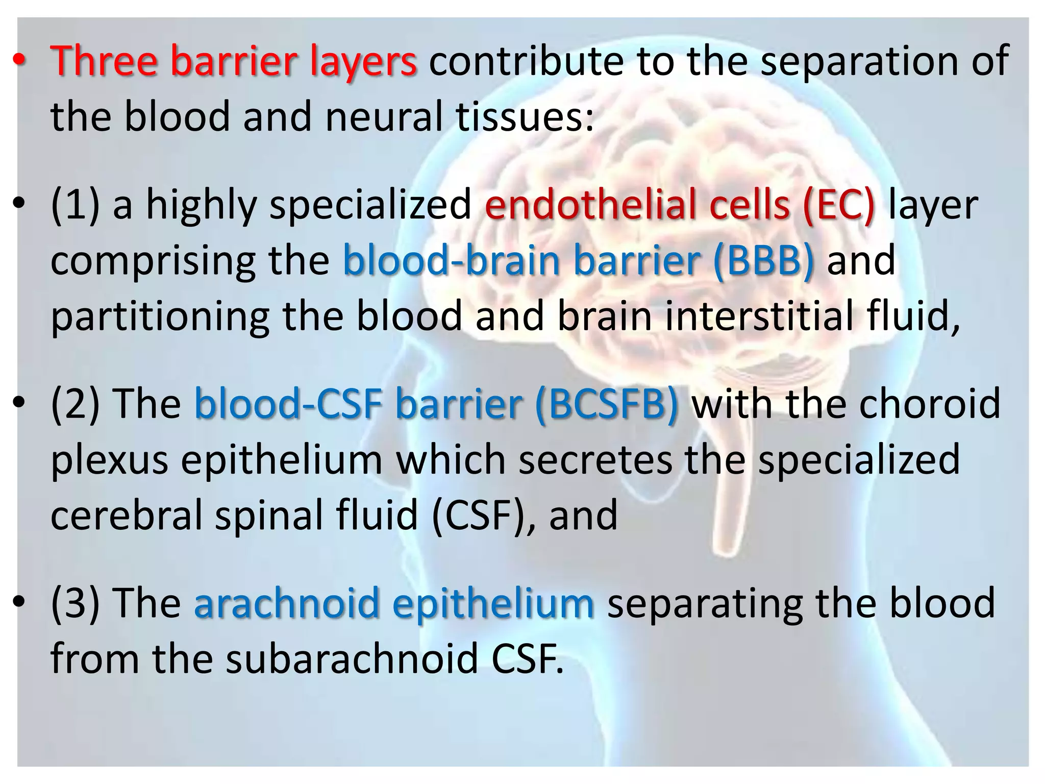
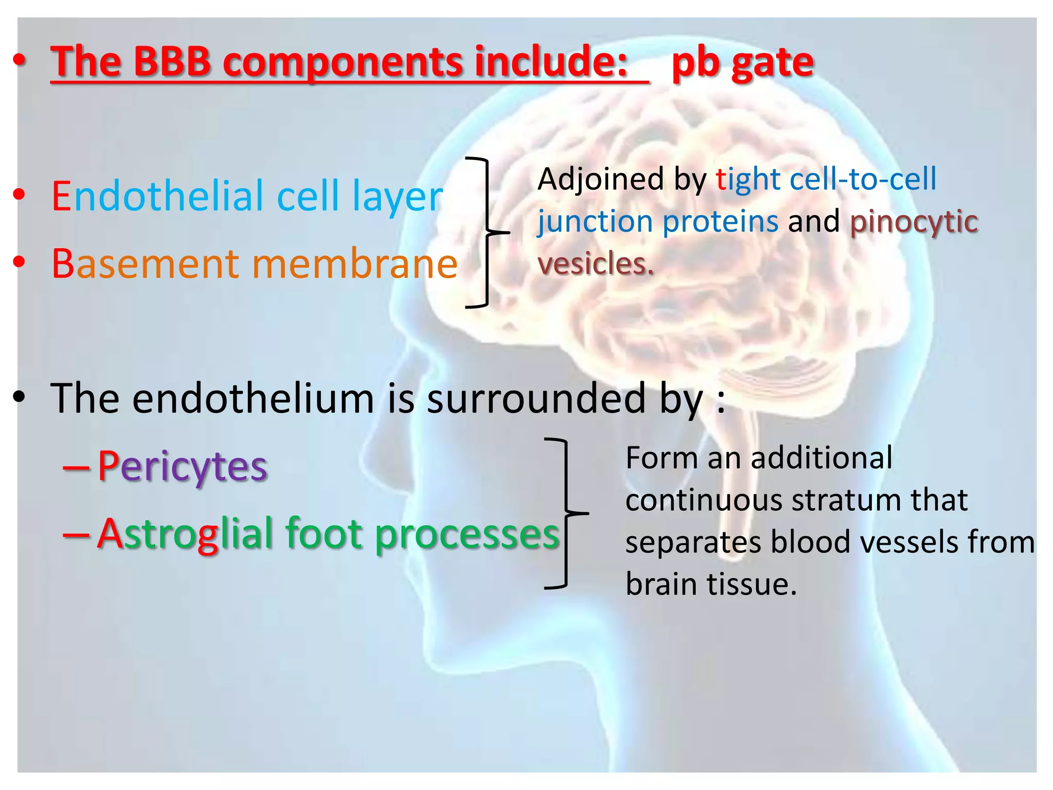


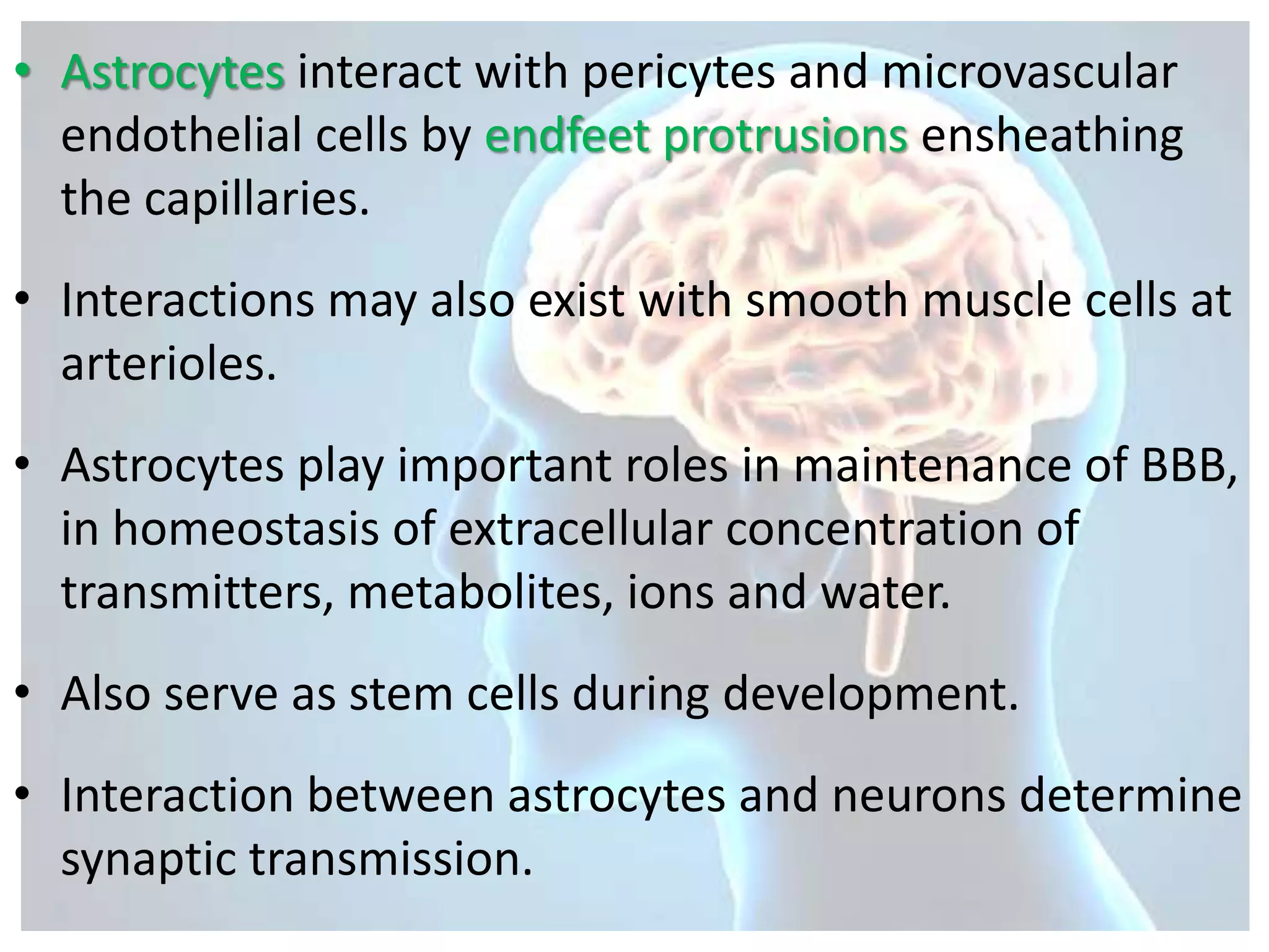


The blood-brain barrier (BBB) tightly regulates transportation between the blood and brain to maintain homeostasis. It is formed by endothelial cells, pericytes, astrocytes, and a basement membrane. These structures comprise the neurovascular unit. Three barrier layers exist - the BBB, blood-cerebrospinal fluid barrier, and arachnoid epithelium. The BBB prevents diffusion of polar solutes through tight junctions between endothelial cells. It is selectively permeable to allow passage of water, oxygen, dioxide and lipid-soluble substances while restricting entry of toxins, bacteria, and macromolecules. The BBB physiology regulates glucose, amino acid, water and drug transport critical for brain function and protection.


