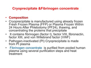
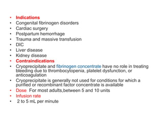
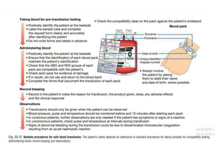
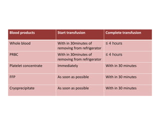
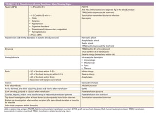
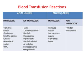
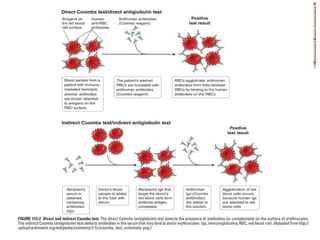
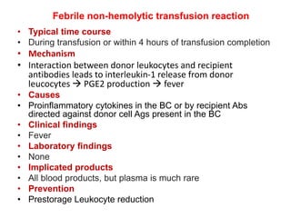
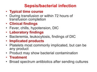
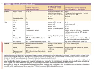
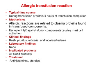
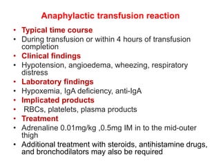
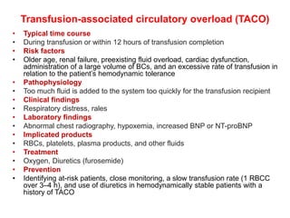
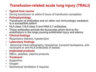
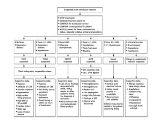
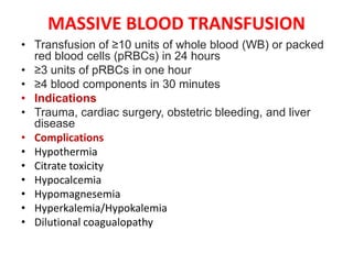
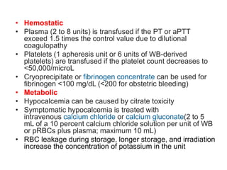
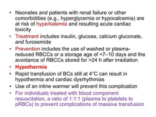
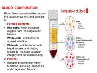
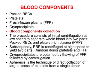
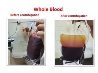
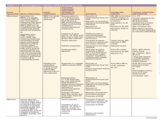
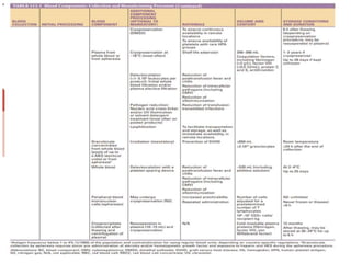
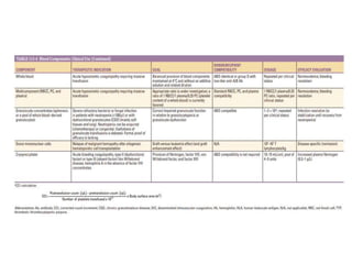
This document discusses blood and blood components. It describes the composition of blood and its formed elements and plasma. It then discusses the various blood components that can be prepared including packed red blood cells, platelets, fresh frozen plasma, cryoprecipitate, and describes the processes involved in collecting and preparing these components. It also covers topics like shelf life, dosing, and indications for transfusion of the various components. It discusses some potential transfusion reactions like acute hemolytic, delayed hemolytic, febrile non-hemolytic, sepsis/bacterial infection, and allergic reactions.
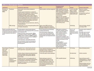
2. BLOOD COMPOSITION
Blood flows throughout the body in
the vascular system, and consists
of:
1. Formed elements
• Red cells, which transport
oxygen from the lungs to the
tissues
• White cells, which defend
against infection
• Platelets, which interact with
blood vessels and clotting
factors to maintain vascular
integrity and prevent bleeding
2. Plasma:
• contains proteins with many
functions, including antibodies
and coagulation factors
3. BLOOD COMPONENTS
• Packed RBCs
• Platelets
• Fresh-frozen plasma (FFP)
• Cryoprecipitate
• Blood components collection
• The procedure consists of initial centrifugation at
low speed to separate whole blood into two parts:
Packed RBCs and platelet-rich plasma (PRP)
• Subsequently, PRP is centrifuged at high speed to
yield two parts: Random donor platelets and FFP
• Cryoprecipitates are obtained by thawing of FFP
followed by centrifugation
• Apheresis is the technique of direct collection of
large excess of platelets from a single donor
9. Anti-coagulants in blood bag
ANTI- COAGULANTS FULL FORM SHELF LIFE
ACD ACID CITRATE DEXTROSE 21 DAYS
CPD CITRATE PHOSPHATE
DEXTROSE
21 DAYS
CPDA CITRATE PHOSPHATE
DEXTROSE + ADENOSINE
35 DAYS
SAGM SODIUM ADENINE
GLUCOSE MANNITOL
(Longest shelf life)
42 days
10. Functions of the anticoagulants
COMPONENT FUCNTION
CITRATE Anti-coagulant by chelating calcium
PHOSPHATE Acts as BUFFER & helps in maintaining
pH
DEXTROSE Provides nutrition
ADENINE Provides substrate for ATP synthesis
(increased shelf life)
11. Whole blood
• Shelf life 35 days
• Average unit of blood
350+49 or 450ml+63ml
anticoagulant
• Both red cell and plasma
component can be prepared
• Indication – acute
hypovolemic coagulopathy
requiring massive transfusion
• Rarely used now a days
12. Packed RBCs
• Types
• Leukoreduced
• Irradiated
• Saline washed
• Leuko reduced RBC
• Prepared by removing a proportion of the plasma from
leucocyte depleted whole blood
• Chances of a febrile reaction can be reduced
• Indications
• Chronically transfused patients
• Potential and transplant recipients
• Patients with previous febrile non hemolytic transfusion
reactions
13. • Irradiation
• RBC units can be irradiated prior to transfusion by
subjecting them to 2500 cGy of irradiation, targeted to
the central portion of the component, with a minimum
dose of 1500 cGy delivered to any part of the
component
• Irradiation sources include gamma rays from either a
cesium-137 or cobalt-60 blood irradiator or x-rays
using a standalone machine
• The 2500 cGy dose is sufficient to inactivate
lymphocytes in the product
• Viable donor lymphocytes can attack recipient cells in
individuals who are unable to mount an immune
response against them, causing transfusion-
associated graft-versus-host disease (ta-GVHD)
• Ta-GVHD can target all hematopoietic cells as well as
other tissues, leading to bone marrow aplasia and
other complications that are ultimately fatal
14. • Irradiation is used to prevent ta-GVHD in at-risk individuals
• Patients at most risk include those who are severely
immunocompromised and those who would fail to recognize the
transfused lymphocytes as foreign, due to human leukocyte antigen
(HLA) homology
• Indications
• Recipients of intrauterine or neonatal exchange transfusion;
premature neonates
• Individuals with congenital cell-mediated immunodeficiency states
• Individuals treated with specific types of potent immunosuppressive
therapies (purine analogs, antithymocyte globulin [ATG], certain
monoclonal antibodies); this may include those being treated for
non-Hodgkin lymphoma (NHL) or other hematologic malignancies
• Recipients of hematopoietic stem cell transplant (autologous or
allogeneic)
• Individuals with Hodgkin lymphoma (any stage of disease)
• Individuals at risk for partial HLA matching with the donor due to
directed donations, HLA-matched products, or genetically
homogeneous populations
15. • Washed red cells
• Units of RBCs can be washed to reduce or eliminate
complications associated with the small amount of residual
plasma in the unit
• Indications
• Severe or recurrent allergic reactions (eg, hives) associated
with red cell transfusion
• IgA deficiency with circulating anti-IgA antibodies that react
with IgA in the donor plasma, when an IgA-deficient donor is
not available. Frozen deglycerolized red cells may be the
component of choice for these individuals
• Individuals at risk for hyperkalemia
• Washing is done in an automated system using 0.9 percent
sodium chloride immediately before infusion
• The shelf-life of washed blood is four hours at 20 to 24°C or
24 hours if stored at 1 to 6°C
• Frozen deglycerolized RBC units also undergo extensive
washing and can be used for the same indications
16. PLATELETS
• Storage
• Room temperature, because cold induces clustering of von
Willebrand factor (WVF) receptors on the platelet surface and
morphological changes of the platelets, leading to enhanced
clearance by hepatic macrophages and reduced platelet survival in
the recipient
• Shelf life: 5 days
• Dose Adults one WBD unit per 10 kg of body weight, which
translates to four to six units of WBD platelets or one apheresis unit
• Pediatric dose is 5 to 10 mL/kg
• Infusion rate For an average-sized adult, six units of WBD
platelets or one unit of apheresis platelets are transfused over
approximately 20 to 30 minutes
• Patients at risk for transfusion-associated circulatory overload
(TACO) can be transfused at a slower rate as long as the transfusion
is completed within four hours of issuance from the blood bank
17. INDICATIONS FOR PLATELET TRANSFUSION
• Actively bleeding patients with thrombocytopenia
• Preparation for an invasive procedure if the thrombocytopenia is severe
• Neurosurgery or ocular surgery – <100,000/microL
• Most other major surgery – <50,000/microL
• Endoscopic procedures – <50,000/microL for therapeutic procedures;
20,000/microL for low risk diagnostic procedures
• Bronchoscopy with bronchoalveolar lavage (BAL) – <20,000 to 30,000/microL
• Central line placement – <20,000/microL
• Lumbar puncture
• <10,000 to 20,000/microL in patients with hematologic malignancies
• <40,000 to 50,000 in patients without hematologic malignancies
• Neuraxial analgesia/anesthesia – <80,000/microL
• Bone marrow aspiration/biopsy – <20,000/microL
• Prevention of spontaneous bleeding
• Afebrile patients with platelet counts <10,000/microL due to bone marrow
suppression
• For patients with fever, infection, or inflammation,transfuse at a platelet count
≤15,000 to 20,000/microL due to the increased risk of bleeding
• Patients with acute promyelocytic leukemia (APL) have a coexisting
coagulopathy, transfuse at a platelet count ≤30,000 to 50,000/microL
18. • Apheresis (single donor) platelets
• Collected from volunteer donors in a one to two-hour apheresis
procedure
• Platelets are selectively removed along with some white blood cells
(WBCs) and plasma, and most red blood cells (RBCs) and plasma
are returned to the donor
• Some systems will also remove the majority of WBCs, resulting in a
leukodepleted product
• A typical apheresis platelet unit provides the equivalent of four to six
units of platelets from whole blood (3 to 4 x 10¹¹ platelets)
• It is common in larger donors with high platelet counts for an
apheresis procedure to collect enough platelets to split the collection
into two separate transfusable apheresis units ("doubles," or
"splits"), and sometimes three separate units ("triples") can be
obtained from a single donor, increasing the operational efficiency of
the collection facility and the availably platelet supply
• Advantages
• Exposure of the recipient to a single donor rather than multiple
donors
• Ability to match donor and recipient characteristics such as human
leukocyte antigen (HLA) type, cytomegalovirus (CMV) status, and
blood type
19. • Whole blood derived (WBD) pooled platelets
• A single unit of platelets can be isolated from a whole
blood donation by centrifuging the blood within the
closed collection system to separate the platelets from
the red blood cells (RBCs)
• Two manufacturing methods are used
• The platelet rich plasma (RPR) method
• It involves the centrifugation of the whole blood first
under a sufficient g-force to pellet the RBCs (the "soft
spin") but leaves most of the platelets in suspension in
the RPR
• The PRP is then centrifuged again in a second
container at a higher g-force to pellet the platelets (the
"hard spin")
• The supernatant platelet-poor plasma is removed
• The platelets are then resuspended in the residual
plasma and stored
20. • The buffy coat method
• In this method, whole blood is first subjected to a hard spin to allow
plasma to be expressed off the top and the sedimented RBCs to be
expressed off the bottom, leaving behind the platelet-containing
buffy coat
• Several buffy coat units can then be pooled together with plasma or
platelet additive solution, and the pool is subjected to a soft spin to
sediment the residual RBCs
• The pooled platelet concentrate is then expressed off the RBCs and
store
• Advantages of WBD platelets
• Lower cost and ease of collection and processing
• Disadvantage of WBD platelets
• Recipient exposure to multiple donors in a single transfusion and the
increased work of bacterial testing
• In addition, it is more labor intensive and time consuming to perform
the required bacterial testing
21. Fresh Frozen Plasma (FFP)
• Prepared from single units of whole blood or from
plasma collected by apheresis techniques.
• Frozen at -18 to -30°C within eight hours of
collection and, when appropriately stored, is
usable for one year from the date of collection
• Volume of approximately 200 to 250 mL
• Jumbo units - 600 mL
• Contains all of the coagulation factors and other
proteins present in the original unit of blood,
slightly diluted by the citrate-containing
anticoagulant solution used to collect the blood
22. • Indications
• Massive transfusion
• Severe liver disease or DIC
• Rare clotting factor deficiencies
• Therapeutic plasma exchange inTTP
• Warfarin overdose if not corrected by vitamin k
and/or PCC ,depending on the clinical setting
• DOSE AND INFUSION RATE
• 10 to 15 mL/kg (ie, approximately three to five
units) in most adults
• Healthy individual – 2 to 3 mL/kg/hour
• Individual with volume overload or heart failure – 1
mL/kg/hour
• Individual undergoing plasma exchange – 60
mL/minute
23. Cryoprecipitate &Fibrinogen concentrate
• Composition
• Cryoprecipitate is manufactured using already frozen
Fresh Frozen Plasma (FFP) or Plasma Frozen Within
24 Hours After Phlebotomy (PF24), thawing, and
concentrating the proteins that precipitate
• It contains fibrinogen (factor I), factor VIII, fibronectin,
factor XIII, and von Willebrand factor (VWF)
• Pathogen-inactivated (PI) Cryoprecipitate is made
from PI plasma
• Fibrinogen concentrate is purified from pooled human
plasma using several purification steps and heat
treatment
24. • Indications
• Congenital fibrinogen disorders
• Cardiac surgery
• Postpartum hemorrhage
• Trauma and massive transfusion
• DIC
• Liver disease
• Kidney disease
• Contraindications
• Cryoprecipitate and fibrinogen concentrate have no role in treating
bleeding due to thrombocytopenia, platelet dysfunction, or
anticoagulation
• Cryoprecipitate is generally not used for conditions for which a
purified or recombinant factor concentrate is available
• Dose For most adults,between 5 and 10 units
• Infusion rate
• 2 to 5 mL per minute
27. Screening of blood
• Donated blood should be screened for:
1. HIV
2. HEPATITIS B
3. HEPATITIS C
4. MALARIA
5. SYPHILIS
Hepatitis C: Most common transfusion
transmitted infection
Malaria can be transmitted by all blood
products
28.
29.
30.
31.
32. Blood products Start transfusion Complete transfusion
Whole blood With in 30minutes of
removing from refrigerator
≤ 4 hours
PRBC With in 30minutes of
removing from refrigerator
≤ 4 hours
Platelet concentrate Immediately With in 30 minutes
FFP As soon as possible With in 30 minutes
Cryoprecipitate As soon as possible With in 30 minutes
35. Acute hemolytic transfusion reaction
• Typical time course
• During transfusion or within 24 hours of transfusion completion
• Mechanism:
• ABO incompatibility (Type 2 Hypersensitivity)
• Pre existing high affinity IgM antibodies rapidly induce competent
mediated lysis, intra vascular hemolysis, and hemoglobinuria
• Clinical findings
• Fever,chills,chest pain,back pain tachypnea,tachycardia,
hypotension, DIC , acute renal failure
• Laboratory findings
• Hemoglobinemia, hemoglobinuria, positive direct antiglobulin
(Coombs) test (may be negative if all cells have hemolyzed),
findings of DIC (prolonged PT, prolonged aPTT, low fibrinogen,
thrombocytopenia)
36. • Implicated products
• RBCs, plasma (much less common), rarely platelets
• Incompatible blood product (typically ABO incompatible due to
clerical error)
• Treatment
• Supportive
• Vigorous hydration with isotonic saline and diuretics to maintain
urine output
• In patients with DIC and severe bleeding, PC, plasma, and
cryoprecipitate or fibrinogen may be required
37. Delayed Hemolytic Transfusion Reaction
• Onset
• After 24hrs ( usually 2 weeks)
• Mechanism
• Recipients have prior exposure to RBC antigens
• After the transfusion, anamnestic response results
in antibody production
• Typically caused by IgG antibodies to foreign protein
antigens and are associated with laboratory features
of hemolysis
• Presentation
• Mild fever, hemolytic anemia, Coomb’s test-positive.
• Management
• Supportive care
38.
39.
40. Febrile non-hemolytic transfusion reaction
• Typical time course
• During transfusion or within 4 hours of transfusion completion
• Mechanism
• Interaction between donor leukocytes and recipient
antibodies leads to interleukin-1 release from donor
leucocytes PGE2 production fever
• Causes
• Proinflammatory cytokines in the BC or by recipient Abs
directed against donor cell Ags present in the BC
• Clinical findings
• Fever
• Laboratory findings
• None
• Implicated products
• All blood products, but plasma is much rare
• Prevention
• Prestorage Leukocyte reduction
41. Sepsis/bacterial infection
• Typical time course
• During transfusion or within 72 hours of
transfusion completion
• Clinical findings
• Fever, chills, hypotension, DIC
• Laboratory findings
• Bacteremia, leukocytosis, findings of DIC
• Implicated products
• Platelets most commonly implicated, but can be
any product
• Product may show bacterial contamination
• Treatment
• Broad spectrum antibiotics after sending cultures
42.
43. Allergic transfusion reaction
• Typical time course
• During transfusion or within 4 hours of transfusion completion
• Mechanism:
• Allergic reactions are related to plasma proteins found
in transfused components.
• Reciepient IgE against donor components causing mast cell
activation
• Clinical findings
• Rash, pruritus, urticaria, and localized edema
• Laboratory findings
• None
• Implicated products
• All blood products
• Treatment
• Antihistamines, steroids
44. Anaphylactic transfusion reaction
• Typical time course
• During transfusion or within 4 hours of transfusion
completion
• Clinical findings
• Hypotension, angioedema, wheezing, respiratory
distress
• Laboratory findings
• Hypoxemia, IgA deficiency, anti-IgA
• Implicated products
• RBCs, platelets, plasma products
• Treatment
• Adrenaline 0.01mg/kg ,0.5mg IM in to the mid-outer
thigh
• Additional treatment with steroids, antihistamine drugs,
and bronchodilators may also be required
45. Transfusion-associated circulatory overload (TACO)
• Typical time course
• During transfusion or within 12 hours of transfusion completion
• Risk factors
• Older age, renal failure, preexisting fluid overload, cardiac dysfunction,
administration of a large volume of BCs, and an excessive rate of transfusion in
relation to the patient’s hemodynamic tolerance
• Pathophysiology
• Too much fluid is added to the system too quickly for the transfusion recipient
• Clinical findings
• Respiratory distress, rales
• Laboratory findings
• Abnormal chest radiography, hypoxemia, increased BNP or NT-proBNP
• Implicated products
• RBCs, platelets, plasma products, and other fluids
• Treatment
• Oxygen, Diuretics (furosemide)
• Prevention
• Identifying at-risk patients, close monitoring, a slow transfusion rate (1 RBCC
over 3–4 h), and use of diuretics in hemodynamically stable patients with a
history of TACO
46. Transfusion-related acute lung injury (TRALI)
• Typical time course
• During transfusion or within 6 hours of transfusion completion
• Pathophysiology
• Transfusion of antibodies and or/ other non immunologic mediators
to a susceptible patient
• HLA class I,HLA class II and HNA 5,7 antibodies
• These antibodies activate the leucocytes,which bind to the
endothelium in the lungs,causing endothelial injury and edema
• Clinical findings
• Respiratory distress, hypotension
• Laboratory findings
• Abnormal chest radiography, hypoxemia, transient leukopenia, anti-
neutrophil or anti-HLA antibodies (if tested)
• Implicated products
• RBCs, platelets, plasma products
• Treatment
• Supportive
• Oxygen
• Mechanical Ventilation if required
47.
48. • Iron Overload
• Each unit of RBCs contains 200–250 mg of iron
• In frequently transfused recipients, iron
accumulation that is left untreated will affect
endocrine, hepatic, and cardiac function
• Death may occur from cardiac failure or
arrhythmia
• Iron overload can be assessed by means of serum
ferritin measurements, magnetic resonance
imaging, and liver biopsy
• Prevention and treatment
• Careful monitoring and iron chelation
49. • Post transfusion Purpura
• Rare reaction (~1/105 BCs)
• Thrombocytopenia-related bleeding disorder developing 5–12 days
after PC (and more rarely RBCC) transfusion, predominantly in
women
• Platelet-specific alloAbs are found in the recipient, most frequently
anti-HPA-1a in HPA-1a-negative alloimmunized individuals
• The delayed thrombocytopenia is due to a secondary increased
production of alloAbs
• The mechanisms for the destruction of the patient’s own platelets
remain unclear
• Management
• Supportive
• Polyvalent immunoglobulin
• Steroids
• Plasma exchange
• Additional platelet transfusions may worsen the thrombocytopenia or
be associated with poor increments
• Prevention
• Use of washed BCs or BCs from HPA-compatible donors
50. • Graft-Versus-Host Disease
• Extremely rare adverse reaction
• Mediated by engrafted donor T lymphocytes in a recipient
unable to reject such allogenic lymphocytes (as in severely
immunosuppressed patients or patients homozygous for an
HLA haplotype shared with the donor)
• Such donor T lymphocytes interact with host HLA Ags and
mount an immune response, which is manifested clinically by
the development, of cytopenia, fever, a characteristic skin
rash, diarrhea, and liver function abnormalities 5–10 days
after transfusion
• Highly resistant to treatment with immunosuppressive
therapies as well as ablative therapy followed by allogeneic
bone marrow transplantation and is fatal in >90% of cases.
• Prevention in at-risk patients
• Irradiation of cellular BCs (minimum of 25 Gy) or treating BCs
with pathogen reduction technology that will deplete all living
cells in the component
51. MASSIVE BLOOD TRANSFUSION
• Transfusion of ≥10 units of whole blood (WB) or packed
red blood cells (pRBCs) in 24 hours
• ≥3 units of pRBCs in one hour
• ≥4 blood components in 30 minutes
• Indications
• Trauma, cardiac surgery, obstetric bleeding, and liver
disease
• Complications
• Hypothermia
• Citrate toxicity
• Hypocalcemia
• Hypomagnesemia
• Hyperkalemia/Hypokalemia
• Dilutional coagualopathy
52. • Hemostatic
• Plasma (2 to 8 units) is transfused if the PT or aPTT
exceed 1.5 times the control value due to dilutional
coagulopathy
• Platelets (1 apheresis unit or 6 units of WB-derived
platelets) are transfused if the platelet count decreases to
<50,000/microL
• Cryoprecipitate or fibrinogen concentrate can be used for
fibrinogen <100 mg/dL (<200 for obstetric bleeding)
• Metabolic
• Hypocalcemia can be caused by citrate toxicity
• Symptomatic hypocalcemia is treated with
intravenous calcium chloride or calcium gluconate(2 to 5
mL of a 10 percent calcium chloride solution per unit of WB
or pRBCs plus plasma; maximum 10 mL)
• RBC leakage during storage, longer storage, and irradiation
increase the concentration of potassium in the unit
53. • Neonates and patients with renal failure or other
comorbidities (e.g., hyperglycemia or hypocalcemia) are
at risk of hyperkalemia and resulting acute cardiac
toxicity
• Treatment includes insulin, glucose, calcium gluconate,
and furosemide
• Prevention includes the use of washed or plasma-
reduced RBCCs or a storage age of <7–10 days and the
avoidance of RBCCs stored for >24 h after irradiation
• Hypothermia
• Rapid transfusion of BCs still at 4°C can result in
hypothermia and cardiac dysrhythmias
• Use of an inline warmer will prevent this complication
• For individuals treated with blood component
resuscitation, a ratio of 1:1:1 (plasma to platelets to
pRBCs) to prevent complications of massive transfusion