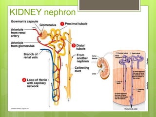
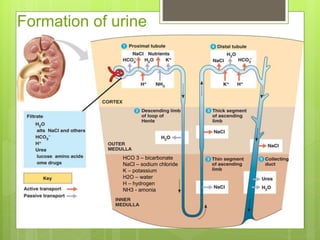
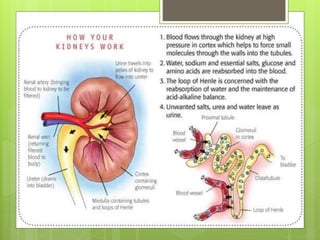
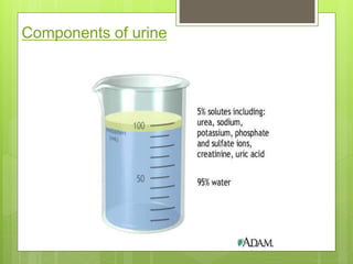
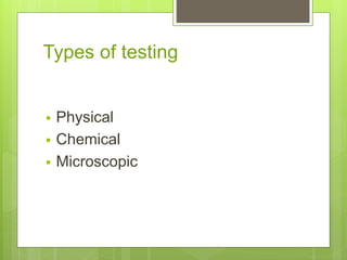
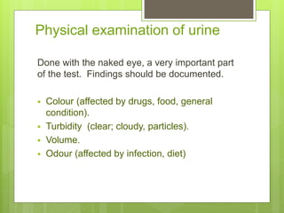
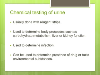
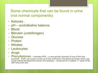
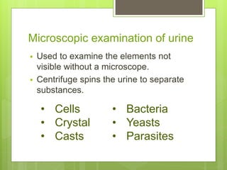
Lec No 1 (contd):
Urinalysis
Dr.K.Parameswari
INTRODUCTION
URINALYSIS is a simple non-invasive
diagnostic test which can provide a glimpse into
a person’s health
Functions of the Kidneys
Homeostatic Functions
1. Waste excretion (urine formation)
a. Nitrogenous end products: urea, creatinine, uric
acid, etc.
b. Metabolic degradation of peptide hormones:
glucagon, insulin, PTH, growth hormone, FSH, and
gastrin.
2. Fluid/electrolyte balance (Na+, K+, water)
3. Acid/base regulation:
kidneys generate and reclaim filtered bicarbonate, as
well as secrete excess acid to maintain balance.
4. Balance of other electrolytes (Ca++, Mg++, Phosphate
PO4
3-)
Objectives
 Give an overview of the anatomy and
physiology of the urinary system
 Explain how urine is produced and its
components
 Describe the types of urine samples and tests
 Describe the requirements and procedure for
dipstick urinalysis using the manual method
The urinary system
Organs of the urinary
system
 Kidneys
 Ureters
 Urinary bladder
 Urethra
The function of the urinary
system
 The kidneys regulate: acid-base balance;
electrolyte concentration; extracellular fluid volume
(homeostasis).
 The kidneys remove waste & water from the blood
stream and reabsorb vital nutrients.
 The kidneys regulate the blood pressure.
 The urinary bladder stores urine.
KIDNEY nephron
Formation of urine
HCO 3 – bicarbonate
NaCl – sodium chloride
K – potassium
H2O – water
H – hydrogen
NH3 - amonia
Components of urine
Components of urine
Types of testing
 Physical
 Chemical
 Microscopic
Physical examination of urine
Done with the naked eye, a very important part
of the test. Findings should be documented.
 Colour (affected by drugs, food, general
condition).
 Turbidity (clear; cloudy, particles).
 Volume.
 Odour (affected by infection, diet)
Chemical testing of urine
 Usually done with reagent strips.
 Used to determine body processes such as
carbohydrate metabolism, liver or kidney function.
 Used to determine infection.
 Can be used to determine presence of drug or toxic
environmental substances.
Some chemicals that can be found in urine
(not normal components)
 Ketones .
 pH – acid/alkaline balance.
 Blood
 Bilirubin (urobilinogen)
 Glucose
 Protein
 Nitrates
 Leukocytes
 drugs
 Phenylketones – indicates PKU – a rare genetic disorder of one of the liver
enzymes. If left, can cause a build up of the chemical in the blood and brain which can
cause mental development issues and epilepsy – screened for in babies 1st week of life
with heel prick test.
Microscopic examination of urine
 Used to examine the elements not
visible without a microscope.
 Centrifuge spins the urine to separate
substances.
• Cells
• Crystal
• Casts
• Bacteria
• Yeasts
• Parasites
Other tests
 Pregnancy tests – EIA (enzyme immunoassay
test) used to detect human chorionic
gonadotrophin (hCG), secreted by the
placenta.
 STIs - chlamydia
Visual significance of urinalysis
 Colour: The colour and clarity of the urine has significant
implications and should always be noted. The colour of normal
urine varies with its concentration, from deep yellow to almost
clear. In disease, the colour may be abnormal due to excretion
of the endogenous pigments as well as drugs and their
metabolites.
 Odour: Odour in the urine of patients who have a urinary tract
infection, is often due to the urea-splitting organisms. This
makes it smell ammonia. The presence of urinary ketones, as
in diabetic ketoacidosis, leads to an acetone smell. The
presence of malodorous urine does not indicate the presence
of infection and does not negate the need for testing.
Clinical significance of test results
 Glucose - is found when its concentration in plasma
exceed the renal threshold (may indicate diabetes)
 Bilirubin/urobilinogen – indicates an excess in the
plasma. Commonest cause of positive results is liver
cell injury e.g. hepatitis, paracetamol overdose, late-
stage cirrhosis.
 Ketones – due to excessive breakdown of body fat.
Common in fasting, may indicate low carbohydrate
diet, vomiting & fever, present in starvation
Clinical significance of test results
(cont.)
 Specific gravity – a measure of solute concentration.
High values can be found in dehydration. Low values
found in high fluid intake. Diabetes insipidus; chronic
renal failure; hypercalcaemia; hypokalaemia.
 Blood – menstruation, kidney disorders; urinary tract
disorders (e.g. tumours, prostatic enlargement).
 pH – high values - commonest cause of high vales is
stale urine; large intake of antacids;UTI with ammonia
forming organisms. Low values – acidosis (diabetic &
lactic); starvation; potassium depletion.
UTI testing pathway
Urine sample Visual appearance
clear
Test with reagent
strip
If all Negative- nitrite,
leucocytes, blood,
protein - discard
If any Positive –
nitrates, leucocytes,
blood, protein = UTI
Obviously infected Send for C&S / treat
Clinical significance of test results
(cont.)
 Protein – excess albumen in the urine is unusually due
increased permeability in the glomeruli. Positive results in
acute and chronic kidney disease, pre-eclampsia.
 Nitrite – UTI – most of the organisms which infect the
urinary tract contains an enzyme that convers nitrate
(normally found in urine) to nitrite which is not found in
urine in the absence of infection. Some organisms do not
convert nitrate to nitrite (false negative).
 Leucocytes – leucocytes enter inflamed tissue from the
blood and are shed into the urine. UTI is commonest cause
of positive results.
Any questions?

Clinical Chemistry urinalysis

  • 1.
    Lec No 1(contd): Urinalysis Dr.K.Parameswari
  • 2.
    INTRODUCTION URINALYSIS is asimple non-invasive diagnostic test which can provide a glimpse into a person’s health
  • 3.
    Functions of theKidneys Homeostatic Functions 1. Waste excretion (urine formation) a. Nitrogenous end products: urea, creatinine, uric acid, etc. b. Metabolic degradation of peptide hormones: glucagon, insulin, PTH, growth hormone, FSH, and gastrin. 2. Fluid/electrolyte balance (Na+, K+, water) 3. Acid/base regulation: kidneys generate and reclaim filtered bicarbonate, as well as secrete excess acid to maintain balance. 4. Balance of other electrolytes (Ca++, Mg++, Phosphate PO4 3-)
  • 4.
    Objectives  Give anoverview of the anatomy and physiology of the urinary system  Explain how urine is produced and its components  Describe the types of urine samples and tests  Describe the requirements and procedure for dipstick urinalysis using the manual method
  • 5.
    The urinary system Organsof the urinary system  Kidneys  Ureters  Urinary bladder  Urethra
  • 6.
    The function ofthe urinary system  The kidneys regulate: acid-base balance; electrolyte concentration; extracellular fluid volume (homeostasis).  The kidneys remove waste & water from the blood stream and reabsorb vital nutrients.  The kidneys regulate the blood pressure.  The urinary bladder stores urine.
  • 7.
  • 8.
    Formation of urine HCO3 – bicarbonate NaCl – sodium chloride K – potassium H2O – water H – hydrogen NH3 - amonia
  • 10.
  • 11.
  • 13.
    Types of testing Physical  Chemical  Microscopic
  • 14.
    Physical examination ofurine Done with the naked eye, a very important part of the test. Findings should be documented.  Colour (affected by drugs, food, general condition).  Turbidity (clear; cloudy, particles).  Volume.  Odour (affected by infection, diet)
  • 15.
    Chemical testing ofurine  Usually done with reagent strips.  Used to determine body processes such as carbohydrate metabolism, liver or kidney function.  Used to determine infection.  Can be used to determine presence of drug or toxic environmental substances.
  • 16.
    Some chemicals thatcan be found in urine (not normal components)  Ketones .  pH – acid/alkaline balance.  Blood  Bilirubin (urobilinogen)  Glucose  Protein  Nitrates  Leukocytes  drugs  Phenylketones – indicates PKU – a rare genetic disorder of one of the liver enzymes. If left, can cause a build up of the chemical in the blood and brain which can cause mental development issues and epilepsy – screened for in babies 1st week of life with heel prick test.
  • 17.
    Microscopic examination ofurine  Used to examine the elements not visible without a microscope.  Centrifuge spins the urine to separate substances. • Cells • Crystal • Casts • Bacteria • Yeasts • Parasites
  • 18.
    Other tests  Pregnancytests – EIA (enzyme immunoassay test) used to detect human chorionic gonadotrophin (hCG), secreted by the placenta.  STIs - chlamydia
  • 19.
    Visual significance ofurinalysis  Colour: The colour and clarity of the urine has significant implications and should always be noted. The colour of normal urine varies with its concentration, from deep yellow to almost clear. In disease, the colour may be abnormal due to excretion of the endogenous pigments as well as drugs and their metabolites.  Odour: Odour in the urine of patients who have a urinary tract infection, is often due to the urea-splitting organisms. This makes it smell ammonia. The presence of urinary ketones, as in diabetic ketoacidosis, leads to an acetone smell. The presence of malodorous urine does not indicate the presence of infection and does not negate the need for testing.
  • 20.
    Clinical significance oftest results  Glucose - is found when its concentration in plasma exceed the renal threshold (may indicate diabetes)  Bilirubin/urobilinogen – indicates an excess in the plasma. Commonest cause of positive results is liver cell injury e.g. hepatitis, paracetamol overdose, late- stage cirrhosis.  Ketones – due to excessive breakdown of body fat. Common in fasting, may indicate low carbohydrate diet, vomiting & fever, present in starvation
  • 21.
    Clinical significance oftest results (cont.)  Specific gravity – a measure of solute concentration. High values can be found in dehydration. Low values found in high fluid intake. Diabetes insipidus; chronic renal failure; hypercalcaemia; hypokalaemia.  Blood – menstruation, kidney disorders; urinary tract disorders (e.g. tumours, prostatic enlargement).  pH – high values - commonest cause of high vales is stale urine; large intake of antacids;UTI with ammonia forming organisms. Low values – acidosis (diabetic & lactic); starvation; potassium depletion.
  • 22.
    UTI testing pathway Urinesample Visual appearance clear Test with reagent strip If all Negative- nitrite, leucocytes, blood, protein - discard If any Positive – nitrates, leucocytes, blood, protein = UTI Obviously infected Send for C&S / treat
  • 23.
    Clinical significance oftest results (cont.)  Protein – excess albumen in the urine is unusually due increased permeability in the glomeruli. Positive results in acute and chronic kidney disease, pre-eclampsia.  Nitrite – UTI – most of the organisms which infect the urinary tract contains an enzyme that convers nitrate (normally found in urine) to nitrite which is not found in urine in the absence of infection. Some organisms do not convert nitrate to nitrite (false negative).  Leucocytes – leucocytes enter inflamed tissue from the blood and are shed into the urine. UTI is commonest cause of positive results.
  • 24.