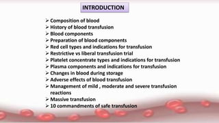
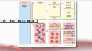
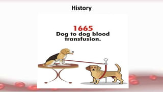
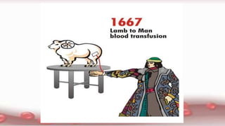
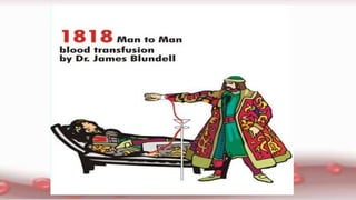
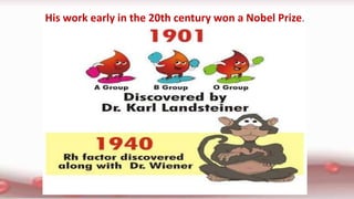
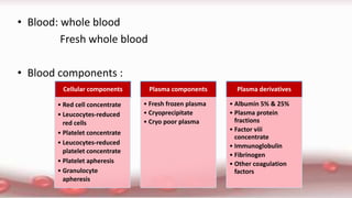
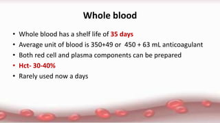
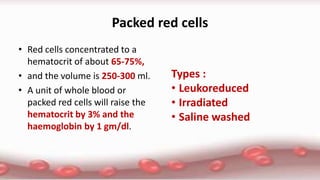
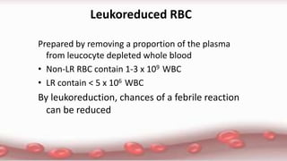
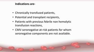
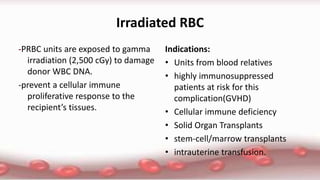
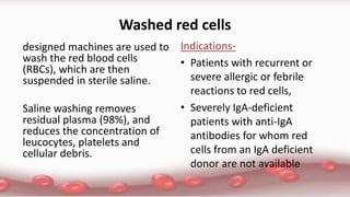
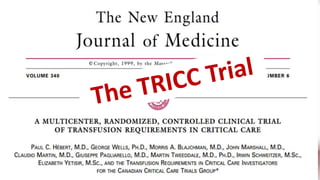
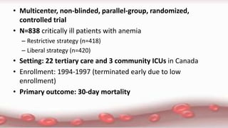
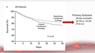
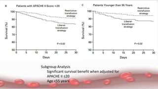
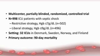
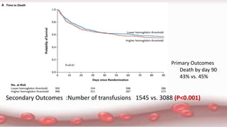
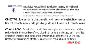
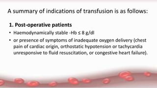
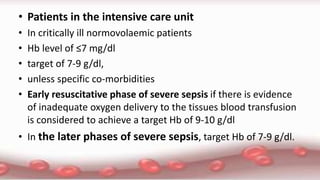
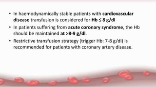
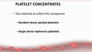
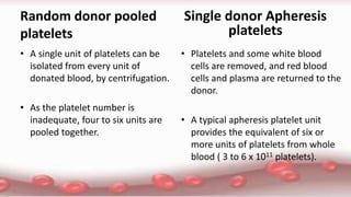
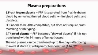
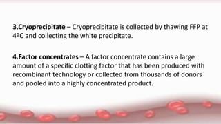
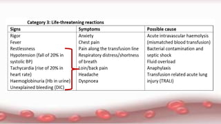
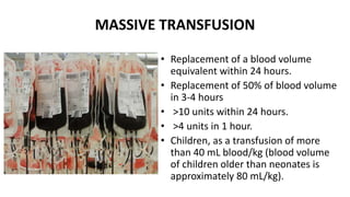
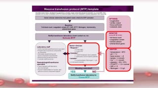
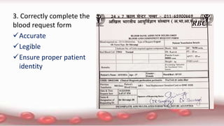
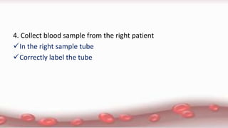
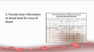
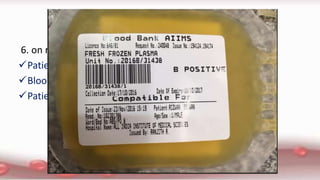
Blood Component
Therapy
Presenter: Dr Shivangi
Moderator: Dr Kamal Kataria
Dr Kabita Chatterjee
INTRODUCTION
Composition of blood
History of blood transfusion
Blood components
Preparation of blood components
Red cell types and indications for transfusion
Restrictive vs liberal transfusion trial
Platelet concentrate types and indications for transfusion
Plasma components and indications for transfusion
Changes in blood during storage
Adverse effects of blood transfusion
Management of mild , moderate and severe transfusion
reactions
Massive transfusion
10 commandments of safe transfusion
COMPOSITION OF BLOOD
Blood volume
• Premature baby:90-100 ml/kg
• Full term baby:80-90 ml/kg
• Infant :70-75 ml/kg
• Children :70-80 ml/kg
• Adult :70-90 ml/kg
History
His work early in the 20th century won a Nobel Prize.
• Blood: whole blood
Fresh whole blood
• Blood components :
Cellular components
• Red cell concentrate
• Leucocytes-reduced
red cells
• Platelet concentrate
• Leucocytes-reduced
platelet concentrate
• Platelet apheresis
• Granulocyte
apheresis
Plasma components
• Fresh frozen plasma
• Cryoprecipitate
• Cryo poor plasma
Plasma derivatives
• Albumin 5% & 25%
• Plasma protein
fractions
• Factor viii
concentrate
• Immunoglobulin
• Fibrinogen
• Other coagulation
factors
Blood donation
Blood donation Triple bag
• CPD and CP2D- 21 days
• CPDA- 35 days
• Shelf life can be extended to 42 days with addition of an additive
solution (SAGM) to red cells within 72 hours of collection
Preservatives
• supports ATP generation by glycolytic
pathways.Glucose
• synthesizes ATPAdenine
• prevents coagulation by chelating calcium.Citrate
• prevents fall in pH
Sodium di-phosphate
Heavy spin,4o
C(within 6 hrs)
Fresh Whole Blood
Packed Red Cells
Stored in 1- 6o
C
Fresh Plasma
Freeze -80o
C immediately
Stored at < -18o
C
Protocol for preparation of Red Cells and FFP1.
• If plasma is frozen within 6 hours  FFP
• If plasma frozen within 24 hours after phlebotomy  Plasma
frozen within 24 hours (PF24)
• Otherwise the plasma is removed within the 72 hour time period
and sent for fractionation into gamma globulin and albumin
• Plasma used for fractionation of coagulation factors must be of
same quality of FFP or atleast PF24
Fresh Whole Blood
Packed Red Cells
Light spin, 22o
C(within 6 hrs)
Platelet Rich Plasma
Platelet Concentrate Fresh Plasma
Store at 22o
C Freeze(FFP)
Heavy spin,22o
C
Preparation of Platelets from whole blood
Thaw at 4o
C & heavy spin
Fresh Frozen Plasma
Cryoprecipitate
-Refrozen within 1 hr
-Store at < - 18
o
C
Cryoremoved Plasma
Freeze -80o
C immediately
Stored at < -18o
C
Preparation of Cryoprecipitate
Cryoprecipitate
–If a unit of FFP is allowed to thaw at 4
degrees for 24 hours, a milky white
flocculum consisting of several cold
insoluble globulins can be separated from
liquid plasma
–Contain Fibrinogen (I), Factor VIII, Factor
XIII and Von Willebrand factor
–Volume -10-15ml
–Refrozen to – 18 C : can be used upto a
year
• Several patients can benefit from single unit of blood
• Transfusion of specific component that patient requires
• Allows optimal survival of each constituent
Advantages of preparing blood components
Whole blood
• Whole blood has a shelf life of 35 days
• Average unit of blood is 350+49 or 450 + 63 mL anticoagulant
• Both red cell and plasma components can be prepared
• Hct- 30-40%
• Rarely used now a days
Packed red cells
• Red cells concentrated to a
hematocrit of about 65-75%,
• and the volume is 250-300 ml.
• A unit of whole blood or
packed red cells will raise the
hematocrit by 3% and the
haemoglobin by 1 gm/dl.
Types :
• Leukoreduced
• Irradiated
• Saline washed
Leukoreduced RBC
Prepared by removing a proportion of the plasma
from leucocyte depleted whole blood
• Non-LR RBC contain 1-3 x 10⁹ WBC
• LR contain < 5 x 10⁶ WBC
By leukoreduction, chances of a febrile reaction
can be reduced
Indications are-
• Chronically transfused patients,
• Potential and transplant recipients,
• Patients with previous febrile non hemolytic
transfusion reactions,
• CMV-seronegative at-risk patients for whom
seronegative components are not available.
Irradiated RBC
-PRBC units are exposed to gamma
irradiation (2,500 cGy) to damage
donor WBC DNA.
-prevent a cellular immune
proliferative response to the
recipient’s tissues.
Indications:
• Units from blood relatives
• highly immunosuppressed
patients at risk for this
complication(GVHD)
• Cellular immune deficiency
• Solid Organ Transplants
• stem-cell/marrow transplants
• intrauterine transfusion.
Washed red cells
designed machines are used to
wash the red blood cells
(RBCs), which are then
suspended in sterile saline.
Saline washing removes
residual plasma (98%), and
reduces the concentration of
leucocytes, platelets and
cellular debris.
Indications-
• Patients with recurrent or
severe allergic or febrile
reactions to red cells,
• Severely IgA-deficient
patients with anti-IgA
antibodies for whom red
cells from an IgA deficient
donor are not available
• Multicenter, non-blinded, parallel-group, randomized,
controlled trial
• N=838 critically ill patients with anemia
– Restrictive strategy (n=418)
– Liberal strategy (n=420)
• Setting: 22 tertiary care and 3 community ICUs in Canada
• Enrollment: 1994-1997 (terminated early due to low
enrollment)
• Primary outcome: 30-day mortality
Primary Outcome
30-day mortality
18.7% vs. 23.3%
(P=0.11)
Subgroup Analysis
Significant survival benefit when adjusted for
APACHE II ≤20
Age <55 years
The TRISS Trial
• Multicenter, partially blinded, randomized, controlled trial
• N=998 ICU patients with septic shock
–Restrictive strategy, Hgb ≤7g/dL (n=502)
–Liberal strategy, Hgb ≤9g/dL (n=496)
• Setting: 32 ICUs in Denmark, Sweden, Norway, and Finland
• Primary outcome: 90-day mortality
Primary Outcomes
Death by day 90
43% vs. 45%
Secondary Outcomes :Number of transfusions 1545 vs. 3088 (P<0.001)
OBJECTIVE: To compare the benefit and harm of restrictive versus
liberal transfusion strategies to guide red blood cell transfusions.
CONCLUSIONS: Restrictive transfusion strategies were associated with a
reduction in the number of red blood cell units transfused, but mortality,
overall morbidity, and myocardial infarction seemed to be unaltered.
Restrictive transfusion strategies are safe in most clinical settings.
-BMJ 2015
A summary of indications of transfusion is as follows:
1. Post-operative patients
• Haemodynamically stable -Hb ≤ 8 g/dl
• or presence of symptoms of inadequate oxygen delivery (chest
pain of cardiac origin, orthostatic hypotension or tachycardia
unresponsive to fluid resuscitation, or congestive heart failure).
• Patients in the intensive care unit
• In critically ill normovolaemic patients
• Hb level of ≤7 mg/dl
• target of 7-9 g/dl,
• unless specific co-morbidities
• Early resuscitative phase of severe sepsis if there is evidence
of inadequate oxygen delivery to the tissues blood transfusion
is considered to achieve a target Hb of 9-10 g/dl
• In the later phases of severe sepsis, target Hb of 7-9 g/dl.
• In haemodynamically stable patients with cardiovascular
disease transfusion is considered for Hb ≤ 8 g/dl
• In patients suffering from acute coronary syndrome, the Hb
should be maintained at >8-9 g/dl.
• Restrictive transfusion strategy (trigger Hb: 7-8 g/dl) is
recommended for patients with coronary artery disease.
PLATELET CONCENTRATES
• Two methods to collect this component
–Random donor pooled platelets
–Single donor Apheresis platelets
Random donor pooled
platelets
• A single unit of platelets can be
isolated from every unit of
donated blood, by centrifugation.
• As the platelet number is
inadequate, four to six units are
pooled together.
• Platelets and some white blood
cells are removed, and red blood
cells and plasma are returned to the
donor.
• A typical apheresis platelet unit
provides the equivalent of six or
more units of platelets from whole
blood ( 3 to 6 x 10¹¹ platelets).
Single donor Apheresis
platelets
Unit of PC
• Volume: 70-90 ml.
• Shelf Life - 5 days
• Stored at a core temperature of 22°C +/- 2°C
with continuous gentle agitation
Indications for the transfusion of platelets
.
• platelet count <10 X
103 platelets/microliter
• platelet count <50 X
103 platelets/microliter
• platelet count <100 X
103 platelets/microliter
• normal platelet count
• prevent spontaneous haemorrhage
• actively bleeding,
• scheduled to undergo an invasive
procedure
• have a qualitative intrinsic platelet
disorder.
• who have a central nervous system
injury,
• have multisystem trauma,
• are undergoing neurosurgery,
• or require an intrathecal catheter for
anesthesia
• ongoing active bleeding congenital
platelet disorder, chronic antiplatelet
therapy, or uremia
Special considerations —
• Heparin-induced thrombocytopenia (HIT)
• Thrombotic thrombocytopenia purpura (TTP)
• Hemolytic-uremic syndromes (HUS)
• Disseminated intravascular coagulation (DIC)
Dosage
• One random unit of platelets raises the platelet count in an
adult by 5,000–8,000/cu mm.
• In children, 0.1–0.2 units/kg will increase the platelet count by
30–50,000/cu mm.
• The expected increase will be less if the patient has sepsis,
splenomegaly, platelet auto- or allo-antibodies or is receiving
chemotherapy.
Plasma preparations
1.Fresh frozen plasma – FFP is separated from freshly drawn
blood by removing the red blood cells, white blood cells, and
platelets.
FFP needs to be ABO-compatible, but does not require cross
matching or Rh typing.
2.Thawed plasma – FFP becomes "thawed plasma" if it is not
transfused within 24 hours of being thawed.
Thawed plasma can be transfused up to five days after being
thawed, if stored at refrigerator temperature (1 to 6ºC).
3.Cryoprecipitate – Cryoprecipitate is collected by thawing FFP at
4ºC and collecting the white precipitate.
4.Factor concentrates – A factor concentrate contains a large
amount of a specific clotting factor that has been produced with
recombinant technology or collected from thousands of donors
and pooled into a highly concentrated product.
5.Liquid plasma – Liquid plasma is prepared by removing the red
blood cells, white blood cells, and platelets from previously
stored whole blood. Its use is extremely rare because it has low
levels of factor V.
6.Intravenous immunoglobulin – Intravenous immunoglobulin is
the purified immunoglobulin fraction of plasma pooled from
several thousand donors. It is useful in the treatment of some
autoimmune and immunodeficiency states.
Indications for transfusion :
• Less well established than the indications for red blood cell
transfusion.
• Active bleeding in the setting of known or strongly suspected
coagulation abnormalities
• Massive transfusion of packed red blood cells due to dilutional
deficiency of coagulation factors.
• Prior to invasive and surgical procedures for which there is a
HIGH risk of bleeding complications ANY potentially significant
abnormality of their coagulation tests.
• Prior to invasive procedures for which there is a LOW risk of
bleeding complications , if the patient has a SEVERE
abnormality of their coagulation tests
• Plasma should NOT be transfused to reverse excessive
warfarin effects, unless there is active bleeding or an urgent
invasive or surgical procedure is required.
Cryoprecipitate
• From one donor usually contains
• 100 antihemophilic units (AHU) and
• 250 mg of fibrinogen;
• it is thawed at 37°C and administered through a standard
blood filter.
• contains factor VIII, fibrinogen, fibronectin, von Willebrand's
factor and factor XIII, is used for the correction of inherited and
acquired coagulopathies.
Recommendations
• Prophylaxis in non bleeding perioperative patients with
congenital fibrinogen deficiencies or vW disease
• Bleeding patients with vW disease
• Correction of microvascular bleeding in massively transfused
patients with low fibrinogen
CHANGES DURING STORAGE OF BLOOD
CLINICAL IMPLICATIONS: DURATION OF BLOOD
STORAGE
• Although conclusions cannot be definitive, logic
dictates that blood less than 14 days of storage
should be better than older storage.
• The Red Cell Storage Duration Study (RECESS).
• The INFORM TRIAL
Among patients in a general hospital population, there was no
significant difference in the rate of death among those who
underwent transfusion with the freshest available blood and
those who underwent transfusion according to the standard
practice of transfusing the oldest available blood.
The INFORM Trial (Oct 2016)
• In an unconscious or anaesthetized patient, hypotension and
uncontrolled bleeding may be the only sign of an incompatible
(mismatched) transfusion.
• In a conscious patient undergoing an acute severe haemolytic
transfusion reaction, signs and symptoms may appear within
minutes of transfusion of 5 to 10 mL of blood.
• Close observation at the start of the transfusion of each unit is
therefore essential.
• With the exception of urticarial allergic and febrile non‐
haemolytic reactions, all are potentially fatal and require urgent treat
ment.
•
MASSIVE TRANSFUSION
• Replacement of a blood volume
equivalent within 24 hours.
• Replacement of 50% of blood volume
in 3‐4 hours
• >10 units within 24 hours.
• >4 units in 1 hour.
• Children, as a transfusion of more
than 40 mL blood/kg (blood volume
of children older than neonates is
approximately 80 mL/kg).
2..
1.. 3..
4..
Mitigated
by massive
transfusion
protocol
• A
10 commandments of safe transfusion
1. assess the patient’s clinical need for blood and when it is required
inform the patient and relative
2. Record the indications for transfusion in the patient’s notes
select the blood product and quality required
3. Correctly complete the
blood request form
Accurate
Legible
Ensure proper patient
identity
4. Collect blood sample from the right patient
In the right sample tube
Correctly label the tube
5. Provide clear information
to blood bank for issue of
blood
6. on receiving the bag check identity on :
Patients
Blood product
Patient’s documentation
7. Administration of blood product :
• Recording
• Monitoring
8.Record in the patient’s notes :
 Type and volume of each product
 Unique donation number
 Blood grp of each unit
 Time of transfusion
 Signature of administering person
9. monitor the patient before during
and after completion of transfusion.
 Severe reactions most commonly prese
nt during the first 15 minutes of a trans
fusion. All patients and in particular, un
conscious patients should be monitore
d during this period and for the first 15
minutes of each subsequent unit.
10. identify and respond immediately to any adverse effect of
transfusion and record in file .
Thank you

blood products

  • 1.
    Blood Component Therapy Presenter: DrShivangi Moderator: Dr Kamal Kataria Dr Kabita Chatterjee
  • 2.
    INTRODUCTION Composition of blood Historyof blood transfusion Blood components Preparation of blood components Red cell types and indications for transfusion Restrictive vs liberal transfusion trial Platelet concentrate types and indications for transfusion Plasma components and indications for transfusion Changes in blood during storage Adverse effects of blood transfusion Management of mild , moderate and severe transfusion reactions Massive transfusion 10 commandments of safe transfusion
  • 3.
  • 4.
    Blood volume • Prematurebaby:90-100 ml/kg • Full term baby:80-90 ml/kg • Infant :70-75 ml/kg • Children :70-80 ml/kg • Adult :70-90 ml/kg
  • 5.
  • 8.
    His work earlyin the 20th century won a Nobel Prize.
  • 9.
    • Blood: wholeblood Fresh whole blood • Blood components : Cellular components • Red cell concentrate • Leucocytes-reduced red cells • Platelet concentrate • Leucocytes-reduced platelet concentrate • Platelet apheresis • Granulocyte apheresis Plasma components • Fresh frozen plasma • Cryoprecipitate • Cryo poor plasma Plasma derivatives • Albumin 5% & 25% • Plasma protein fractions • Factor viii concentrate • Immunoglobulin • Fibrinogen • Other coagulation factors
  • 10.
  • 11.
    • CPD andCP2D- 21 days • CPDA- 35 days • Shelf life can be extended to 42 days with addition of an additive solution (SAGM) to red cells within 72 hours of collection Preservatives • supports ATP generation by glycolytic pathways.Glucose • synthesizes ATPAdenine • prevents coagulation by chelating calcium.Citrate • prevents fall in pH Sodium di-phosphate
  • 12.
    Heavy spin,4o C(within 6hrs) Fresh Whole Blood Packed Red Cells Stored in 1- 6o C Fresh Plasma Freeze -80o C immediately Stored at < -18o C Protocol for preparation of Red Cells and FFP1.
  • 13.
    • If plasmais frozen within 6 hours  FFP • If plasma frozen within 24 hours after phlebotomy  Plasma frozen within 24 hours (PF24) • Otherwise the plasma is removed within the 72 hour time period and sent for fractionation into gamma globulin and albumin • Plasma used for fractionation of coagulation factors must be of same quality of FFP or atleast PF24
  • 14.
    Fresh Whole Blood PackedRed Cells Light spin, 22o C(within 6 hrs) Platelet Rich Plasma Platelet Concentrate Fresh Plasma Store at 22o C Freeze(FFP) Heavy spin,22o C Preparation of Platelets from whole blood
  • 15.
    Thaw at 4o C& heavy spin Fresh Frozen Plasma Cryoprecipitate -Refrozen within 1 hr -Store at < - 18 o C Cryoremoved Plasma Freeze -80o C immediately Stored at < -18o C Preparation of Cryoprecipitate
  • 16.
    Cryoprecipitate –If a unitof FFP is allowed to thaw at 4 degrees for 24 hours, a milky white flocculum consisting of several cold insoluble globulins can be separated from liquid plasma –Contain Fibrinogen (I), Factor VIII, Factor XIII and Von Willebrand factor –Volume -10-15ml –Refrozen to – 18 C : can be used upto a year
  • 17.
    • Several patientscan benefit from single unit of blood • Transfusion of specific component that patient requires • Allows optimal survival of each constituent Advantages of preparing blood components
  • 18.
    Whole blood • Wholeblood has a shelf life of 35 days • Average unit of blood is 350+49 or 450 + 63 mL anticoagulant • Both red cell and plasma components can be prepared • Hct- 30-40% • Rarely used now a days
  • 19.
    Packed red cells •Red cells concentrated to a hematocrit of about 65-75%, • and the volume is 250-300 ml. • A unit of whole blood or packed red cells will raise the hematocrit by 3% and the haemoglobin by 1 gm/dl. Types : • Leukoreduced • Irradiated • Saline washed
  • 20.
    Leukoreduced RBC Prepared byremoving a proportion of the plasma from leucocyte depleted whole blood • Non-LR RBC contain 1-3 x 10⁹ WBC • LR contain < 5 x 10⁶ WBC By leukoreduction, chances of a febrile reaction can be reduced
  • 21.
    Indications are- • Chronicallytransfused patients, • Potential and transplant recipients, • Patients with previous febrile non hemolytic transfusion reactions, • CMV-seronegative at-risk patients for whom seronegative components are not available.
  • 22.
    Irradiated RBC -PRBC unitsare exposed to gamma irradiation (2,500 cGy) to damage donor WBC DNA. -prevent a cellular immune proliferative response to the recipient’s tissues. Indications: • Units from blood relatives • highly immunosuppressed patients at risk for this complication(GVHD) • Cellular immune deficiency • Solid Organ Transplants • stem-cell/marrow transplants • intrauterine transfusion.
  • 23.
    Washed red cells designedmachines are used to wash the red blood cells (RBCs), which are then suspended in sterile saline. Saline washing removes residual plasma (98%), and reduces the concentration of leucocytes, platelets and cellular debris. Indications- • Patients with recurrent or severe allergic or febrile reactions to red cells, • Severely IgA-deficient patients with anti-IgA antibodies for whom red cells from an IgA deficient donor are not available
  • 25.
    • Multicenter, non-blinded,parallel-group, randomized, controlled trial • N=838 critically ill patients with anemia – Restrictive strategy (n=418) – Liberal strategy (n=420) • Setting: 22 tertiary care and 3 community ICUs in Canada • Enrollment: 1994-1997 (terminated early due to low enrollment) • Primary outcome: 30-day mortality
  • 26.
  • 27.
    Subgroup Analysis Significant survivalbenefit when adjusted for APACHE II ≤20 Age <55 years
  • 28.
  • 29.
    • Multicenter, partiallyblinded, randomized, controlled trial • N=998 ICU patients with septic shock –Restrictive strategy, Hgb ≤7g/dL (n=502) –Liberal strategy, Hgb ≤9g/dL (n=496) • Setting: 32 ICUs in Denmark, Sweden, Norway, and Finland • Primary outcome: 90-day mortality
  • 30.
    Primary Outcomes Death byday 90 43% vs. 45% Secondary Outcomes :Number of transfusions 1545 vs. 3088 (P<0.001)
  • 31.
    OBJECTIVE: To comparethe benefit and harm of restrictive versus liberal transfusion strategies to guide red blood cell transfusions. CONCLUSIONS: Restrictive transfusion strategies were associated with a reduction in the number of red blood cell units transfused, but mortality, overall morbidity, and myocardial infarction seemed to be unaltered. Restrictive transfusion strategies are safe in most clinical settings. -BMJ 2015
  • 32.
    A summary ofindications of transfusion is as follows: 1. Post-operative patients • Haemodynamically stable -Hb ≤ 8 g/dl • or presence of symptoms of inadequate oxygen delivery (chest pain of cardiac origin, orthostatic hypotension or tachycardia unresponsive to fluid resuscitation, or congestive heart failure).
  • 33.
    • Patients inthe intensive care unit • In critically ill normovolaemic patients • Hb level of ≤7 mg/dl • target of 7-9 g/dl, • unless specific co-morbidities • Early resuscitative phase of severe sepsis if there is evidence of inadequate oxygen delivery to the tissues blood transfusion is considered to achieve a target Hb of 9-10 g/dl • In the later phases of severe sepsis, target Hb of 7-9 g/dl.
  • 34.
    • In haemodynamicallystable patients with cardiovascular disease transfusion is considered for Hb ≤ 8 g/dl • In patients suffering from acute coronary syndrome, the Hb should be maintained at >8-9 g/dl. • Restrictive transfusion strategy (trigger Hb: 7-8 g/dl) is recommended for patients with coronary artery disease.
  • 35.
    PLATELET CONCENTRATES • Twomethods to collect this component –Random donor pooled platelets –Single donor Apheresis platelets
  • 36.
    Random donor pooled platelets •A single unit of platelets can be isolated from every unit of donated blood, by centrifugation. • As the platelet number is inadequate, four to six units are pooled together. • Platelets and some white blood cells are removed, and red blood cells and plasma are returned to the donor. • A typical apheresis platelet unit provides the equivalent of six or more units of platelets from whole blood ( 3 to 6 x 10¹¹ platelets). Single donor Apheresis platelets
  • 37.
    Unit of PC •Volume: 70-90 ml. • Shelf Life - 5 days • Stored at a core temperature of 22°C +/- 2°C with continuous gentle agitation
  • 38.
    Indications for thetransfusion of platelets . • platelet count <10 X 103 platelets/microliter • platelet count <50 X 103 platelets/microliter • platelet count <100 X 103 platelets/microliter • normal platelet count • prevent spontaneous haemorrhage • actively bleeding, • scheduled to undergo an invasive procedure • have a qualitative intrinsic platelet disorder. • who have a central nervous system injury, • have multisystem trauma, • are undergoing neurosurgery, • or require an intrathecal catheter for anesthesia • ongoing active bleeding congenital platelet disorder, chronic antiplatelet therapy, or uremia
  • 39.
    Special considerations — •Heparin-induced thrombocytopenia (HIT) • Thrombotic thrombocytopenia purpura (TTP) • Hemolytic-uremic syndromes (HUS) • Disseminated intravascular coagulation (DIC)
  • 40.
    Dosage • One randomunit of platelets raises the platelet count in an adult by 5,000–8,000/cu mm. • In children, 0.1–0.2 units/kg will increase the platelet count by 30–50,000/cu mm. • The expected increase will be less if the patient has sepsis, splenomegaly, platelet auto- or allo-antibodies or is receiving chemotherapy.
  • 41.
    Plasma preparations 1.Fresh frozenplasma – FFP is separated from freshly drawn blood by removing the red blood cells, white blood cells, and platelets. FFP needs to be ABO-compatible, but does not require cross matching or Rh typing. 2.Thawed plasma – FFP becomes "thawed plasma" if it is not transfused within 24 hours of being thawed. Thawed plasma can be transfused up to five days after being thawed, if stored at refrigerator temperature (1 to 6ºC).
  • 42.
    3.Cryoprecipitate – Cryoprecipitateis collected by thawing FFP at 4ºC and collecting the white precipitate. 4.Factor concentrates – A factor concentrate contains a large amount of a specific clotting factor that has been produced with recombinant technology or collected from thousands of donors and pooled into a highly concentrated product.
  • 43.
    5.Liquid plasma –Liquid plasma is prepared by removing the red blood cells, white blood cells, and platelets from previously stored whole blood. Its use is extremely rare because it has low levels of factor V. 6.Intravenous immunoglobulin – Intravenous immunoglobulin is the purified immunoglobulin fraction of plasma pooled from several thousand donors. It is useful in the treatment of some autoimmune and immunodeficiency states.
  • 44.
    Indications for transfusion: • Less well established than the indications for red blood cell transfusion. • Active bleeding in the setting of known or strongly suspected coagulation abnormalities • Massive transfusion of packed red blood cells due to dilutional deficiency of coagulation factors. • Prior to invasive and surgical procedures for which there is a HIGH risk of bleeding complications ANY potentially significant abnormality of their coagulation tests.
  • 45.
    • Prior toinvasive procedures for which there is a LOW risk of bleeding complications , if the patient has a SEVERE abnormality of their coagulation tests • Plasma should NOT be transfused to reverse excessive warfarin effects, unless there is active bleeding or an urgent invasive or surgical procedure is required.
  • 46.
    Cryoprecipitate • From onedonor usually contains • 100 antihemophilic units (AHU) and • 250 mg of fibrinogen; • it is thawed at 37°C and administered through a standard blood filter. • contains factor VIII, fibrinogen, fibronectin, von Willebrand's factor and factor XIII, is used for the correction of inherited and acquired coagulopathies.
  • 47.
    Recommendations • Prophylaxis innon bleeding perioperative patients with congenital fibrinogen deficiencies or vW disease • Bleeding patients with vW disease • Correction of microvascular bleeding in massively transfused patients with low fibrinogen
  • 48.
  • 49.
    CLINICAL IMPLICATIONS: DURATIONOF BLOOD STORAGE • Although conclusions cannot be definitive, logic dictates that blood less than 14 days of storage should be better than older storage. • The Red Cell Storage Duration Study (RECESS). • The INFORM TRIAL
  • 50.
    Among patients ina general hospital population, there was no significant difference in the rate of death among those who underwent transfusion with the freshest available blood and those who underwent transfusion according to the standard practice of transfusing the oldest available blood. The INFORM Trial (Oct 2016)
  • 52.
    • In anunconscious or anaesthetized patient, hypotension and uncontrolled bleeding may be the only sign of an incompatible (mismatched) transfusion. • In a conscious patient undergoing an acute severe haemolytic transfusion reaction, signs and symptoms may appear within minutes of transfusion of 5 to 10 mL of blood. • Close observation at the start of the transfusion of each unit is therefore essential. • With the exception of urticarial allergic and febrile non‐ haemolytic reactions, all are potentially fatal and require urgent treat ment.
  • 54.
  • 56.
    MASSIVE TRANSFUSION • Replacementof a blood volume equivalent within 24 hours. • Replacement of 50% of blood volume in 3‐4 hours • >10 units within 24 hours. • >4 units in 1 hour. • Children, as a transfusion of more than 40 mL blood/kg (blood volume of children older than neonates is approximately 80 mL/kg).
  • 57.
  • 58.
  • 59.
  • 60.
    10 commandments ofsafe transfusion
  • 61.
    1. assess thepatient’s clinical need for blood and when it is required inform the patient and relative
  • 62.
    2. Record theindications for transfusion in the patient’s notes select the blood product and quality required
  • 63.
    3. Correctly completethe blood request form Accurate Legible Ensure proper patient identity
  • 64.
    4. Collect bloodsample from the right patient In the right sample tube Correctly label the tube
  • 65.
    5. Provide clearinformation to blood bank for issue of blood
  • 66.
    6. on receivingthe bag check identity on : Patients Blood product Patient’s documentation
  • 67.
    7. Administration ofblood product : • Recording • Monitoring
  • 69.
    8.Record in thepatient’s notes :  Type and volume of each product  Unique donation number  Blood grp of each unit  Time of transfusion  Signature of administering person
  • 70.
    9. monitor thepatient before during and after completion of transfusion.  Severe reactions most commonly prese nt during the first 15 minutes of a trans fusion. All patients and in particular, un conscious patients should be monitore d during this period and for the first 15 minutes of each subsequent unit.
  • 71.
    10. identify andrespond immediately to any adverse effect of transfusion and record in file .
  • 72.

Editor's Notes

  • #3 pH range of blood is 7.35 to 7.45, slightly alkaline. The venous blood normally has a lower pH 38°C(100.4°F), about 1° higher than oral or rectal body temperature. about 20% of extracellular fluid, amounting to 8% of total body mass. The blood volume is 5L to 6L in average sized adult male & 4L to 5L(1.2gal) in an average sized adult female.
  • #6 The first animal-to-animal (dog) blood transfusion at Oxford in 1665 by Richard Lower
  • #7 In 1667, Jean-Baptiste Denis who was physician to King Louis XIV, performed the transfusion of blood from an animal to a human. Denis transfused the blood from a sheep to a 15-year old boy and later to a labourer, both of whom survived the transfusions.
  • #13 The centrifugal speed depends on the diameter of the rotor. The speed can vary based on the centrifuge and/or the rotor and has to be standardised. Mention should be made about the refrigerated centrifuge attaining required temperature before centrifugation Heavy Spin- 3500 rpm x 5 minutes Light Spin – 1000 rpm x 3 minutes Plasma : FFP and PF24 refer to plasma that is separated and frozen at -18 C within 8 hrs/ 24 hrs of collection of whole blood and can be stored for 1 year Documented deficiencies of Factors V VII XI XIII can be corrected by FFP/ PF24 though concentrates for Factors XI and XIII are available Thawed plasma, which is FFP and PF24 thawed and then stored at 1-6 C for 5 days is used routinely
  • #15 For preparation of RDP centrifugation involves a soft spin followed by a hard spin Platelets derived from whole blood: Approx. 20% platelet transfusion doses are provided by platelet concentrates prepared from individual whole blood units within 8 hours of collection These whole blood derived platelet concentrates are administered in groups of 4-8, sometimes pooled in a single pack prior to transfusion Can be LR by use of filters designed esp for platelets Following Production of PRPs  they are subjected to hard spin following which they separate into plasma and platelet concentrate Most platelet concentrates contain – 7000 /micro litre in volume of 50 ml
  • #16 Cryoprecipitate should be prepared from Fresh Frozen Plasma anytime during it’s storage period, but preferably as early as possible in it’s shelf life as coagulation factors slowly deteriorate over a period of time
  • #21 , especially (alloimmunized from pregnancy, risk for HLA alloimmunization from blood transfusions can be reduced)
  • #25 Red blood cell transfusions were previously given routinely whenever the hemoglobin level was <10 g/dL. This practice was based upon unproven physiological and clinical assumptions. However, a hemoglobin concentration of <7 g/dL became the accepted threshold after the multicenter Transfusion Requirements in Critical Care (TRICC) trial. Hebert PC, et al. "A multicenter, randomized, controlled clinical trial of transfusion requirements in critical care". The New England Journal of Medicine. 1999. 340(6):409-417. Among critically ill patients, how does a restrictive transfusion strategy (hemoglobin goal of 7-9 g/dL) compare with a liberal transfusion strategy (hemoglobin goal of 10-12 g/dL) in decreasing mortality? In critically ill patients, restrictive transfusion (Hgb >7 g/dL) is associated with better survival compared to liberal strategy (Hgb >10). The Transfusion Requirements in Critical Care (TRICC) trial randomized 838 patients admitted to the ICU without evidence of active bleeding to a restrictive transfusion strategy (transfusion to maintain hemoglobin >7 g/dL) versus a liberal strategy (transfusion to maintain hemoglobin ≥10 g/dL). The study enrolled patients who were euvolemic after initial fluid resuscitation. There was adequate separation between the two groups, with an average hemoglobin 8.5 vs. 10.7 g/dL, respectively. The restrictive transfusion strategy was associated with decreased rates of in-hospital mortality compared to those seen with the liberal transfusion strategy. This benefit was most prominent among the less critically ill patients (APACHE II score ≤20) and <55 years old. However, in the restrictive arm, there was a trend towards improved outcomes in patients with active cardiac ischemia. TRICC's results were met favorably among blood bank specialists and other guardians of the red blood cell supply,  but many questioned the external validity of the study. In particular, some criticisms included that only 13% of patients screened were ultimately randomized, that the study's results were unlikely to apply to patients at high altitude, and that red cell transfusions should not be restricted among patients with traumatic brain injury. Before the publication of TRICC, ICU patients were routinely transfused for hemoglobin <10 g/dL. Modern practice guidelines  now recommend a Hgb transfusion goal of 7-9 g/dL, except in choice patients. A large non-blinded, randomized trial, Transfusion Strategies for Acute Upper Gastrointestinal Bleeding (2013) studied restrictive versus liberal transfusion strategies in patients with upper GI bleeding, a group that was notably excluded from TRICC. It found a similar reduction in mortality with restrictive transfusion goals. More recently, the 2014 TRISS trial  found no difference 90 day all-cause mortality for Hgb transfusion goals of 7 vs. 9 g/dL in patients with septic shock.
  • #27 Primary Outcomes 30-day mortality 18.7% vs. 23.3% (ARR 4.7%; P=0.11)
  • #33 uidelines for the use of blood transfusion have been published by many scientific societies. Some of the recent ones are from the American Society of Anesthesiologists,[10] the Society of Critical Care  Medicine,[11] the American Association of Blood Banks (AABB),[12] the American College of Physicians[13] and the British Committee for Standards in Haematology.[14] Most recommend the use of restrictive transfusion strategy. However, high-quality evidence is available for very few clinical settings. 
  • #35 Indian Journal of Anaesthesia Medknow Publications Indications for blood and blood product transfusion Sandhya Yaddanapudi and LN Yaddanapudi
  • #38 Platelets are stored at room temperature, because cold induces clustering of von Willebrand factor receptors on the platelet surface and morphological changes of the platelets, leading to enhanced clearance by hepatic macrophages and reduced platelet survival in the recipient.
  • #39 therapeutic (ie, stop active bleeding) or prophylactic (ie, prevent bleeding platelet count <10 X 103 platelets/microliter. The purpose is to prevent spontaneous hemorrhage. ●Patients with a platelet count <50 X 103 platelets/microliter who are actively bleeding, are scheduled to undergo an invasive procedure, or have a qualitative intrinsic platelet disorder. ●Patients with a platelet count <100 X 103 platelets/microliter who have a central nervous system injury, have multisystem trauma, are undergoing neurosurgery, or require an intrathecal catheter for anesthesia Patients with a normal platelet count who have ongoing active bleeding and a reason for platelet dysfunction, such as a congenital platelet disorder, chronic antiplatelet therapy, or uremia
  • #45 (eg, thoracic, abdominal, spinal, intracranial, and some urologic surgeries), if the patient has
  • #46 eg, endoscopy, cardiac catheterization, central venous catheter insertion (ie, prothrombin time >2-times control, INR >2, or partial thromboplastin time >2-times control). Transfusion of plasma is not indicated prior to such procedures if the coagulation tests are only mildly abnormal.
  • #48 Earlier, recommended in bleeding patients with hypofibrinogenemia, von Willebrand's disease and haemophilia A (when factor VIII concentrate is not available).
  • #49 During storage, RBCs metabolize glucose to lactate, hydrogen ions accumulate, and plasma pH decreases. The storage temperatures of 1° to 6° C stimulate the sodium-potassium pump, and RBCs lose K+ and gain Na. The osmotic fragility of RBCs increases during storage, and some cells undergo lysis, resulting in increased plasma Hb levels. Progressive decreases in RBC concentrations of ATP and 2,3-diphosphoglycerate (2,3- DPG) occur during storage.
  • #50 CONCLUSIONS
  • #54 Slow the transfusion.   Administer antihistamine IM.    If no clinical improvement within 30 minutes or if signs and symptoms worsen, treat as Category 2.  If improved, restart transfusion slowly.  
  • #55 Stop the transfusion and keep IV line open with normal saline in another site.  Return the blood unit with transfusion administration set, freshly collected urine and new blood  samples (1 clotted and 1 anticoagulated), drawn from a vein opposite to the transfusion site, to  the blood transfusion centre for laboratory investigations.  Administer antihistamine IM and oral or rectal antipyretic.  Give IV corticosteroids and bronchodilators if there are anaphylactoid features (e.g. bronchospasm, stridor).  If clinical improvement occurs, restart transfusion slowly with new blood unit and observe  carefully.  If no clinical improvement within 15 minutes or if signs and symptoms worsen, treat as Category 3.  Collect urine for next 24 hours for evidence of haemolysis and send for laboratory investigations 
  • #56 Stop the transfusion and keep IV line open with normal saline in another site.   Infuse normal saline to maintain systolic BP.    Maintain airway and give high flow oxygen by mask.   Give adrenaline (as 1:1000 solution) 0.01 mg/kg body weight by slow intramuscular injection.   Give IV corticosteroids and bronchodilators if there are anaphylactoid features.   Give diuretic: e.g. frusemide 1 mg/kg IV or equivalent.   Check a fresh urine specimen visually for signs of haemoglobinuria.    Notify the superior or senior doctor attending the patient, and the blood centre immediately.   Send blood unit with transfusion set, fresh urine sample and new blood samples (1 clotted and 1  anticoagulated), drawn from a vein opposite the infusion site, with the appropriate request form  to the blood transfusion centre for investigation.   Start a 24‐hour urine collection and record all intake and output. Maintain fluid balance chart.   Assess for bleeding from puncture sites or wounds. If there is clinical or laboratory evidence of  DIC, give platelets (adult: 4‐6 units) and either cryoprecipitate (adult: 12 units) or FFP (adult: 3  units).   Reassess. If hypotensive:  – Give further saline.   – Give inotrope, if available.   If urine output falls or there is laboratory evidence of acute renal failure (rising K+, urea,  creatinine):  – Maintain fluid balance accurately.  – Give further diuretic: e.g. frusemide 1 mg/kg IV or equivalent.  – Consider dopamine infusion, if available.  – Seek expert help: the patient may need renal dialysis.    If bacteraemia is suspected (rigor, fever, collapse, no evidence of a haemolytic reaction), start a  broad‐spectrum antibiotic IV. 
  • #58 The decision to start, depends on the physiological state of patient evidence of amount of blood loss potential for ongoing hemorrhage Trauma, crush injury Orthopedic surgery Liver surgery- transplantation
  • #60  Recently, MTPs have shifted toward predetermined blood product administration in an effort to mitigate and treat coagulopathy by adequate coagulation factor administration early in the resuscitation, decreasing the amount of crystalloid administered and removing the delay in ordering, preparing, and subsequent administration of blood products.20 This is in contrast to the laboratory-based approach where there is a delay in the ordering and subsequent administration of platelet, plasma, and cryoprecipitate products because of delays due to laboratory turnaround time and product preparation time. Most MTPs using the predetermined approach have preset transfusion packages that are delivered consecutively until the patient either dies or their bleeding is under control (Fig. 2). In addition, predetermined blood product administration clearly defines the proportion of blood components to transfuse, decreasing the chances of less than optimal transfusion therapy (i.e., large volumes of RBC products with little or no transfusion of plasma or platelet products).48
  • #64 When blood is required for transfusion, the prescribing clinician should complete and sign a blood  request form that is designed to provide all necessary information. All details requested on the  blood request form must be completed accurately and legibly.      The blood request form should always be accompanied by the patient’s blood sample. The  sample is placed in a sample tube that is correctly labelled and is uniquely identifiable with the  patient.   The  blood  sample  shall  not  be  submitted  in  a  syringe,  as  this  could  lead  to  errors  when  transferring to a test tube for grouping and compatibility testing. It may also cause haemolysis.    For  a  routine  case,  the  sample  and  request  form  should  be  submitted  to  the  transfusion  department at least 24 hours before required, to make sure of the availability of blood.   Physicians may request those, who accompany the patient, to consider becoming blood donors  if they are healthy and lead a healthy lifestyle.  
  • #65 The taking of a blood sample from the patient needs supervision. If the patient is conscious at the  time of taking the sample, ask him/her to identify himself/herself by given name and all other  appropriate information.    A 5 mL blood sample should be collected into a dry test tube and then correctly and clearly labelled  with the patient’s details, and submitted to the blood centre for testing. The specimen label must  include the following information:     Patient’s full name, age and sex.   Registration number.   Ward/bed number.   Date and time specimen taken.   Phlebotomist’s signature/initials.  20  – Use positive patient identification to identify the patient. NEVER pre‐label the sample  tube before phlebotomy.  – Use the blood product request form, write legibly and fill in all appropriate details.  – When taking a blood sample for cross match, complete the whole procedure before any  other task is undertaken – it is important that there are no interruptions during the  process.  – The signature of the individual who took the sample must appear on the specimen label.    Retention of blood samples:   Blood samples from recipient and donor(s) must be retained for 7 days at +2°C to +8°C after  each transfusion.    Should another transfusion be necessary 72 hours after the earlier transfusion, a fresh sample  shall be requested for cross match. Collection of a second 5 mL blood sample is required for rechecking and further cross matching and must be retained in case of investigation of transfusion  reaction.  
  • #66 Collection and receipt of blood   ALWAYS take a completed patient documentation label to the issue room of the blood transfusion department when collecting the first unit of blood.   MATCH the details on the blood request form against the blood compatibility label (tag), the bag  unit number and the patient documentation label.   If everything matches, sign out the unit with the date and time.  21   If there is any discrepancy, DO NOT sign out the unit; contact the staff member of the blood  transfusion department immediately.   When receiving the unit of blood in the clinical area, check that it is the right unit for the right  patient.    Always  check  patient/component  compatibility/identity.  Inspect  pack  and  contents  for  signs  of  deterioration or damage.  
  • #69 Monitoring the transfusion      It is essential to take baseline observations and to ensure that the patient is monitored during  the transfusion in order to detect any adverse event as early as possible. Before commencing the  transfusion, it is essential to encourage the patient to notify a nurse or doctor immediately if he  or she becomes aware of any discomfort such as shivering, flushing, pain or shortness of breath  or begins to feel anxious.   Ensure that the patient is in a setting where he or she can be directly observed.   For each unit of blood transfused, monitor the patient:  - Before starting the transfusion (baseline observation).  - 15 minutes after starting the transfusion.  - At least every hour during transfusion.  - Carry out a final set of observations 15 minutes after each unit has been transfused.    
  • #70 Whether the patient and/or relatives were informed about the transfusion.   The reason for transfusion.   Signature of the prescribing clinician.   Pre‐transfusion checks of:  - Patient’s identity.  - Blood bag.  - Compatibility label.  - Signature of individual performing the pre‐transfusion identity check.   The transfusion:  - Type and volume of each product transfused.  - Unique donation number of each unit transfused.  - Blood group of each unit transfused.  - Time at which the transfusion of each unit commenced.  - Signature of the person administering the blood component.  - Monitoring of the patient before, during and on completion of transfusion.  - All other details related to the transfusion process.  - Informed consent.  - Administration of the unit.  - Set‐up time of each unit transfused.  - Time of transfusion. 
  • #71 At each of these stages, record the following information on the patient’s chart:  - Patient’s general appearance.  - Temperature.  - Pulse.  - Blood pressure.  - Respiratory rate.     Make note of the following:  - Time the transfusion started.  - Time the transfusion was completed.  - Volume and type of blood products transfused.  - Unique donation number of all products transfused.  - Any adverse effect.     Record in the patient’s notes:  – Type and volume of each unit transfused.  – Unique donation number of each unit transfused.  – Blood group of each unit transfused.  – Time at which the transfusion of each unit commenced.  – Signature of the individual responsible for administration of the blood.  – Record the time of completion of the transfusion.  – Record the details of transfusion reaction.     Identify and respond immediately to any adverse effect, by stopping the transfusion.   24  Severe reactions most commonly present during the first 15 minutes of a transfusion. All patients and  in particular, unconscious patients should be monitored during this period and for the first 15  minutes of each subsequent unit.    Specific instructions concerning possible adverse events shall be provided to the patient.     The transfusion of each unit of the whole blood or red blood cells should be completed within four  hours of the start of the transfusion. If a unit is not fully transfused within four hours, discontinue its  use and dispose of the remainder through the clinical waste system. 
  • #72 Stop transfusion Keep iv line open with normal saline Inform attending physician and blood bank Recheck all labels Send an edta and clotted sample blood bag and transfusion set Urine sample Severe reactions most commonly present during the first 15 minutes of a transfusion. All patients and  in particular, unconscious patients should be monitored during this period and for the first 15  minutes of each subsequent unit.