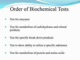
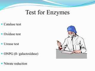
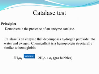
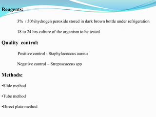
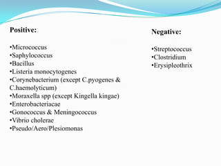
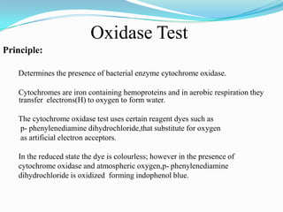
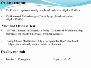
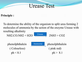
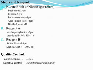
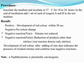
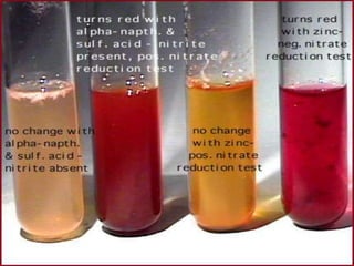
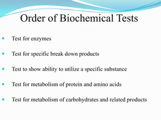
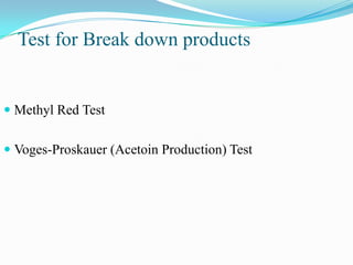
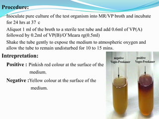
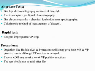
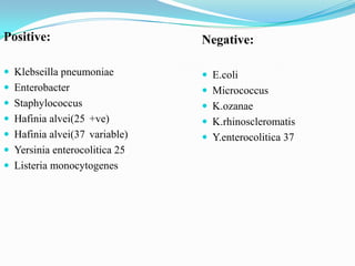
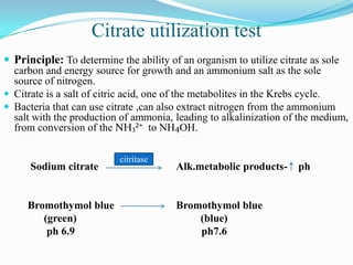
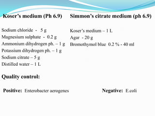
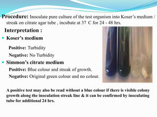
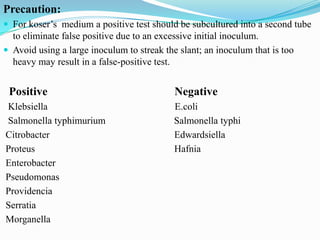
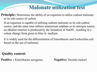
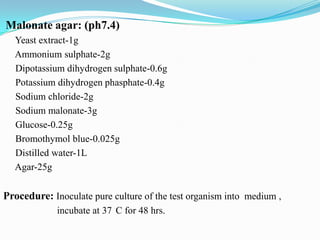
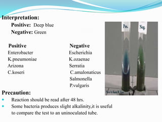
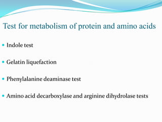
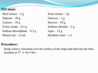
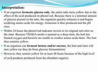
SlideShare a Scribd company logo
1 of 91
Dr. Saumya Singh
Order of Biochemical Tests


Test for enzymes



Test for metabolism of carbohydrates and related
products



Test for specific break down products



Test to show ability to utilize a specific substance



Test for metabolism of protein and amino acids
Test for Enzymes
 Catalase test
 Oxidase test
 Urease test
 ONPG (ß- galactosidase)
 Nitrate reduction
Catalase test
Principle:
Demonstrate the presence of an enzyme catalase.
Catalase is an enzyme that decomposes hydrogen peroxide into
water and oxygen. Chemically,it is a hemoprotein structurally
similar to hemoglobin
2H2o2

catalase

2H2o + o2 (gas bubbles)
• Catalase is present in most cytochrome containing aerobic and

facultative anaerobic bacteria (except streptococcus spp).
Anaerobic bacteria eg. clostridium spp. possess the peroxidase
enzyme in lieu of Catalase.
• Hydrogen peroxide forms as one of the oxidative end product of

aerobic carbohydrate metabolism. If this is allowed to
accumulate in the bacterial cells it becomes lethal to the bacteria.
• Catalase thus helps in converting H2o2 to H2o and o2
• Optimal PH for Catalase action is 7.
Reagents:
3% / 30%hydrogen peroxide stored in dark brown bottle under refrigeration
18 to 24 hrs culture of the organism to be tested

Quality control:
Positive control - Staphylococcus aureus
Negative control – Streptococcus spp

Methods:
•Slide method
•Tube method
•Direct plate method
Slide Method
With an inoculating wire
transfer growth from the
centre of the colony to the
surface of the glass slide.
Add 1 drop of 30% H2O2
and observed for bubble
formation.

Tube Method

Direct Plate Method

Small amount of culture to
be tested is picked from
nutrient agar with a clean
sterile glass rod/capillary
tube and inserted into
H2O2 solution held in a
small clean tube.

1 ml of 3% H2O2 is
poured over a 24 hour
nutrient agar culture of the
test organism directly.

Result: The rapid and sustained appearance of bubbles/effervascence indicates a
positive test.A few tiny bubbles forming after 20 to 30 sec is not considered a positive
test.

Precaution: Avoid colonies from blood agar( contains catalase )
Platinum wire / Nichrome wire should be used with caution.
Positive:

Negative:

•Micrococcus
•Saphylococcus
•Bacillus
•Listeria monocytogenes
•Corynebacterium (except C.pyogenes &
C.haemolyticum)
•Moraxella spp (except Kingella kingae)
•Enterobacteriacae
•Gonococcus & Meningococcus
•Vibrio cholerae
•Pseudo/Aero/Plesiomonas

•Streptococcus
•Clostridium
•Erysipleothrix
Oxidase Test
Principle:
Determines the presence of bacterial enzyme cytochrome oxidase.
Cytochromes are iron containing hemoproteins and in aerobic respiration they
transfer electrons(H) to oxygen to form water.

The cytochrome oxidase test uses certain reagent dyes such as
p- phenylenediamine dihydrochloride,that substitute for oxygen
as artificial electron acceptors.
In the reduced state the dye is colourless; however in the presence of
cytochrome oxidase and atmospheric oxygen,p- phenylenediamine
dihydrochloride is oxidized forming indophenol blue.
Oxidase reagent:
•

1% Kovac’s reagent(tetra methyl -p-phenylenediamide dihydrochloride )

•

1% Gordons & Mcleods reagent(Dimethly - p- phenylenediamide
dihydrochloride)

Modified Oxidase Test:
• 6%TMPD Reagent in Dimethly sulfoxide (DMSO) used for differentiating
micrococci spp (positive in 30 secs) from staphylococci.
•

Ewing Johnson Modification ( 0.1gm Îą-naphthol in 10ml95% ethanol,
0.1gm p-aminodimethylaniline oxalate in 10ml d.w).

Quality control:
•

Positive:

P.aeruginosa

Negative: E.coli
Direct plate method

Wet filter paper method
(KOVAC’S METHOD)

Dry filter paper method

2 to 3 drops of reagent is added
directly to colonies growing on
medium.
(1gm in100ml distilled water)

A strip of Whatman’s No. 1
filter paper soaked in freshly
prepared 1% Oxidase reagent is
smeared with test bacterium.

A strip of Whatman’s No. 1
filter paper is soaked in
freshly prepared 1% Oxidase
reagent,drained for 30 sec,
freez dried and stored in dark
bottle tightly. For use, strip is
removed, laid in Petri dish ,
moistened with distilled
water and smeared with test
bacterium.

Result:

A positive reaction is indicated by a Deep blue colour appearing within 10 sec.

Precautions: 1) Stainless steel or Nichrome inoculating loop and wires should not be used.
2) Tetra methyl derivative of pPD is recommended.
3) Do not perform the test from colonies growing in Macconkey medium.
Positive
•Neisseria spp
•Haemophillus sps
•Pseudomonas spp(except P.maltophilia)
•Aeromonas spp
•Alcaligenes
•Vibrio
•Campylobacter
•Plesiomonas shigelloides
•Micrococcus spp
•Moraxella

Negative
•Enterobacteriaceae
•Acenitobacter spp
•Brucella canis
•Bordetella parapertusis
•Franscisella tularensis
•Gardenella vaginalis
Urease Test
Principle :
To determine the ability of the organism to split urea forming 2
molecules of ammonia by the action of the enzyme Urease with
resulting alkalinity
Urease
NH2.CO.NH2 + H2O
2NH3 + CO2
phenolphthalein
( Colourless)
ph < 8.1

Ammonia

phenolphthalein
( pink red)
ph > 8.1
Stuarts Urea broth (pH 6.8)
• Monopotassium phosphate-9.1gm
•Disodium phosphate-9.5gm
•Urea-20gm
•Phenol red indicator-0 .02gm
•Yeast extract-0.1gm
•Distilled water- 1l

Christensens urea agar (pH 6.8)
•Peptone-1gm
•Glucose-1gm
•Sodium chloride-5gm
•Monopotassium phosphate-2gm
•Urea-20gm
•Phenol red indicator-0.012gm
•Agar-15gm
•Distilled water-1l

Quality control
•

Positive – Proteus spp & Klebsiella spp(weak positive)

•

Negative – E.coli
Procedure:
The surface of the agar slant is streaked with the test organism / the broth
medium is inoculated with the loopful of pure culture of the test organism
and incubated at 37 c for 18-24 hrs.

Interpretation:
Rapid hydrolysers gives +ve results in 1-2 hrs, less active sp. require 3 or more days.
Stuarts broth –

Positive red colour through out the broth.

Christensens agar–Rapid urea splitters red through out the medium,
Slow urea splitters red colour initially in slant only
& then gradually entire tube.
No urea hydrolysis-Negative yellow or straw colour
Precaution:
Both the Urease test medium depend upon the demonstration of alkalinity i.e.
not specific for Urease i.e. even protein hydrolysis may result in alkalinity
hence false positive may be seen in Pseudomonas . To eliminate such false
positivity control test using the same medium without urea should be tested.
Elek’s test:
Reagent :
• Urea-4gm
• KH2PO4-50ML
• NaOH-35ML
• Distilled water -115ml
• Nessler’s reagent(50gm KI in 50ml of H2O,add Hgcl2 sol.until a

perm.precipitate appears.add 200ml NaOH and make upto 1lt with water).

Method :
Emulsify the organism in 0.5ml of substrate tube,place the tube in water bath
at 37 c for 3hr.Remove the tube and add 0.1ml Nessler’s reagent and read
after 3mins.

Positive-yellow to dark brown precipitate.
Negative –control and negative tubes are colourless.
Alternate tests:
Reagent impregnated urease strip test (2hrs)
Key urease tablets test
Urea discs
Ewings Urea R broth (pH 6.9)

Positive
Proteus
Klebsiella pneumoniae
Actinobacillus sp.
Bacillus lentus
Enterobacter
Cryptococcus
Helicobacter pylori
Bordetella bronchoseptica

Negative
Escherichia
Providencia
Aeromonas
Edwarseilla
S.typhi
0-Nitrophenyl-ß-D-Galactopyranoside (ONPG) Test
Principle:
To demonstrate the presence or absence of the enzyme ß Galactosidase
Biochemistry:
O – Nitrophenyl – ß – D – galactopyranoside is structurally similar to lactose except
that orthonitrophenyl has been substituted for glucose
H2o
ONPG (colorless)

Galactose+ Orthonitrophenol(yellow)
galactosidase
H20

Lactose

Galactose + Glucose

galactosidase
It differentiate late lactose fermenting organism and is of particular use in
identification of enterobacteria
• Disaccharide compound composed of glucose and galactose connected through an
oxygen linkage known as galactoside bond.
• Lactose fermentation depends upon the 2 enzymes ß-galactoside permease and
ß-galactosidase.
• ß-galactoside permease permits the transport of ß-galactoside such as lactose into the
bacterial cell wall.

• ß-galactosidase cleave the ß-galactoside bond after it enters into the bacterial cell
liberating glucose and galactose.
•Non lactose fermenting bacteria are devoid of both the enzymes.

•Late lactose fermenters lack the enzyme permease but have ß- galactosidase activity and
so the permeases activity occurs sluggishly and lactose fermentation is delayed by
2-8 days. In these instances a +ve ONPG test may provide a rapid identification of
delayed lactose fermentors .
Media and reagents:
pH 7.0-7.5
Sodium phosphate buffer-(0.01mol/lt,ph7.5) 100ml
O-Nitrophenyl- ß-D-galactopyranoside(ONPG)-0.6gm
Toluene
Physiologic saline 0.85%
Quality control:
Positive – E.coli
Negative – Proteus spp
TUBE METHOD
A loopful bacterial growth (KIA,TSI agar) is
emulsified in 0.5 ml of physiologic saline. 1
drop of toluene is added and vigorously mix.
An equal quantity of buffer ONPG solution is
added and mixture is placed at 37 waterbath.

DISC METHOD
A loopful of bacterial suspension is added to
the ONPG substrate ( 1 ml of distilled water to
a tablet in test tube , placed in 37 waterbath.

Interpretation:
Yellow color within 5-10 mins - Positive
Colorless after 24 hrs
- Negative

Precaution:
•Glucose containing medium should not be used.
•Do not use if medium yellow.
•Store away from direct sunlight, ONPG is light sensitive
Nitrate reduction
Principle:
To determine the ability of the organism to reduce nitrate to
nitrites or free nitrogen gas in the presence of enzyme nitrate
reductase.
Purpose:
Aid in the identification of Enterobacteriaceae(+) except certain
biotypes of Pantoa agglomerans and certain species of Serratia
and Yersinia(-).
Also aids in identifying members of
Haemophilus,Neisseria,Moraxella species.
Media and Reagent:
A. Nitrate Broth or Nitrate agar (Slant)
Beef extract-3gm
Peptone-5gm
Potassium nitrate-1gm
Agar (nitrite-free)-12gm
Distilled water -1lt

B. Reagent A
α – Naphthylamine -5gm
Acetic acid (5N), 30%-1lt

C. Reagent B
Sulfanilic acid-8gm
Acetic acid (5N) , 30%-1lt

Quality Control:
Positive control - E.coli
Negative control - Acinetobacter baumannii
Procedure:
Inoculate the medium and incubate at 37 C for 18 to 24 hours.At the
end of incubation add 1 ml of each of reagent A and B to the test
medium.

Result:
Positive – Development of red colour within 30 sec.
Negative-No colour change
* Negative reaction(True)- Nitrates not reduced
* Negative reaction(False)- Reduction of products other than
nitrites ( test reaction detects only nitrites).
Development of red colour after adding of zinc dust indicates the
presence of residual nitrates and confirms true negative reactions.
Note- Îą Naphthylamine is potentially carcinogenic.
Order of Biochemical Tests


Test for enzymes



Test for specific break down products



Test to show ability to utilize a specific substance



Test for metabolism of protein and amino acids



Test for metabolism of carbohydrates and related products
Test for Break down products

 Methyl Red Test

 Voges-Proskauer (Acetoin Production) Test
Methyl Red Test
Principle:
To test the ability of the organism to produce and maintain stable acid end
products (lactic,acetic,formic) from glucose fermentation and to overcome
the buffering capacity of the system. This is a qualitative test for acid
production.

Biochemistry:
 Methyl red is a pH indicator with a range between 6(Y) and 4.4(R).
 The pH at which the MR detects acid is considerably lower than the pH of

other indicators.
 Thus to produce a colour change the test organism must produce a large
quantity of acid from the substrate being used.
Media & Reagents :
 MR/VP Broth (glucose phosphate peptone water):
Peptone-5 gm
K2HPO4-4 gm
Water-1 L
Glucose 10%-50 ml
 MR pH indicator : MR 0.1 g in 300ml of 95% Ethanol & Distilled water 200ml.

Quality Control:
Positive : E.coli

Negative : E. aerogenes
Procedure:
Inoculate the MR/VP broth with a pure culture of the test organism and
incubate at 37 for 48 to 72 hrs
Add 5 drops of MR reagent to the broth.

Interpretation:
Positive : red color (ph 4.4 or less) at the surface of the
medium.

Negative : Yellow color (pH 6.0) at the surface of
the medium.
Positive

Negative:

E.coli

Klebseilla

Yersinia spp

Enterobacter aerogenes
Enterobacter cloacae

Listeria monocytogenes
Gram negative non enteric bacilli

Precaution: orange colour reaction is considered negative.
Voges Proskauer Test
Principle :
To determine the ability of the organisms to produce neutral end product
acetyl methyl carbinol (acetoin) from glucose fermentation. In presence of
atmospheric oxygen and KOH, acetoin is converted to Diacetyl and Îąnaphthol (serves as a catalyst) along with arginine to produce red complex.

Quality control :
Positive : Enterobacter aerogenes

Negative : E.coli
Media & Reagents:
MR/VP Broth : pH 6.9
Peptone – 1.5 g
Glucose 10% solution – 50 ml
K2HPO4-5 g
water-1 L

0’Meara reagent:(40g KOH & 0.3g creatine in 100ml distilled water)/
VP (A) : Îą naphthol 5% (color intensifier)
( Îą- naphthol 5g in 100ml Absolute ethyl alcohol)

VP (B) : 40% potassium hydroxide (oxidising agent)
( KOH 40g in 100ml Distilled water)
Procedure:
Inoculate pure culture of the test organism into MR/VP broth and incubate
for 24 hrs at 37 c
Aliquot 1 ml of the broth to a sterile test tube and add 0.6ml of VP(A)
followed by 0.2ml of VP(B)/O’Meara rg(0.5ml)
Shake the tube gently to expose the medium to atmospheric oxygen and
allow the tube to remain undisturbed for 10 to 15 mins.

Intrepretation:
Positive : Pinkish red colour at the surface of the
medium.

Negative :Yellow colour at the surface of the
medium.
Alternate Tests:
 Gas liquid chromatography measure of diacetyl.
 Electron capture gas liquid chromatography.
 Gas chromatography – chemical ionization mass spectrography.
 Calorimetric method of measurement of diacetyl.

Rapid test:
 Reagent impregnated VP strip.

Precautions:
 Organism like Hafnia alvei & Proteus mirabilis may give both MR & VP

positive results although VP reaction is delayed.
 Excess KOH may mask a weak VP positive reactions.
 The test should not be read after 1hr.
Positive:

Negative:

 Klebseilla pneumoniae

 E.coli

 Enterobacter

 Micrococcus

 Staphylococcus

 K.ozanae

 Hafinia alvei(25 +ve)

 K.rhinoscleromatis

 Hafinia alvei(37 variable)

 Y.enterocolitica 37

 Yersinia enterocolitica 25
 Listeria monocytogenes
Test to show ability to utilize a specific
substrate.
 Citrate utilization Test
 Malonate utilization Test
Citrate utilization test
 Principle: To determine the ability of an organism to utilize citrate as sole
carbon and energy source for growth and an ammonium salt as the sole
source of nitrogen.
 Citrate is a salt of citric acid, one of the metabolites in the Krebs cycle.
 Bacteria that can use citrate ,can also extract nitrogen from the ammonium
salt with the production of ammonia, leading to alkalinization of the medium,
from conversion of the NH3²⁺ to NH4OH.

Sodium citrate

Bromothymol blue
(green)
ph 6.9

citritase

Alk.metabolic products-

Bromothymol blue
(blue)
ph7.6

ph
Koser’s medium (Ph 6.9)

Simmon’s citrate medium (ph 6.9)

Sodium chloride - 5 g
Magnesium sulphate - 0.2 g
Ammonium dihydrogen ph. – 1 g
Potassium dihydrogen ph. – 1 g
Sodium citrate – 5 g
Distilled water – 1 L

Koser’s medium – 1 L
Agar - 20 g
Bromothymol blue 0.2 % - 40 ml

Quality control:
Positive: Enterobacter aerogenes

Negative: E.coli
Procedure: Inoculate pure culture of the test organism into Koser’s medium /
streak on citrate agar tube , incubate at 37 C for 24 - 48 hrs.

Interpretation :
 Koser’s medium
Positive: Turbidity
Negative: No Turbidity

 Simmon’s citrate medium
Positive: Blue colour and streak of growth.
Negative: Original green colour and no colour.
A positive test may also be read without a blue colour if there is visible colony
growth along the inoculation streak line & it can be confirmed by inoculating
tube for additional 24 hrs.
Precaution:
 For koser’s medium a positive test should be subcultured into a second tube

to eliminate false positive due to an excessive initial inoculum.
 Avoid using a large inoculum to streak the slant; an inoculum that is too
heavy may result in a false-positive test.

Positive
Klebsiella
Salmonella typhimurium
Citrobacter
Proteus
Enterobacter
Pseudomonas
Providencia
Serratia
Morganella

Negative
E.coli
Salmonella typhi
Edwardsiella
Hafnia
Malonate utilization test
Principle: Determines the ability of an organism to utilize sodium malonate
as its sole source of carbon.
If an organism is capable of utilizing sodium malonate as its sole carbon
source, and the same time utilizes ammonium sulphate as its nitrogen source,
an alkaline reaction is produced by the formation of NaOH , resulting in a
colour change from green to blue in medium.
It is widely used for the differentiation of Enterobacter and Escherichia coli
based on the use of malonate.

Quality control:
Positive : Enterobacter aerogenes

Negative: Yersinia ruckeri
Malonate agar: (ph7.4)
Yeast extract-1g
Ammonium sulphate-2g
Dipotassium dihydrogen sulphate-0.6g
Potassium dihydrogen phasphate-0.4g
Sodium chloride-2g
Sodium malonate-3g
Glucose-0.25g
Bromothymol blue-0.025g
Distilled water-1L
Agar-25g

Procedure: Inoculate pure culture of the test organism into medium ,
incubate at 37 C for 48 hrs.
Interpretation:
Positive: Deep blue
Negative: Green
Positive

Negative

Enterobacter
K.pneumoniae
Arizona
C.koseri

Escherichia
K.ozaenae
Serratia
C.amalonaticus
Salmonella
P.vulgaris

Precaution:



Reaction should be read after 48 hrs.
Some bacteria produces slight alkalinity,it is useful
to compare the test to an uninoculated tube.
Test for metabolism of protein and amino acids
 Indole test
 Gelatin liquefaction
 Phenylalanine deaminase test

 Amino acid decarboxylase and arginine dihydrolase tests
Indole test
Principle:
To determine the ability of the organism to split Indole from the tryptophan
molecule.

Biochemistry:
Indole is one of the metabolic degradation product of the amino acid
tryptophan.
Bacteria that possess the enzyme tryptophanase are capable of hydrolyzing
and deaminating tryptophan with the production of Indole, Pyruvic acid and
ammonia.
Indole is then tested for by a colorimetric reaction with p-dimethylaminobenzaldehyde (active chemical in Kovac / Ehrlich reagent) resulting in
formation of red complex.
Media & Reagents:
Tryptophan 1%
Peptone – 2g
Sodium chloride – 0.5 g
Distilled water – 100 ml

Ehrlich’s(other GNB)
p - dimethyl aminobenzaldehyde - 2g
Abs. ethyl alcohol – 190 ml
Conc. HCL – 40 ml

Kovac’s(Enterobacteriaceae)
p - dimethyl aminobenzaldehyde – 2g
Pure amyl or Iso amyl alcohol – 150 ml
Conc.HCL – 50 ml

Quality control:
Positive – Elizabethkingia meningoseptica
Negative – CDC group EO-2

Positive – E.coli
Negative – K.pneumoniae
Procedure:
Inoculate tryptophan broth with the test organism
and incubate for 18 to 24 hrs at 37 c.
Add 15 drops of Indole reagent down the inner
wall of the tube.
If Ehrlich reagent is used it should be preceded
by addition of 1 ml of xylene, this is not
necessary for Kovac reagent.

Intrepretation:
Positive:Development of bright fuchsia red color
at the interface of the interface of the
reagent and the broth within seconds after
adding the reagent .

Negative : No colour change after the addition
of the reagent
Positive

Negative

 E.coli

 Salmonella

 P.vulgaris

 K.pneumoniae

 P.penneri

 Enterobacter

 K.oxytoca

 P.mirablis

 K.ornitholytica

 Pseudomonas

 Citrobacter diversus

 Hafnia

 Citrobacter amalonaticus

 Serratia

 E.tarda

 Providencia

 Morganella morgagni

 Morgenella

 S.dysenteriae sero type 2

 S.sonnei
 S.dysenteriae sero type 1
Other media which can be used for indole production
Sulphide indole motility agar
Motility indole ornithine agar

Indole rapid tests
Indole spot test(filter paper)
Indole spot test for anaerobic bacteria
Indole microtechnique(indole test strip)

Precautions
Medium containing glucose should not be used
Gelatin liquefaction / Hydrolysis
Principle:
 Determines the ability of the organism to produce the proteolytic enzyme

(gelatinases) that liquefy gelatin.
 Gelatinases hydrolyses gelatin into polypeptides and individual amino acids.
 It destroy the structure of gelatin(incorporated into the media ,is free of
preservatives and heavy metals) and converts semisolid agar into a liquid.

Quality control :
Positve : P.vulgaris

Negative: Enterobacter aerogenes
Nutrient gelatin (ph-6.8)
Pancreatic digest of gelatin – 5 g
Beef extract – 3 g
Gelatin – 120 g
Distilled water – 1 L

Gelatin,0.4 %(ph-7.0)
Gelatin – 40 g
Distilled water – 1 L

Procedure :





Inoculate a tube of solidified (refrigerated) nutrient gelatin by stab
technique.
Incubate the inoculated tube and a control gelatin tube at 37Âş C at which
temperature ,the gelatin will liquefy.
At the end of each 24-hour incubation period, place the inoculated tube and
the control tube of liquefied gelatin in a refrigerator, for a sufficient length
of time (2 week) to determine whether digestion of gelatin has occurred.
Interpretation:
Positive test : If a portion of gelatin liquefies.
Negative test : If the gelatin remains solid.
Positive
Negative
Bacillus spp.
Staphylococcus
P.vulgaris
P.mirabilis
Enterobacter cloacae
Clostridium perfringens
Pseudomonas
Serratia
Salmonella

B.fastidiosus
Morganella
Providentia spp.
E.coli
Klebsiella

Precaution:
Tube should not be disturbed.
A gelatin conc. of 12-15 % may inhibit growth of some microorganisms .
Phenylalanine deaminase test
Principle: Determines the ability of the organism to deaminate phenylalanine
with the production of phenyl pyruvic acid , which will react with
ferric salt to give green colour.
Ferric chloride acts a chelating agent and combines with the
phenyl pyruvic acid ( end product of deamination ).
PPA is a keto acid and forms a green colour in the presence of
ferric chloride in the tube.

Quality control :
Positive: P.vulgaris

Negative : E.coli
Phenylalanine agar medium
(ph-7.4)

10%ferric chloride,acidified

Yeast extract – 3 g
DL -phenylalanine – 2g
Disodium hydrogen phosphate – 1 g
Sodium chloride – 5 g
Agar – 12 g
Distilled water – 1 L

D.w -97.5ml
Conc.HCL-2.5ml

Ferric chloride-12g

Procedure :
Inoculate medium with the test organism (BHI broth) and incubate for 1824hrs at 37 c.
Add 4-5 drops of aqueous ferric chloride to the slant.
Interpretation:
Positive: Green colour develops on slant.
Negative: Slant remains original colour.
Positive

Negative

Proteus
Morganella
Providencia
E.Cloacae
Serratia
Hafnia

E.coli
Klebsiella
Salmonella
Shigella

Precaution: Test should be interpreted within 5 min.
Decarboxylase Test (Moeller’s method)
Principle:
Determines the enzymatic ability of an organism to decarboxylate or hydrolyze
an amino acid to form an amine with the liberation of carbon dioxide.
The production of these decarboxylases is induced by a low ph and as
result of their reaction ph rises to neutrality or above .
The lysine and ornithine reaction are truly decarboxylases test but the arginine
reaction is more correctly recognized now as a dihydrolase test.

Quality control:
Positive

Negative

Lysine –
Ornithine Arginine Base -

E.cloacae
K.pneumoniae
K.pneumoniae
K.pneumoniae

K.pneumoniae
E.cloacae
E.cloacae
1) Moeller Decarboxylase Broth (Control) (pH 5.2) / Agar (3 g)
Peptic Digest of Animal Tissue - 5 g
Beef Extract - 5 g
Bromcresol Purple - 5 ml
Glucose - 0.5 g
Cresol Red – 2.5 ml
Pyridoxal - 5 g
Distilled water – 1 L

(2) Moeller Arginine Dihydrolase Broth:
Same as (1) with the addition of 10.0 g of L-Arginine HCl.

(3) Moeller Lysine Decarboxylase Broth:
Same as (1) with the addition of 10.0 g of L-Lysine HCl.

(4) Moeller Ornithine Decarboxylase Broth:
Same as (1) with the addition of 10.0 g of L-Ornithine HCl.
Procedure :
Inoculate each of the three decarboxylase
broths / agar medium with the test organism
(BHI broth), add 4 mm layer of sterile mineral
oil and incubate for 4 days (glucose fermenting
organisms), 7 days (glucose non-fermenting
organisms) at 37 c.

Interpretation :
Positive: Alkaline (Purple) colour change
compared with the colour tube.(ph 6.8)

Negative : No colour change or acid ( yellow)
colour in test and control tube.(ph 5.2)
Positive

Negative

Lysine -

E.coli , Klebsiella
Hafnia , E.aerogenes
Salmonella ,Serratia .

Citrobacter
E.cloacae, E.amnigenus
Proteus, Morgenella,
Providentia,Yersinia.

Ornithine -

E.coli , Hafnia
Morgenella, P.mirabilis

Citrobacter, Providentia
P.vulgaris , P.penneri

Arginine –

E.cloacae , P.aeruginosa
P.fluorescens , P.putida
S.anginosus , S.sanguis
Plesiomonas,E.faecalis

Proteus, Providentia
Morgenella , Serratia
Hafnia , E.aerogenes
E.avium

Precaution: Mineral oil is applied to surface of each inoculated broth medium .
Test for metabolism of carbohydrates and related products
 Oxidation and Fermentation test (Hugh & Leifson)
 Fermentation test
Oxidation / fermentation Test (Hugh & Leifson)
Principle :
To determine the oxidative or fermentative metabolism of a carbohydrate or
its non utilization

Biochemistry:
 This test employs a semi-solid medium in tubes containing the carbohydrate

under test (usually glucose) and a pH indicator.If acid is produced only at the
medium surface (aerobic), attack on the sugar is oxidative.
 Oxidation is a aerobic process and bacterial oxidisers are usually strict
aerobes.
 If acid is found through out the tube including the lower layer (anaerobic)
,breakdown is fermentative.
 Fermentation is a anaerobic process and bacterial fermenters of
carbohydrates are usually facultative anaerobes.
Medium: (ph – 7.1)
Peptone – 2g
Sodium chloride – 5 g
Di potassium hydrogen phosphate – 0.3 g
Bromothymol blue – 3 ml
Agar – 3g
Water – 1 L
The carbohydrate to be added is sterilized separately and added to
give final conc. of 1 %.

Quality control:
Fermenter

Oxidizer

E.coli

P.aeruginosa

Non utilizer
A.faecalis
Procedure:
Duplicate tubes of medium are inoculated by stabbing, one tube is covered with
layer of sterile melted petroleum jelly to a depth of 5 – 10 ml ;
Second left uncovered and both are incubated for 7 days at 37 C.

Results:
 Oxidising organisms, eg Pseudomonas species, produce an acid reaction in

the open tube only
 Fermenting organisms, eg Enterobacteriaceae, produce an acid reaction
throughout the medium in both tubes
 Organisms that cannot break down the carbohydrate aerobically or
anaerobically (Non utilizer), eg Alcaligenes faecalis, produce an alkaline
reaction in the open tube and no change in the covered tube
Hugh and Leifson’s medium can also be used for recording gas production
and motility
Fermenters
E.Coli
Staphylococcus
Stomatococcus
Vibrio
Aeromonas

Oxidizers
Pseudomonas
Micrococcus
Fermentation test
Principle: It determines the ability of an organism to ferment a specific
carbohydrate that is incorporated in a basal medium , there by producing acid
with or without visible gas.

Quality control:
A. Peptone medium with andrade’s indicator

Dextrose
Positive ,with gas
Positive , No gas
Negative
E.coli
S.flexneri
P.aeruginosa
B. Heart infusion broth with bromocresol purple indicator
Sorbitol
Positive
Negative
S.mutans
S.mitis
Fermentation Broth:
Trypticase: 1g
Carbohydrate: 0.5 g
Sodium Chloride: 0.5 g
Phenol red : 0.0189 mg

Procedure:
Inoculate each labeled carbohydrate broth with
bacterial culture.(keep uninoculated tubes as control
tubes) ,Incubate for 24 hours at 37oC.

Interpretation:
Acid production: Yellow color(ph 6.8)
Acid and Gas production: Yellow colour and gas.
Gas production can be detected by the presence of
small bubbles in the inverted Durham’s tubes.
Absence of fermentation: The broth retains the red
colour(ph8.4).
Precautions:
After inoculation into a particular sugar, sterilize the loop in order to
avoid cross contamination of the tube with other sugars.
Do not overheat the Phenol red Carbohydrate fermentation broth. The
overheating will result in breaking down of the molecules and form
compounds with a characteristic colour and flavour. The process is
known as caramelization of sugar (the browning of sugar).
Peptone medium with andrade’s
indicator (for enteric and coryneforms).
Positive – Pink with or without gas
formation in Durham tube.(ph5.5).
Negative – Growth but no change in colour.

Heart infusion broth with
bromocresol purple indicator
(streptococci and enterococci ).

Positive – yellow(ph 5.2)
Negative – Growth, but no change in
colour (ph6.8)
Combined tests
 Egg yolk agar; Test for lipase and lecithinase.
 Triple sugar iron agar test for H2S production/Kligler iron agar

test.
Egg yolk agar test
Principle :
 Lecithinase are enzyme released by bacteria that destroy animal tissues.

Lecithin is a normal component of egg yolk in EYA .
lecithinase

 Lecithovitellin





phosphorylcholine + diglyceride
Diglyceride forms a ppt in the medium, this ppt appears as a white opaque
halo surrounding the colony that produce lecithinase.
Bacterial lipase hydrolyze the breakdown of triglyceride into glycerol and
free fatty acids .
Fatty acids are insoluble and cause opacity in EYA , producing an iridescent
sheen on the colonies and surface.
Lipase is not diffusible and the reaction occurs on the surface of agar in the
immediate vicinity of the colony.
Quality control:
Positive – B.cereus ( lecithinase) , S.aureus (lipase)
Negative – E.coli
Egg yolk agar medium:
Nutrient agar – 85 ml
Egg yolk suspension – 15 ml

Procedure:
Streak an area of agar surface to obtain isolated colonies examine for halo
and iridescence. Incubate for 24 to 48 hrs /72 hrs for anaerobes at 37 C.
Interpretation:
Lecithin: use transmitted light to observe
halo.
Positive: development of a milky white
opaque halo around colony.
Negative: no halo in the medium or around
the colony.

Lipase: hold the plate on an angle with
good lightening.
Positive: development of an iridescent sheen
on the surface of the colony and the
surrounding.
Negative: no change in the medium.
Lecithinase positive:

Lipase positive:

B.cereus
C.perfringens
A.haemolyticum
P.fluorescens
Burkhholderia

C.sporogenes
G.vaginalis

Lecithinase negative :

Lipase negative:

C.difficile
P.putida

C.difficile
C.perfringens

Precaution:
Incubation can be required upto 2 weeks.
Triple sugar iron agar test
Principle:
 Bacteria that ferment any of the three sugars in the medium will produce by
products .These by-products are usually acids, which will change
the colour of the red pH-sensitive dye (phenol red) to a yellow colour.
 Triple Sugar Iron Agar (TSI) are used to determine if bacteria can ferment
glucose,sucrose or lactose and if they can produce hydrogen sulfide or other
gases.
 TSI contains three sugars: glucose, lactose and sucrose. Lactose and sucrose
occur in 10 times the concentration of glucose (1.0% versus 0.1%).
 Some bacteria utilize thiosulphate anion as a terminal electron acceptor,
reducing it to sulphide.
 If this occurs, the newly-formed hydrogen sulphide (H2S) reacts with ferrous

sulphate in the medium to form ferrous sulphide, which is visible as a black
precipitate.
 Blackening of the butt due to H2S production may mask the acid reaction
(yellow) in the butt. .
 Production of gases other than hydrogen suphide is indicated either by cracks
or bubbles in the media or the media being pushed away from the bottom of
the tube

Quality control:
A/A :
E.coli
K / A H2S⁺ : S.typhi
K / NC :
P.aeruginosa
TSI slant:
Beef extract – 3 g
Peptone – 20 g
Lactose – 10 g
Ferric citrate – 0.3 g
Sodium thiosulphate – 0.3 g
Phenol red – 12 ml

Yeast extract – 3g
Glucose – 1 g
Sucrose – 10 g
Sodium chloride – 5 g
Agar – 12 g
Distilled water – 1 L

Procedure:
Steak a heavy inoculum over the surface of the slope and stab into the butt ,
incubate at 37 C for 4 hrs.
Interpretation:
 If an organism ferments glucose only, the entire tube turns yellow due to the

effect of the acid produced on phenol red. Because there is a minimal amount
of glucose present in the tube, the organism quickly exhausts it and begins
oxidizing amino acids for energy. Ammonia is thus produced and the pH
rises.
 Within 24 hours the phenol red indicator reverts to its original red color on
the slant. Because TSI/KIA media is poured as a deep slant, the butt has
limited oxygen and bacteria are unable to oxidize amino acids there. The butt
thus remains yellow.
 If an organism can ferment lactose and/or sucrose, the butt and slant will
turn yellow (as they do from glucose fermentation).
 However, they remain yellow for at least 48 hours because of the high level
of acid products produced from the abundant sugar(s).
Reading the results on TSI enables to identify the several pathogens
Results (slant/butt)

Symbol

Interpretation

Red/yellow

K/A

Glucose fermentation only;
Peptone catabolized

Yellow/yellow

A/A

Glucose and lactose and/or sucrose
fermentation

Red/red

K/K

No fermentation; Peptone
catabolized

Red/no color change

K/NC

No fermentation; Peptone used
aerobically

Yellow/yellow with bubbles

A/A,G

Glucose and lactose and/or sucrose
fermentation; Gas produced

Red/yellow with bubbles

K/A,G

Glucose fermentation only; Gas
produced

Red/yellow with bubbles and
black precipitate

K/A,G, H2S

Glucose fermentation only; Gas
produced; H2S produced

Red/yellow with black precipitate

K/A, H2S

Glucose fermentation only; H2S
produced

Yellow/yellow with black
precipitate

A/A, H2S

Glucose and lactose and/or sucrose
fermentation; H2S produced

No change/no change

NC/NC

No fermentation

A=acid production; K=alkaline reaction; G=gas production; H2S=sulfur reduction
Organisms

ATCC™

Slant

*E.coli
*S.enterica
*S.flexneri
*P.aeruginosa

25922
14028
12022
27853

Acid
Alkaline
Alkaline
Alkaline

Butt

Gas

H2S

Acid
Acid
Acid
Alkaline

+
+/–
–
–

–
+
–
–
Miscellaneous test
 Detection of motility – Semi-solid agar
 Coagulase test
Motility testing
Principle:
Motility agar is a differential medium used to determine whether an organism
is equipped with flagella and thus capable of swimming away from a stab
mark. The results of motility agar are often difficult to interpret. Generally, if
the entire tube is turbid, this indicates that the bacteria have moved away
from the stab mark (are motile).

Quality control:
Positive : E.coli

Method:
 Hanging drop

 Semisolid agar deep

Negative:

K.pneumoniae
Procedure:
Hanging drop
Place 1 drop of broth culture in centre of
cover slip. Place small drop of immersion
oil on cover slip corners. Invert the cover
slip over the concavity of depression slide.
Examine with 40 X objective.

Semi-solid agar deep
Stab a culture growing on agar medium to a
depth of only 1/3rd to ½ inch in the middle
of the tube. incubate at 37 C for 7 days.

Interpretation:
Hanging drop:
Positive: organism change position with the respect to each other.
Negative : organism remain in the same relative position to other organism.
Semi-solid agar deep:
Positive: Motile organism will spread out into the medium from the site of inoculation.
Negative: Non motile organism remain at the site of inoculation .
Motile

Non-motile

E.coli
Pseudomonas
Salmonella spp.
Proteus
Citrobacter
Planococcus
Rhodococcus

Klebsiella
Shigella
Acinetobacter
S.gallinarum-pullorum
Coagulase test
Principle:
 Coagulase is an enzyme that clots blood plasma by catalyzing the conversion

of a soluble protein (fibrinogen) to an insoluble protein (fibrin), resulting in a
visible clot.
 Coagulase is present in two forms bound and free (different property)
 Bound coagulase (CF) : Is attached to bacterial cell wall and not present in
culture filtrate, fibrin strands are formed between the bacterial cells when
suspended in plasma, causing them to clump to visible aggregates.
 Free coagulase: Is a thrombin like substance present in culture filtrate. When
a suspension of coagulase producer is prepared in plasma , a visible clot
forms as the result of coagulase reacting with a serum substance (CRF) to
form the complex that in turn reacts with fibrinogen to produce the fibrin
clot.

Quality control:
Positive: S.aureus

Negative: S.epidermidis
Method:
Slide test

Tube test

•Place a drop of plasma (rabbit
plasma with EDTA) on a glass slide.
•Place a drop of distilled water / saline next
to the plasma drop as a control.
•Emulsify a colony in each drop, mix well.
•Rock the slide for 5-10 sec.

1)Emulsify several colonies in 0.5 ml of
rabbit plasma to give a milky suspension,
incubate at 37 C for 4 hrs.
2) Prepare 1:6 dilution of the plasma in
saline and place 1 ml diluted plasma in
test tube , emulsify a colony, incubate at
37 C upto 4 hrs.
Examine for clot formation.

Interpretation:
Slide test:
Positive : Macroscopic clumping in 10 sec or less in plasma drop and no
clumping in control drop.
Negative: No clumping in either drop.

Tube test:
Positive: Clot of any degree.
Negative: No clot.
Coagulase:
Positive
S.aureus
S.intermedius

Negative
S.saprophyticus
S.epidermidis
S.hominis
S.capitis

Precaution:
 Rabbit plasma is preferably used over

human plasma.
 Plasma should be sterilized.
Biochemical     reactions

More Related Content

What's hot

Medical Microbiology Laboratory (biochemical tests - iii)
Medical Microbiology Laboratory (biochemical tests - iii)Medical Microbiology Laboratory (biochemical tests - iii)
Medical Microbiology Laboratory (biochemical tests - iii)Hussein Al-tameemi
 
CITRATE UTILIZATION TEST.pptx
CITRATE UTILIZATION TEST.pptxCITRATE UTILIZATION TEST.pptx
CITRATE UTILIZATION TEST.pptxcrazybabe29
 
Osmotic fragility test
Osmotic fragility testOsmotic fragility test
Osmotic fragility testSivaranjini N
 
Culture of fungus
Culture of fungusCulture of fungus
Culture of fungusviveksanjumt
 
Lab diagnosis of fungal infections, Dr Naveen Reddy
Lab diagnosis of fungal infections, Dr Naveen ReddyLab diagnosis of fungal infections, Dr Naveen Reddy
Lab diagnosis of fungal infections, Dr Naveen ReddyNaveen Parvathareddy
 
Biochemical tests for bacterial identification
Biochemical tests for bacterial identificationBiochemical tests for bacterial identification
Biochemical tests for bacterial identificationSuprakash Das
 
Biochemical tests (1st part)
Biochemical tests (1st part)Biochemical tests (1st part)
Biochemical tests (1st part)Aman Ullah
 
ISOLATION AND IDENTIFICATION OF NLF BACTERIA IN VARIOUS SAMPLES.
ISOLATION AND IDENTIFICATION OF NLF BACTERIA IN VARIOUS SAMPLES.ISOLATION AND IDENTIFICATION OF NLF BACTERIA IN VARIOUS SAMPLES.
ISOLATION AND IDENTIFICATION OF NLF BACTERIA IN VARIOUS SAMPLES.Daisy Saini
 
laboratory diagnosis of staphylococcus
laboratory diagnosis of staphylococcuslaboratory diagnosis of staphylococcus
laboratory diagnosis of staphylococcusSHALINI BISHT
 
Microbiology (laboratory diagnosis of respiratory tract infections)
Microbiology (laboratory diagnosis of respiratory tract infections)Microbiology (laboratory diagnosis of respiratory tract infections)
Microbiology (laboratory diagnosis of respiratory tract infections)Osama Al-Zahrani
 
Sugar fermentation tests, Cetrimide agar medium, Hugh Leifson medium
Sugar fermentation tests, Cetrimide agar medium, Hugh Leifson medium Sugar fermentation tests, Cetrimide agar medium, Hugh Leifson medium
Sugar fermentation tests, Cetrimide agar medium, Hugh Leifson medium Shivam kumar Sriwas
 
Biochemical Reaction - Prac. Microbiology
Biochemical Reaction - Prac. MicrobiologyBiochemical Reaction - Prac. Microbiology
Biochemical Reaction - Prac. MicrobiologyCU Dentistry 2019
 
Laboratory diagnosis of fungal infections
Laboratory diagnosis of fungal infectionsLaboratory diagnosis of fungal infections
Laboratory diagnosis of fungal infectionsDr.Dinesh Jain
 
Automation in Biochemistry (Autoanalyzers)
Automation in Biochemistry (Autoanalyzers)Automation in Biochemistry (Autoanalyzers)
Automation in Biochemistry (Autoanalyzers)Pradeep Singh Narwat
 
Special stains in cytology
Special stains in cytologySpecial stains in cytology
Special stains in cytologySuma Venugopal
 

What's hot (20)

Medical Microbiology Laboratory (biochemical tests - iii)
Medical Microbiology Laboratory (biochemical tests - iii)Medical Microbiology Laboratory (biochemical tests - iii)
Medical Microbiology Laboratory (biochemical tests - iii)
 
CITRATE UTILIZATION TEST.pptx
CITRATE UTILIZATION TEST.pptxCITRATE UTILIZATION TEST.pptx
CITRATE UTILIZATION TEST.pptx
 
Catalase test
Catalase testCatalase test
Catalase test
 
stains
stainsstains
stains
 
Osmotic fragility test
Osmotic fragility testOsmotic fragility test
Osmotic fragility test
 
Culture of fungus
Culture of fungusCulture of fungus
Culture of fungus
 
Lab diagnosis of fungal infections, Dr Naveen Reddy
Lab diagnosis of fungal infections, Dr Naveen ReddyLab diagnosis of fungal infections, Dr Naveen Reddy
Lab diagnosis of fungal infections, Dr Naveen Reddy
 
AEC COUNT
AEC COUNTAEC COUNT
AEC COUNT
 
Biochemical tests for bacterial identification
Biochemical tests for bacterial identificationBiochemical tests for bacterial identification
Biochemical tests for bacterial identification
 
Biochemical tests (1st part)
Biochemical tests (1st part)Biochemical tests (1st part)
Biochemical tests (1st part)
 
ISOLATION AND IDENTIFICATION OF NLF BACTERIA IN VARIOUS SAMPLES.
ISOLATION AND IDENTIFICATION OF NLF BACTERIA IN VARIOUS SAMPLES.ISOLATION AND IDENTIFICATION OF NLF BACTERIA IN VARIOUS SAMPLES.
ISOLATION AND IDENTIFICATION OF NLF BACTERIA IN VARIOUS SAMPLES.
 
laboratory diagnosis of staphylococcus
laboratory diagnosis of staphylococcuslaboratory diagnosis of staphylococcus
laboratory diagnosis of staphylococcus
 
Le cell
Le cellLe cell
Le cell
 
DECALCIFICATION
DECALCIFICATIONDECALCIFICATION
DECALCIFICATION
 
Microbiology (laboratory diagnosis of respiratory tract infections)
Microbiology (laboratory diagnosis of respiratory tract infections)Microbiology (laboratory diagnosis of respiratory tract infections)
Microbiology (laboratory diagnosis of respiratory tract infections)
 
Sugar fermentation tests, Cetrimide agar medium, Hugh Leifson medium
Sugar fermentation tests, Cetrimide agar medium, Hugh Leifson medium Sugar fermentation tests, Cetrimide agar medium, Hugh Leifson medium
Sugar fermentation tests, Cetrimide agar medium, Hugh Leifson medium
 
Biochemical Reaction - Prac. Microbiology
Biochemical Reaction - Prac. MicrobiologyBiochemical Reaction - Prac. Microbiology
Biochemical Reaction - Prac. Microbiology
 
Laboratory diagnosis of fungal infections
Laboratory diagnosis of fungal infectionsLaboratory diagnosis of fungal infections
Laboratory diagnosis of fungal infections
 
Automation in Biochemistry (Autoanalyzers)
Automation in Biochemistry (Autoanalyzers)Automation in Biochemistry (Autoanalyzers)
Automation in Biochemistry (Autoanalyzers)
 
Special stains in cytology
Special stains in cytologySpecial stains in cytology
Special stains in cytology
 

Similar to Biochemical reactions

MICROBIOLOGY biochemical test detailed.pptx
MICROBIOLOGY biochemical test detailed.pptxMICROBIOLOGY biochemical test detailed.pptx
MICROBIOLOGY biochemical test detailed.pptxabhijeetpadhi001
 
Biochemical tests and physiological tests for various groups of Bacteria
Biochemical tests and physiological tests for various groups of BacteriaBiochemical tests and physiological tests for various groups of Bacteria
Biochemical tests and physiological tests for various groups of BacteriaHARISH J
 
Chapter 10 Biochemical tests.pptx bacteriology
Chapter 10 Biochemical tests.pptx bacteriologyChapter 10 Biochemical tests.pptx bacteriology
Chapter 10 Biochemical tests.pptx bacteriologybizuayehutafese2
 
Morphological and Selective Biochemical characterization of some specimen
Morphological and Selective Biochemical characterization of some specimenMorphological and Selective Biochemical characterization of some specimen
Morphological and Selective Biochemical characterization of some specimenAtifa Ambreen
 
Basic of microbiology part -1
Basic of microbiology   part -1Basic of microbiology   part -1
Basic of microbiology part -1mohamed younus
 
⁨فحوصات كيميائيه_بكتيريه_240920220229⁩
 ⁨فحوصات كيميائيه_بكتيريه_240920220229⁩ ⁨فحوصات كيميائيه_بكتيريه_240920220229⁩
⁨فحوصات كيميائيه_بكتيريه_240920220229⁩Osman Guure
 
BIOCHEMICAL REACTIONS – PART 1.pptx
BIOCHEMICAL REACTIONS – PART 1.pptxBIOCHEMICAL REACTIONS – PART 1.pptx
BIOCHEMICAL REACTIONS – PART 1.pptxSiva Kkumar
 
biochemical reactions(gnb)-4.pptx
biochemical reactions(gnb)-4.pptxbiochemical reactions(gnb)-4.pptx
biochemical reactions(gnb)-4.pptxVaisHali822687
 
catalase test presented by dr mohan singh dhakad
catalase test presented by dr mohan singh dhakadcatalase test presented by dr mohan singh dhakad
catalase test presented by dr mohan singh dhakadMohanSinghDhakad1
 
Biochemical tests for gram positive cocci
Biochemical tests for gram positive cocciBiochemical tests for gram positive cocci
Biochemical tests for gram positive cocciAman Ullah
 
biochemicalreactionsnew-190412162327.pdf
biochemicalreactionsnew-190412162327.pdfbiochemicalreactionsnew-190412162327.pdf
biochemicalreactionsnew-190412162327.pdfssuser668f10
 
Biochemical reactions new
Biochemical reactions newBiochemical reactions new
Biochemical reactions newRockstarvj009
 
Biochemical reactions PPT.pptx
Biochemical reactions PPT.pptxBiochemical reactions PPT.pptx
Biochemical reactions PPT.pptxMonishabasavaraj
 
New Microsoft PowerPoint Presentation.pptx
New Microsoft PowerPoint Presentation.pptxNew Microsoft PowerPoint Presentation.pptx
New Microsoft PowerPoint Presentation.pptxPratikShinde189184
 
6th lab 2nd part biochemical tests
6th lab 2nd part  biochemical tests6th lab 2nd part  biochemical tests
6th lab 2nd part biochemical testsAreej Abu Hanieh
 
URINE culture and sensitivity.pptx
URINE culture and sensitivity.pptxURINE culture and sensitivity.pptx
URINE culture and sensitivity.pptxMagAhmed
 

Similar to Biochemical reactions (20)

MICROBIOLOGY biochemical test detailed.pptx
MICROBIOLOGY biochemical test detailed.pptxMICROBIOLOGY biochemical test detailed.pptx
MICROBIOLOGY biochemical test detailed.pptx
 
Biochemical tests and physiological tests for various groups of Bacteria
Biochemical tests and physiological tests for various groups of BacteriaBiochemical tests and physiological tests for various groups of Bacteria
Biochemical tests and physiological tests for various groups of Bacteria
 
Chapter 10 Biochemical tests.pptx bacteriology
Chapter 10 Biochemical tests.pptx bacteriologyChapter 10 Biochemical tests.pptx bacteriology
Chapter 10 Biochemical tests.pptx bacteriology
 
Morphological and Selective Biochemical characterization of some specimen
Morphological and Selective Biochemical characterization of some specimenMorphological and Selective Biochemical characterization of some specimen
Morphological and Selective Biochemical characterization of some specimen
 
Basic of microbiology part -1
Basic of microbiology   part -1Basic of microbiology   part -1
Basic of microbiology part -1
 
⁨فحوصات كيميائيه_بكتيريه_240920220229⁩
 ⁨فحوصات كيميائيه_بكتيريه_240920220229⁩ ⁨فحوصات كيميائيه_بكتيريه_240920220229⁩
⁨فحوصات كيميائيه_بكتيريه_240920220229⁩
 
BIOCHEMICAL REACTIONS – PART 1.pptx
BIOCHEMICAL REACTIONS – PART 1.pptxBIOCHEMICAL REACTIONS – PART 1.pptx
BIOCHEMICAL REACTIONS – PART 1.pptx
 
biochemical reactions(gnb)-4.pptx
biochemical reactions(gnb)-4.pptxbiochemical reactions(gnb)-4.pptx
biochemical reactions(gnb)-4.pptx
 
catalase test presented by dr mohan singh dhakad
catalase test presented by dr mohan singh dhakadcatalase test presented by dr mohan singh dhakad
catalase test presented by dr mohan singh dhakad
 
Water Analysis for Drinking Water
Water Analysis for Drinking WaterWater Analysis for Drinking Water
Water Analysis for Drinking Water
 
Biochemical tests for gram positive cocci
Biochemical tests for gram positive cocciBiochemical tests for gram positive cocci
Biochemical tests for gram positive cocci
 
biochemicalreactionsnew-190412162327.pdf
biochemicalreactionsnew-190412162327.pdfbiochemicalreactionsnew-190412162327.pdf
biochemicalreactionsnew-190412162327.pdf
 
Biochemical reactions new
Biochemical reactions newBiochemical reactions new
Biochemical reactions new
 
IMVIC TEST.pptx
IMVIC TEST.pptxIMVIC TEST.pptx
IMVIC TEST.pptx
 
Biochemical reactions PPT.pptx
Biochemical reactions PPT.pptxBiochemical reactions PPT.pptx
Biochemical reactions PPT.pptx
 
Microbiology
MicrobiologyMicrobiology
Microbiology
 
Biochemical test
Biochemical testBiochemical test
Biochemical test
 
New Microsoft PowerPoint Presentation.pptx
New Microsoft PowerPoint Presentation.pptxNew Microsoft PowerPoint Presentation.pptx
New Microsoft PowerPoint Presentation.pptx
 
6th lab 2nd part biochemical tests
6th lab 2nd part  biochemical tests6th lab 2nd part  biochemical tests
6th lab 2nd part biochemical tests
 
URINE culture and sensitivity.pptx
URINE culture and sensitivity.pptxURINE culture and sensitivity.pptx
URINE culture and sensitivity.pptx
 

Recently uploaded

Influencing policy (training slides from Fast Track Impact)
Influencing policy (training slides from Fast Track Impact)Influencing policy (training slides from Fast Track Impact)
Influencing policy (training slides from Fast Track Impact)Mark Reed
 
Computed Fields and api Depends in the Odoo 17
Computed Fields and api Depends in the Odoo 17Computed Fields and api Depends in the Odoo 17
Computed Fields and api Depends in the Odoo 17Celine George
 
Procuring digital preservation CAN be quick and painless with our new dynamic...
Procuring digital preservation CAN be quick and painless with our new dynamic...Procuring digital preservation CAN be quick and painless with our new dynamic...
Procuring digital preservation CAN be quick and painless with our new dynamic...Jisc
 
Difference Between Search & Browse Methods in Odoo 17
Difference Between Search & Browse Methods in Odoo 17Difference Between Search & Browse Methods in Odoo 17
Difference Between Search & Browse Methods in Odoo 17Celine George
 
Full Stack Web Development Course for Beginners
Full Stack Web Development Course  for BeginnersFull Stack Web Development Course  for Beginners
Full Stack Web Development Course for BeginnersSabitha Banu
 
Introduction to AI in Higher Education_draft.pptx
Introduction to AI in Higher Education_draft.pptxIntroduction to AI in Higher Education_draft.pptx
Introduction to AI in Higher Education_draft.pptxpboyjonauth
 
Types of Journalistic Writing Grade 8.pptx
Types of Journalistic Writing Grade 8.pptxTypes of Journalistic Writing Grade 8.pptx
Types of Journalistic Writing Grade 8.pptxEyham Joco
 
Introduction to ArtificiaI Intelligence in Higher Education
Introduction to ArtificiaI Intelligence in Higher EducationIntroduction to ArtificiaI Intelligence in Higher Education
Introduction to ArtificiaI Intelligence in Higher Educationpboyjonauth
 
HỌC TỐT TIẾNG ANH 11 THEO CHƯƠNG TRÌNH GLOBAL SUCCESS ĐÁP ÁN CHI TIẾT - CẢ NĂ...
HỌC TỐT TIẾNG ANH 11 THEO CHƯƠNG TRÌNH GLOBAL SUCCESS ĐÁP ÁN CHI TIẾT - CẢ NĂ...HỌC TỐT TIẾNG ANH 11 THEO CHƯƠNG TRÌNH GLOBAL SUCCESS ĐÁP ÁN CHI TIẾT - CẢ NĂ...
HỌC TỐT TIẾNG ANH 11 THEO CHƯƠNG TRÌNH GLOBAL SUCCESS ĐÁP ÁN CHI TIẾT - CẢ NĂ...Nguyen Thanh Tu Collection
 
How to Configure Email Server in Odoo 17
How to Configure Email Server in Odoo 17How to Configure Email Server in Odoo 17
How to Configure Email Server in Odoo 17Celine George
 
ACC 2024 Chronicles. Cardiology. Exam.pdf
ACC 2024 Chronicles. Cardiology. Exam.pdfACC 2024 Chronicles. Cardiology. Exam.pdf
ACC 2024 Chronicles. Cardiology. Exam.pdfSpandanaRallapalli
 
Romantic Opera MUSIC FOR GRADE NINE pptx
Romantic Opera MUSIC FOR GRADE NINE pptxRomantic Opera MUSIC FOR GRADE NINE pptx
Romantic Opera MUSIC FOR GRADE NINE pptxsqpmdrvczh
 
Framing an Appropriate Research Question 6b9b26d93da94caf993c038d9efcdedb.pdf
Framing an Appropriate Research Question 6b9b26d93da94caf993c038d9efcdedb.pdfFraming an Appropriate Research Question 6b9b26d93da94caf993c038d9efcdedb.pdf
Framing an Appropriate Research Question 6b9b26d93da94caf993c038d9efcdedb.pdfUjwalaBharambe
 
Atmosphere science 7 quarter 4 .........
Atmosphere science 7 quarter 4 .........Atmosphere science 7 quarter 4 .........
Atmosphere science 7 quarter 4 .........LeaCamillePacle
 
Roles & Responsibilities in Pharmacovigilance
Roles & Responsibilities in PharmacovigilanceRoles & Responsibilities in Pharmacovigilance
Roles & Responsibilities in PharmacovigilanceSamikshaHamane
 
Planning a health career 4th Quarter.pptx
Planning a health career 4th Quarter.pptxPlanning a health career 4th Quarter.pptx
Planning a health career 4th Quarter.pptxLigayaBacuel1
 

Recently uploaded (20)

Influencing policy (training slides from Fast Track Impact)
Influencing policy (training slides from Fast Track Impact)Influencing policy (training slides from Fast Track Impact)
Influencing policy (training slides from Fast Track Impact)
 
Rapple "Scholarly Communications and the Sustainable Development Goals"
Rapple "Scholarly Communications and the Sustainable Development Goals"Rapple "Scholarly Communications and the Sustainable Development Goals"
Rapple "Scholarly Communications and the Sustainable Development Goals"
 
Computed Fields and api Depends in the Odoo 17
Computed Fields and api Depends in the Odoo 17Computed Fields and api Depends in the Odoo 17
Computed Fields and api Depends in the Odoo 17
 
Model Call Girl in Tilak Nagar Delhi reach out to us at 🔝9953056974🔝
Model Call Girl in Tilak Nagar Delhi reach out to us at 🔝9953056974🔝Model Call Girl in Tilak Nagar Delhi reach out to us at 🔝9953056974🔝
Model Call Girl in Tilak Nagar Delhi reach out to us at 🔝9953056974🔝
 
Procuring digital preservation CAN be quick and painless with our new dynamic...
Procuring digital preservation CAN be quick and painless with our new dynamic...Procuring digital preservation CAN be quick and painless with our new dynamic...
Procuring digital preservation CAN be quick and painless with our new dynamic...
 
Difference Between Search & Browse Methods in Odoo 17
Difference Between Search & Browse Methods in Odoo 17Difference Between Search & Browse Methods in Odoo 17
Difference Between Search & Browse Methods in Odoo 17
 
Raw materials used in Herbal Cosmetics.pptx
Raw materials used in Herbal Cosmetics.pptxRaw materials used in Herbal Cosmetics.pptx
Raw materials used in Herbal Cosmetics.pptx
 
Full Stack Web Development Course for Beginners
Full Stack Web Development Course  for BeginnersFull Stack Web Development Course  for Beginners
Full Stack Web Development Course for Beginners
 
Introduction to AI in Higher Education_draft.pptx
Introduction to AI in Higher Education_draft.pptxIntroduction to AI in Higher Education_draft.pptx
Introduction to AI in Higher Education_draft.pptx
 
Types of Journalistic Writing Grade 8.pptx
Types of Journalistic Writing Grade 8.pptxTypes of Journalistic Writing Grade 8.pptx
Types of Journalistic Writing Grade 8.pptx
 
Introduction to ArtificiaI Intelligence in Higher Education
Introduction to ArtificiaI Intelligence in Higher EducationIntroduction to ArtificiaI Intelligence in Higher Education
Introduction to ArtificiaI Intelligence in Higher Education
 
Model Call Girl in Bikash Puri Delhi reach out to us at 🔝9953056974🔝
Model Call Girl in Bikash Puri  Delhi reach out to us at 🔝9953056974🔝Model Call Girl in Bikash Puri  Delhi reach out to us at 🔝9953056974🔝
Model Call Girl in Bikash Puri Delhi reach out to us at 🔝9953056974🔝
 
HỌC TỐT TIẾNG ANH 11 THEO CHƯƠNG TRÌNH GLOBAL SUCCESS ĐÁP ÁN CHI TIẾT - CẢ NĂ...
HỌC TỐT TIẾNG ANH 11 THEO CHƯƠNG TRÌNH GLOBAL SUCCESS ĐÁP ÁN CHI TIẾT - CẢ NĂ...HỌC TỐT TIẾNG ANH 11 THEO CHƯƠNG TRÌNH GLOBAL SUCCESS ĐÁP ÁN CHI TIẾT - CẢ NĂ...
HỌC TỐT TIẾNG ANH 11 THEO CHƯƠNG TRÌNH GLOBAL SUCCESS ĐÁP ÁN CHI TIẾT - CẢ NĂ...
 
How to Configure Email Server in Odoo 17
How to Configure Email Server in Odoo 17How to Configure Email Server in Odoo 17
How to Configure Email Server in Odoo 17
 
ACC 2024 Chronicles. Cardiology. Exam.pdf
ACC 2024 Chronicles. Cardiology. Exam.pdfACC 2024 Chronicles. Cardiology. Exam.pdf
ACC 2024 Chronicles. Cardiology. Exam.pdf
 
Romantic Opera MUSIC FOR GRADE NINE pptx
Romantic Opera MUSIC FOR GRADE NINE pptxRomantic Opera MUSIC FOR GRADE NINE pptx
Romantic Opera MUSIC FOR GRADE NINE pptx
 
Framing an Appropriate Research Question 6b9b26d93da94caf993c038d9efcdedb.pdf
Framing an Appropriate Research Question 6b9b26d93da94caf993c038d9efcdedb.pdfFraming an Appropriate Research Question 6b9b26d93da94caf993c038d9efcdedb.pdf
Framing an Appropriate Research Question 6b9b26d93da94caf993c038d9efcdedb.pdf
 
Atmosphere science 7 quarter 4 .........
Atmosphere science 7 quarter 4 .........Atmosphere science 7 quarter 4 .........
Atmosphere science 7 quarter 4 .........
 
Roles & Responsibilities in Pharmacovigilance
Roles & Responsibilities in PharmacovigilanceRoles & Responsibilities in Pharmacovigilance
Roles & Responsibilities in Pharmacovigilance
 
Planning a health career 4th Quarter.pptx
Planning a health career 4th Quarter.pptxPlanning a health career 4th Quarter.pptx
Planning a health career 4th Quarter.pptx
 

Biochemical reactions

  • 2. Order of Biochemical Tests  Test for enzymes  Test for metabolism of carbohydrates and related products  Test for specific break down products  Test to show ability to utilize a specific substance  Test for metabolism of protein and amino acids
  • 3. Test for Enzymes  Catalase test  Oxidase test  Urease test  ONPG (ß- galactosidase)  Nitrate reduction
  • 4. Catalase test Principle: Demonstrate the presence of an enzyme catalase. Catalase is an enzyme that decomposes hydrogen peroxide into water and oxygen. Chemically,it is a hemoprotein structurally similar to hemoglobin 2H2o2 catalase 2H2o + o2 (gas bubbles)
  • 5. • Catalase is present in most cytochrome containing aerobic and facultative anaerobic bacteria (except streptococcus spp). Anaerobic bacteria eg. clostridium spp. possess the peroxidase enzyme in lieu of Catalase. • Hydrogen peroxide forms as one of the oxidative end product of aerobic carbohydrate metabolism. If this is allowed to accumulate in the bacterial cells it becomes lethal to the bacteria. • Catalase thus helps in converting H2o2 to H2o and o2 • Optimal PH for Catalase action is 7.
  • 6. Reagents: 3% / 30%hydrogen peroxide stored in dark brown bottle under refrigeration 18 to 24 hrs culture of the organism to be tested Quality control: Positive control - Staphylococcus aureus Negative control – Streptococcus spp Methods: •Slide method •Tube method •Direct plate method
  • 7. Slide Method With an inoculating wire transfer growth from the centre of the colony to the surface of the glass slide. Add 1 drop of 30% H2O2 and observed for bubble formation. Tube Method Direct Plate Method Small amount of culture to be tested is picked from nutrient agar with a clean sterile glass rod/capillary tube and inserted into H2O2 solution held in a small clean tube. 1 ml of 3% H2O2 is poured over a 24 hour nutrient agar culture of the test organism directly. Result: The rapid and sustained appearance of bubbles/effervascence indicates a positive test.A few tiny bubbles forming after 20 to 30 sec is not considered a positive test. Precaution: Avoid colonies from blood agar( contains catalase ) Platinum wire / Nichrome wire should be used with caution.
  • 8. Positive: Negative: •Micrococcus •Saphylococcus •Bacillus •Listeria monocytogenes •Corynebacterium (except C.pyogenes & C.haemolyticum) •Moraxella spp (except Kingella kingae) •Enterobacteriacae •Gonococcus & Meningococcus •Vibrio cholerae •Pseudo/Aero/Plesiomonas •Streptococcus •Clostridium •Erysipleothrix
  • 9. Oxidase Test Principle: Determines the presence of bacterial enzyme cytochrome oxidase. Cytochromes are iron containing hemoproteins and in aerobic respiration they transfer electrons(H) to oxygen to form water. The cytochrome oxidase test uses certain reagent dyes such as p- phenylenediamine dihydrochloride,that substitute for oxygen as artificial electron acceptors. In the reduced state the dye is colourless; however in the presence of cytochrome oxidase and atmospheric oxygen,p- phenylenediamine dihydrochloride is oxidized forming indophenol blue.
  • 10. Oxidase reagent: • 1% Kovac’s reagent(tetra methyl -p-phenylenediamide dihydrochloride ) • 1% Gordons & Mcleods reagent(Dimethly - p- phenylenediamide dihydrochloride) Modified Oxidase Test: • 6%TMPD Reagent in Dimethly sulfoxide (DMSO) used for differentiating micrococci spp (positive in 30 secs) from staphylococci. • Ewing Johnson Modification ( 0.1gm Îą-naphthol in 10ml95% ethanol, 0.1gm p-aminodimethylaniline oxalate in 10ml d.w). Quality control: • Positive: P.aeruginosa Negative: E.coli
  • 11. Direct plate method Wet filter paper method (KOVAC’S METHOD) Dry filter paper method 2 to 3 drops of reagent is added directly to colonies growing on medium. (1gm in100ml distilled water) A strip of Whatman’s No. 1 filter paper soaked in freshly prepared 1% Oxidase reagent is smeared with test bacterium. A strip of Whatman’s No. 1 filter paper is soaked in freshly prepared 1% Oxidase reagent,drained for 30 sec, freez dried and stored in dark bottle tightly. For use, strip is removed, laid in Petri dish , moistened with distilled water and smeared with test bacterium. Result: A positive reaction is indicated by a Deep blue colour appearing within 10 sec. Precautions: 1) Stainless steel or Nichrome inoculating loop and wires should not be used. 2) Tetra methyl derivative of pPD is recommended. 3) Do not perform the test from colonies growing in Macconkey medium.
  • 12. Positive •Neisseria spp •Haemophillus sps •Pseudomonas spp(except P.maltophilia) •Aeromonas spp •Alcaligenes •Vibrio •Campylobacter •Plesiomonas shigelloides •Micrococcus spp •Moraxella Negative •Enterobacteriaceae •Acenitobacter spp •Brucella canis •Bordetella parapertusis •Franscisella tularensis •Gardenella vaginalis
  • 13. Urease Test Principle : To determine the ability of the organism to split urea forming 2 molecules of ammonia by the action of the enzyme Urease with resulting alkalinity Urease NH2.CO.NH2 + H2O 2NH3 + CO2 phenolphthalein ( Colourless) ph < 8.1 Ammonia phenolphthalein ( pink red) ph > 8.1
  • 14. Stuarts Urea broth (pH 6.8) • Monopotassium phosphate-9.1gm •Disodium phosphate-9.5gm •Urea-20gm •Phenol red indicator-0 .02gm •Yeast extract-0.1gm •Distilled water- 1l Christensens urea agar (pH 6.8) •Peptone-1gm •Glucose-1gm •Sodium chloride-5gm •Monopotassium phosphate-2gm •Urea-20gm •Phenol red indicator-0.012gm •Agar-15gm •Distilled water-1l Quality control • Positive – Proteus spp & Klebsiella spp(weak positive) • Negative – E.coli
  • 15. Procedure: The surface of the agar slant is streaked with the test organism / the broth medium is inoculated with the loopful of pure culture of the test organism and incubated at 37 c for 18-24 hrs. Interpretation: Rapid hydrolysers gives +ve results in 1-2 hrs, less active sp. require 3 or more days. Stuarts broth – Positive red colour through out the broth. Christensens agar–Rapid urea splitters red through out the medium, Slow urea splitters red colour initially in slant only & then gradually entire tube. No urea hydrolysis-Negative yellow or straw colour
  • 16. Precaution: Both the Urease test medium depend upon the demonstration of alkalinity i.e. not specific for Urease i.e. even protein hydrolysis may result in alkalinity hence false positive may be seen in Pseudomonas . To eliminate such false positivity control test using the same medium without urea should be tested.
  • 17. Elek’s test: Reagent : • Urea-4gm • KH2PO4-50ML • NaOH-35ML • Distilled water -115ml • Nessler’s reagent(50gm KI in 50ml of H2O,add Hgcl2 sol.until a perm.precipitate appears.add 200ml NaOH and make upto 1lt with water). Method : Emulsify the organism in 0.5ml of substrate tube,place the tube in water bath at 37 c for 3hr.Remove the tube and add 0.1ml Nessler’s reagent and read after 3mins. Positive-yellow to dark brown precipitate. Negative –control and negative tubes are colourless.
  • 18. Alternate tests: Reagent impregnated urease strip test (2hrs) Key urease tablets test Urea discs Ewings Urea R broth (pH 6.9) Positive Proteus Klebsiella pneumoniae Actinobacillus sp. Bacillus lentus Enterobacter Cryptococcus Helicobacter pylori Bordetella bronchoseptica Negative Escherichia Providencia Aeromonas Edwarseilla S.typhi
  • 19. 0-Nitrophenyl-ß-D-Galactopyranoside (ONPG) Test Principle: To demonstrate the presence or absence of the enzyme ß Galactosidase Biochemistry: O – Nitrophenyl – ß – D – galactopyranoside is structurally similar to lactose except that orthonitrophenyl has been substituted for glucose H2o ONPG (colorless) Galactose+ Orthonitrophenol(yellow) galactosidase H20 Lactose Galactose + Glucose galactosidase It differentiate late lactose fermenting organism and is of particular use in identification of enterobacteria
  • 20. • Disaccharide compound composed of glucose and galactose connected through an oxygen linkage known as galactoside bond. • Lactose fermentation depends upon the 2 enzymes ß-galactoside permease and ß-galactosidase. • ß-galactoside permease permits the transport of ß-galactoside such as lactose into the bacterial cell wall. • ß-galactosidase cleave the ß-galactoside bond after it enters into the bacterial cell liberating glucose and galactose. •Non lactose fermenting bacteria are devoid of both the enzymes. •Late lactose fermenters lack the enzyme permease but have ß- galactosidase activity and so the permeases activity occurs sluggishly and lactose fermentation is delayed by 2-8 days. In these instances a +ve ONPG test may provide a rapid identification of delayed lactose fermentors .
  • 21. Media and reagents: pH 7.0-7.5 Sodium phosphate buffer-(0.01mol/lt,ph7.5) 100ml O-Nitrophenyl- ß-D-galactopyranoside(ONPG)-0.6gm Toluene Physiologic saline 0.85% Quality control: Positive – E.coli Negative – Proteus spp
  • 22. TUBE METHOD A loopful bacterial growth (KIA,TSI agar) is emulsified in 0.5 ml of physiologic saline. 1 drop of toluene is added and vigorously mix. An equal quantity of buffer ONPG solution is added and mixture is placed at 37 waterbath. DISC METHOD A loopful of bacterial suspension is added to the ONPG substrate ( 1 ml of distilled water to a tablet in test tube , placed in 37 waterbath. Interpretation: Yellow color within 5-10 mins - Positive Colorless after 24 hrs - Negative Precaution: •Glucose containing medium should not be used. •Do not use if medium yellow. •Store away from direct sunlight, ONPG is light sensitive
  • 23. Nitrate reduction Principle: To determine the ability of the organism to reduce nitrate to nitrites or free nitrogen gas in the presence of enzyme nitrate reductase. Purpose: Aid in the identification of Enterobacteriaceae(+) except certain biotypes of Pantoa agglomerans and certain species of Serratia and Yersinia(-). Also aids in identifying members of Haemophilus,Neisseria,Moraxella species.
  • 24. Media and Reagent: A. Nitrate Broth or Nitrate agar (Slant) Beef extract-3gm Peptone-5gm Potassium nitrate-1gm Agar (nitrite-free)-12gm Distilled water -1lt B. Reagent A Îą – Naphthylamine -5gm Acetic acid (5N), 30%-1lt C. Reagent B Sulfanilic acid-8gm Acetic acid (5N) , 30%-1lt Quality Control: Positive control - E.coli Negative control - Acinetobacter baumannii
  • 25. Procedure: Inoculate the medium and incubate at 37 C for 18 to 24 hours.At the end of incubation add 1 ml of each of reagent A and B to the test medium. Result: Positive – Development of red colour within 30 sec. Negative-No colour change * Negative reaction(True)- Nitrates not reduced * Negative reaction(False)- Reduction of products other than nitrites ( test reaction detects only nitrites). Development of red colour after adding of zinc dust indicates the presence of residual nitrates and confirms true negative reactions. Note- Îą Naphthylamine is potentially carcinogenic.
  • 26.
  • 27.
  • 28. Order of Biochemical Tests  Test for enzymes  Test for specific break down products  Test to show ability to utilize a specific substance  Test for metabolism of protein and amino acids  Test for metabolism of carbohydrates and related products
  • 29. Test for Break down products  Methyl Red Test  Voges-Proskauer (Acetoin Production) Test
  • 30. Methyl Red Test Principle: To test the ability of the organism to produce and maintain stable acid end products (lactic,acetic,formic) from glucose fermentation and to overcome the buffering capacity of the system. This is a qualitative test for acid production. Biochemistry:  Methyl red is a pH indicator with a range between 6(Y) and 4.4(R).  The pH at which the MR detects acid is considerably lower than the pH of other indicators.  Thus to produce a colour change the test organism must produce a large quantity of acid from the substrate being used.
  • 31. Media & Reagents :  MR/VP Broth (glucose phosphate peptone water): Peptone-5 gm K2HPO4-4 gm Water-1 L Glucose 10%-50 ml  MR pH indicator : MR 0.1 g in 300ml of 95% Ethanol & Distilled water 200ml. Quality Control: Positive : E.coli Negative : E. aerogenes
  • 32. Procedure: Inoculate the MR/VP broth with a pure culture of the test organism and incubate at 37 for 48 to 72 hrs Add 5 drops of MR reagent to the broth. Interpretation: Positive : red color (ph 4.4 or less) at the surface of the medium. Negative : Yellow color (pH 6.0) at the surface of the medium.
  • 33. Positive Negative: E.coli Klebseilla Yersinia spp Enterobacter aerogenes Enterobacter cloacae Listeria monocytogenes Gram negative non enteric bacilli Precaution: orange colour reaction is considered negative.
  • 34. Voges Proskauer Test Principle : To determine the ability of the organisms to produce neutral end product acetyl methyl carbinol (acetoin) from glucose fermentation. In presence of atmospheric oxygen and KOH, acetoin is converted to Diacetyl and Îąnaphthol (serves as a catalyst) along with arginine to produce red complex. Quality control : Positive : Enterobacter aerogenes Negative : E.coli
  • 35. Media & Reagents: MR/VP Broth : pH 6.9 Peptone – 1.5 g Glucose 10% solution – 50 ml K2HPO4-5 g water-1 L 0’Meara reagent:(40g KOH & 0.3g creatine in 100ml distilled water)/ VP (A) : Îą naphthol 5% (color intensifier) ( Îą- naphthol 5g in 100ml Absolute ethyl alcohol) VP (B) : 40% potassium hydroxide (oxidising agent) ( KOH 40g in 100ml Distilled water)
  • 36. Procedure: Inoculate pure culture of the test organism into MR/VP broth and incubate for 24 hrs at 37 c Aliquot 1 ml of the broth to a sterile test tube and add 0.6ml of VP(A) followed by 0.2ml of VP(B)/O’Meara rg(0.5ml) Shake the tube gently to expose the medium to atmospheric oxygen and allow the tube to remain undisturbed for 10 to 15 mins. Intrepretation: Positive : Pinkish red colour at the surface of the medium. Negative :Yellow colour at the surface of the medium.
  • 37. Alternate Tests:  Gas liquid chromatography measure of diacetyl.  Electron capture gas liquid chromatography.  Gas chromatography – chemical ionization mass spectrography.  Calorimetric method of measurement of diacetyl. Rapid test:  Reagent impregnated VP strip. Precautions:  Organism like Hafnia alvei & Proteus mirabilis may give both MR & VP positive results although VP reaction is delayed.  Excess KOH may mask a weak VP positive reactions.  The test should not be read after 1hr.
  • 38. Positive: Negative:  Klebseilla pneumoniae  E.coli  Enterobacter  Micrococcus  Staphylococcus  K.ozanae  Hafinia alvei(25 +ve)  K.rhinoscleromatis  Hafinia alvei(37 variable)  Y.enterocolitica 37  Yersinia enterocolitica 25  Listeria monocytogenes
  • 39. Test to show ability to utilize a specific substrate.  Citrate utilization Test  Malonate utilization Test
  • 40. Citrate utilization test  Principle: To determine the ability of an organism to utilize citrate as sole carbon and energy source for growth and an ammonium salt as the sole source of nitrogen.  Citrate is a salt of citric acid, one of the metabolites in the Krebs cycle.  Bacteria that can use citrate ,can also extract nitrogen from the ammonium salt with the production of ammonia, leading to alkalinization of the medium, from conversion of the NH3²⁺ to NH4OH. Sodium citrate Bromothymol blue (green) ph 6.9 citritase Alk.metabolic products- Bromothymol blue (blue) ph7.6 ph
  • 41. Koser’s medium (Ph 6.9) Simmon’s citrate medium (ph 6.9) Sodium chloride - 5 g Magnesium sulphate - 0.2 g Ammonium dihydrogen ph. – 1 g Potassium dihydrogen ph. – 1 g Sodium citrate – 5 g Distilled water – 1 L Koser’s medium – 1 L Agar - 20 g Bromothymol blue 0.2 % - 40 ml Quality control: Positive: Enterobacter aerogenes Negative: E.coli
  • 42. Procedure: Inoculate pure culture of the test organism into Koser’s medium / streak on citrate agar tube , incubate at 37 C for 24 - 48 hrs. Interpretation :  Koser’s medium Positive: Turbidity Negative: No Turbidity  Simmon’s citrate medium Positive: Blue colour and streak of growth. Negative: Original green colour and no colour. A positive test may also be read without a blue colour if there is visible colony growth along the inoculation streak line & it can be confirmed by inoculating tube for additional 24 hrs.
  • 43. Precaution:  For koser’s medium a positive test should be subcultured into a second tube to eliminate false positive due to an excessive initial inoculum.  Avoid using a large inoculum to streak the slant; an inoculum that is too heavy may result in a false-positive test. Positive Klebsiella Salmonella typhimurium Citrobacter Proteus Enterobacter Pseudomonas Providencia Serratia Morganella Negative E.coli Salmonella typhi Edwardsiella Hafnia
  • 44. Malonate utilization test Principle: Determines the ability of an organism to utilize sodium malonate as its sole source of carbon. If an organism is capable of utilizing sodium malonate as its sole carbon source, and the same time utilizes ammonium sulphate as its nitrogen source, an alkaline reaction is produced by the formation of NaOH , resulting in a colour change from green to blue in medium. It is widely used for the differentiation of Enterobacter and Escherichia coli based on the use of malonate. Quality control: Positive : Enterobacter aerogenes Negative: Yersinia ruckeri
  • 45. Malonate agar: (ph7.4) Yeast extract-1g Ammonium sulphate-2g Dipotassium dihydrogen sulphate-0.6g Potassium dihydrogen phasphate-0.4g Sodium chloride-2g Sodium malonate-3g Glucose-0.25g Bromothymol blue-0.025g Distilled water-1L Agar-25g Procedure: Inoculate pure culture of the test organism into medium , incubate at 37 C for 48 hrs.
  • 46. Interpretation: Positive: Deep blue Negative: Green Positive Negative Enterobacter K.pneumoniae Arizona C.koseri Escherichia K.ozaenae Serratia C.amalonaticus Salmonella P.vulgaris Precaution:   Reaction should be read after 48 hrs. Some bacteria produces slight alkalinity,it is useful to compare the test to an uninoculated tube.
  • 47. Test for metabolism of protein and amino acids  Indole test  Gelatin liquefaction  Phenylalanine deaminase test  Amino acid decarboxylase and arginine dihydrolase tests
  • 48. Indole test Principle: To determine the ability of the organism to split Indole from the tryptophan molecule. Biochemistry: Indole is one of the metabolic degradation product of the amino acid tryptophan. Bacteria that possess the enzyme tryptophanase are capable of hydrolyzing and deaminating tryptophan with the production of Indole, Pyruvic acid and ammonia. Indole is then tested for by a colorimetric reaction with p-dimethylaminobenzaldehyde (active chemical in Kovac / Ehrlich reagent) resulting in formation of red complex.
  • 49. Media & Reagents: Tryptophan 1% Peptone – 2g Sodium chloride – 0.5 g Distilled water – 100 ml Ehrlich’s(other GNB) p - dimethyl aminobenzaldehyde - 2g Abs. ethyl alcohol – 190 ml Conc. HCL – 40 ml Kovac’s(Enterobacteriaceae) p - dimethyl aminobenzaldehyde – 2g Pure amyl or Iso amyl alcohol – 150 ml Conc.HCL – 50 ml Quality control: Positive – Elizabethkingia meningoseptica Negative – CDC group EO-2 Positive – E.coli Negative – K.pneumoniae
  • 50. Procedure: Inoculate tryptophan broth with the test organism and incubate for 18 to 24 hrs at 37 c. Add 15 drops of Indole reagent down the inner wall of the tube. If Ehrlich reagent is used it should be preceded by addition of 1 ml of xylene, this is not necessary for Kovac reagent. Intrepretation: Positive:Development of bright fuchsia red color at the interface of the interface of the reagent and the broth within seconds after adding the reagent . Negative : No colour change after the addition of the reagent
  • 51. Positive Negative  E.coli  Salmonella  P.vulgaris  K.pneumoniae  P.penneri  Enterobacter  K.oxytoca  P.mirablis  K.ornitholytica  Pseudomonas  Citrobacter diversus  Hafnia  Citrobacter amalonaticus  Serratia  E.tarda  Providencia  Morganella morgagni  Morgenella  S.dysenteriae sero type 2  S.sonnei  S.dysenteriae sero type 1
  • 52. Other media which can be used for indole production Sulphide indole motility agar Motility indole ornithine agar Indole rapid tests Indole spot test(filter paper) Indole spot test for anaerobic bacteria Indole microtechnique(indole test strip) Precautions Medium containing glucose should not be used
  • 53. Gelatin liquefaction / Hydrolysis Principle:  Determines the ability of the organism to produce the proteolytic enzyme (gelatinases) that liquefy gelatin.  Gelatinases hydrolyses gelatin into polypeptides and individual amino acids.  It destroy the structure of gelatin(incorporated into the media ,is free of preservatives and heavy metals) and converts semisolid agar into a liquid. Quality control : Positve : P.vulgaris Negative: Enterobacter aerogenes
  • 54. Nutrient gelatin (ph-6.8) Pancreatic digest of gelatin – 5 g Beef extract – 3 g Gelatin – 120 g Distilled water – 1 L Gelatin,0.4 %(ph-7.0) Gelatin – 40 g Distilled water – 1 L Procedure :    Inoculate a tube of solidified (refrigerated) nutrient gelatin by stab technique. Incubate the inoculated tube and a control gelatin tube at 37Âş C at which temperature ,the gelatin will liquefy. At the end of each 24-hour incubation period, place the inoculated tube and the control tube of liquefied gelatin in a refrigerator, for a sufficient length of time (2 week) to determine whether digestion of gelatin has occurred.
  • 55. Interpretation: Positive test : If a portion of gelatin liquefies. Negative test : If the gelatin remains solid. Positive Negative Bacillus spp. Staphylococcus P.vulgaris P.mirabilis Enterobacter cloacae Clostridium perfringens Pseudomonas Serratia Salmonella B.fastidiosus Morganella Providentia spp. E.coli Klebsiella Precaution: Tube should not be disturbed. A gelatin conc. of 12-15 % may inhibit growth of some microorganisms .
  • 56. Phenylalanine deaminase test Principle: Determines the ability of the organism to deaminate phenylalanine with the production of phenyl pyruvic acid , which will react with ferric salt to give green colour. Ferric chloride acts a chelating agent and combines with the phenyl pyruvic acid ( end product of deamination ). PPA is a keto acid and forms a green colour in the presence of ferric chloride in the tube. Quality control : Positive: P.vulgaris Negative : E.coli
  • 57. Phenylalanine agar medium (ph-7.4) 10%ferric chloride,acidified Yeast extract – 3 g DL -phenylalanine – 2g Disodium hydrogen phosphate – 1 g Sodium chloride – 5 g Agar – 12 g Distilled water – 1 L D.w -97.5ml Conc.HCL-2.5ml Ferric chloride-12g Procedure : Inoculate medium with the test organism (BHI broth) and incubate for 1824hrs at 37 c. Add 4-5 drops of aqueous ferric chloride to the slant.
  • 58. Interpretation: Positive: Green colour develops on slant. Negative: Slant remains original colour. Positive Negative Proteus Morganella Providencia E.Cloacae Serratia Hafnia E.coli Klebsiella Salmonella Shigella Precaution: Test should be interpreted within 5 min.
  • 59. Decarboxylase Test (Moeller’s method) Principle: Determines the enzymatic ability of an organism to decarboxylate or hydrolyze an amino acid to form an amine with the liberation of carbon dioxide. The production of these decarboxylases is induced by a low ph and as result of their reaction ph rises to neutrality or above . The lysine and ornithine reaction are truly decarboxylases test but the arginine reaction is more correctly recognized now as a dihydrolase test. Quality control: Positive Negative Lysine – Ornithine Arginine Base - E.cloacae K.pneumoniae K.pneumoniae K.pneumoniae K.pneumoniae E.cloacae E.cloacae
  • 60. 1) Moeller Decarboxylase Broth (Control) (pH 5.2) / Agar (3 g) Peptic Digest of Animal Tissue - 5 g Beef Extract - 5 g Bromcresol Purple - 5 ml Glucose - 0.5 g Cresol Red – 2.5 ml Pyridoxal - 5 g Distilled water – 1 L (2) Moeller Arginine Dihydrolase Broth: Same as (1) with the addition of 10.0 g of L-Arginine HCl. (3) Moeller Lysine Decarboxylase Broth: Same as (1) with the addition of 10.0 g of L-Lysine HCl. (4) Moeller Ornithine Decarboxylase Broth: Same as (1) with the addition of 10.0 g of L-Ornithine HCl.
  • 61. Procedure : Inoculate each of the three decarboxylase broths / agar medium with the test organism (BHI broth), add 4 mm layer of sterile mineral oil and incubate for 4 days (glucose fermenting organisms), 7 days (glucose non-fermenting organisms) at 37 c. Interpretation : Positive: Alkaline (Purple) colour change compared with the colour tube.(ph 6.8) Negative : No colour change or acid ( yellow) colour in test and control tube.(ph 5.2)
  • 62. Positive Negative Lysine - E.coli , Klebsiella Hafnia , E.aerogenes Salmonella ,Serratia . Citrobacter E.cloacae, E.amnigenus Proteus, Morgenella, Providentia,Yersinia. Ornithine - E.coli , Hafnia Morgenella, P.mirabilis Citrobacter, Providentia P.vulgaris , P.penneri Arginine – E.cloacae , P.aeruginosa P.fluorescens , P.putida S.anginosus , S.sanguis Plesiomonas,E.faecalis Proteus, Providentia Morgenella , Serratia Hafnia , E.aerogenes E.avium Precaution: Mineral oil is applied to surface of each inoculated broth medium .
  • 63. Test for metabolism of carbohydrates and related products  Oxidation and Fermentation test (Hugh & Leifson)  Fermentation test
  • 64. Oxidation / fermentation Test (Hugh & Leifson) Principle : To determine the oxidative or fermentative metabolism of a carbohydrate or its non utilization Biochemistry:  This test employs a semi-solid medium in tubes containing the carbohydrate under test (usually glucose) and a pH indicator.If acid is produced only at the medium surface (aerobic), attack on the sugar is oxidative.  Oxidation is a aerobic process and bacterial oxidisers are usually strict aerobes.  If acid is found through out the tube including the lower layer (anaerobic) ,breakdown is fermentative.  Fermentation is a anaerobic process and bacterial fermenters of carbohydrates are usually facultative anaerobes.
  • 65. Medium: (ph – 7.1) Peptone – 2g Sodium chloride – 5 g Di potassium hydrogen phosphate – 0.3 g Bromothymol blue – 3 ml Agar – 3g Water – 1 L The carbohydrate to be added is sterilized separately and added to give final conc. of 1 %. Quality control: Fermenter Oxidizer E.coli P.aeruginosa Non utilizer A.faecalis
  • 66. Procedure: Duplicate tubes of medium are inoculated by stabbing, one tube is covered with layer of sterile melted petroleum jelly to a depth of 5 – 10 ml ; Second left uncovered and both are incubated for 7 days at 37 C. Results:  Oxidising organisms, eg Pseudomonas species, produce an acid reaction in the open tube only  Fermenting organisms, eg Enterobacteriaceae, produce an acid reaction throughout the medium in both tubes  Organisms that cannot break down the carbohydrate aerobically or anaerobically (Non utilizer), eg Alcaligenes faecalis, produce an alkaline reaction in the open tube and no change in the covered tube Hugh and Leifson’s medium can also be used for recording gas production and motility
  • 68. Fermentation test Principle: It determines the ability of an organism to ferment a specific carbohydrate that is incorporated in a basal medium , there by producing acid with or without visible gas. Quality control: A. Peptone medium with andrade’s indicator Dextrose Positive ,with gas Positive , No gas Negative E.coli S.flexneri P.aeruginosa B. Heart infusion broth with bromocresol purple indicator Sorbitol Positive Negative S.mutans S.mitis
  • 69. Fermentation Broth: Trypticase: 1g Carbohydrate: 0.5 g Sodium Chloride: 0.5 g Phenol red : 0.0189 mg Procedure: Inoculate each labeled carbohydrate broth with bacterial culture.(keep uninoculated tubes as control tubes) ,Incubate for 24 hours at 37oC. Interpretation: Acid production: Yellow color(ph 6.8) Acid and Gas production: Yellow colour and gas. Gas production can be detected by the presence of small bubbles in the inverted Durham’s tubes. Absence of fermentation: The broth retains the red colour(ph8.4).
  • 70. Precautions: After inoculation into a particular sugar, sterilize the loop in order to avoid cross contamination of the tube with other sugars. Do not overheat the Phenol red Carbohydrate fermentation broth. The overheating will result in breaking down of the molecules and form compounds with a characteristic colour and flavour. The process is known as caramelization of sugar (the browning of sugar).
  • 71. Peptone medium with andrade’s indicator (for enteric and coryneforms). Positive – Pink with or without gas formation in Durham tube.(ph5.5). Negative – Growth but no change in colour. Heart infusion broth with bromocresol purple indicator (streptococci and enterococci ). Positive – yellow(ph 5.2) Negative – Growth, but no change in colour (ph6.8)
  • 72. Combined tests  Egg yolk agar; Test for lipase and lecithinase.  Triple sugar iron agar test for H2S production/Kligler iron agar test.
  • 73. Egg yolk agar test Principle :  Lecithinase are enzyme released by bacteria that destroy animal tissues. Lecithin is a normal component of egg yolk in EYA . lecithinase  Lecithovitellin     phosphorylcholine + diglyceride Diglyceride forms a ppt in the medium, this ppt appears as a white opaque halo surrounding the colony that produce lecithinase. Bacterial lipase hydrolyze the breakdown of triglyceride into glycerol and free fatty acids . Fatty acids are insoluble and cause opacity in EYA , producing an iridescent sheen on the colonies and surface. Lipase is not diffusible and the reaction occurs on the surface of agar in the immediate vicinity of the colony.
  • 74. Quality control: Positive – B.cereus ( lecithinase) , S.aureus (lipase) Negative – E.coli Egg yolk agar medium: Nutrient agar – 85 ml Egg yolk suspension – 15 ml Procedure: Streak an area of agar surface to obtain isolated colonies examine for halo and iridescence. Incubate for 24 to 48 hrs /72 hrs for anaerobes at 37 C.
  • 75. Interpretation: Lecithin: use transmitted light to observe halo. Positive: development of a milky white opaque halo around colony. Negative: no halo in the medium or around the colony. Lipase: hold the plate on an angle with good lightening. Positive: development of an iridescent sheen on the surface of the colony and the surrounding. Negative: no change in the medium.
  • 76. Lecithinase positive: Lipase positive: B.cereus C.perfringens A.haemolyticum P.fluorescens Burkhholderia C.sporogenes G.vaginalis Lecithinase negative : Lipase negative: C.difficile P.putida C.difficile C.perfringens Precaution: Incubation can be required upto 2 weeks.
  • 77. Triple sugar iron agar test Principle:  Bacteria that ferment any of the three sugars in the medium will produce by products .These by-products are usually acids, which will change the colour of the red pH-sensitive dye (phenol red) to a yellow colour.  Triple Sugar Iron Agar (TSI) are used to determine if bacteria can ferment glucose,sucrose or lactose and if they can produce hydrogen sulfide or other gases.  TSI contains three sugars: glucose, lactose and sucrose. Lactose and sucrose occur in 10 times the concentration of glucose (1.0% versus 0.1%).  Some bacteria utilize thiosulphate anion as a terminal electron acceptor, reducing it to sulphide.
  • 78.  If this occurs, the newly-formed hydrogen sulphide (H2S) reacts with ferrous sulphate in the medium to form ferrous sulphide, which is visible as a black precipitate.  Blackening of the butt due to H2S production may mask the acid reaction (yellow) in the butt. .  Production of gases other than hydrogen suphide is indicated either by cracks or bubbles in the media or the media being pushed away from the bottom of the tube Quality control: A/A : E.coli K / A H2S⁺ : S.typhi K / NC : P.aeruginosa
  • 79. TSI slant: Beef extract – 3 g Peptone – 20 g Lactose – 10 g Ferric citrate – 0.3 g Sodium thiosulphate – 0.3 g Phenol red – 12 ml Yeast extract – 3g Glucose – 1 g Sucrose – 10 g Sodium chloride – 5 g Agar – 12 g Distilled water – 1 L Procedure: Steak a heavy inoculum over the surface of the slope and stab into the butt , incubate at 37 C for 4 hrs.
  • 80. Interpretation:  If an organism ferments glucose only, the entire tube turns yellow due to the effect of the acid produced on phenol red. Because there is a minimal amount of glucose present in the tube, the organism quickly exhausts it and begins oxidizing amino acids for energy. Ammonia is thus produced and the pH rises.  Within 24 hours the phenol red indicator reverts to its original red color on the slant. Because TSI/KIA media is poured as a deep slant, the butt has limited oxygen and bacteria are unable to oxidize amino acids there. The butt thus remains yellow.  If an organism can ferment lactose and/or sucrose, the butt and slant will turn yellow (as they do from glucose fermentation).  However, they remain yellow for at least 48 hours because of the high level of acid products produced from the abundant sugar(s).
  • 81. Reading the results on TSI enables to identify the several pathogens
  • 82. Results (slant/butt) Symbol Interpretation Red/yellow K/A Glucose fermentation only; Peptone catabolized Yellow/yellow A/A Glucose and lactose and/or sucrose fermentation Red/red K/K No fermentation; Peptone catabolized Red/no color change K/NC No fermentation; Peptone used aerobically Yellow/yellow with bubbles A/A,G Glucose and lactose and/or sucrose fermentation; Gas produced Red/yellow with bubbles K/A,G Glucose fermentation only; Gas produced Red/yellow with bubbles and black precipitate K/A,G, H2S Glucose fermentation only; Gas produced; H2S produced Red/yellow with black precipitate K/A, H2S Glucose fermentation only; H2S produced Yellow/yellow with black precipitate A/A, H2S Glucose and lactose and/or sucrose fermentation; H2S produced No change/no change NC/NC No fermentation A=acid production; K=alkaline reaction; G=gas production; H2S=sulfur reduction
  • 84. Miscellaneous test  Detection of motility – Semi-solid agar  Coagulase test
  • 85. Motility testing Principle: Motility agar is a differential medium used to determine whether an organism is equipped with flagella and thus capable of swimming away from a stab mark. The results of motility agar are often difficult to interpret. Generally, if the entire tube is turbid, this indicates that the bacteria have moved away from the stab mark (are motile). Quality control: Positive : E.coli Method:  Hanging drop  Semisolid agar deep Negative: K.pneumoniae
  • 86. Procedure: Hanging drop Place 1 drop of broth culture in centre of cover slip. Place small drop of immersion oil on cover slip corners. Invert the cover slip over the concavity of depression slide. Examine with 40 X objective. Semi-solid agar deep Stab a culture growing on agar medium to a depth of only 1/3rd to ½ inch in the middle of the tube. incubate at 37 C for 7 days. Interpretation: Hanging drop: Positive: organism change position with the respect to each other. Negative : organism remain in the same relative position to other organism. Semi-solid agar deep: Positive: Motile organism will spread out into the medium from the site of inoculation. Negative: Non motile organism remain at the site of inoculation .
  • 88. Coagulase test Principle:  Coagulase is an enzyme that clots blood plasma by catalyzing the conversion of a soluble protein (fibrinogen) to an insoluble protein (fibrin), resulting in a visible clot.  Coagulase is present in two forms bound and free (different property)  Bound coagulase (CF) : Is attached to bacterial cell wall and not present in culture filtrate, fibrin strands are formed between the bacterial cells when suspended in plasma, causing them to clump to visible aggregates.  Free coagulase: Is a thrombin like substance present in culture filtrate. When a suspension of coagulase producer is prepared in plasma , a visible clot forms as the result of coagulase reacting with a serum substance (CRF) to form the complex that in turn reacts with fibrinogen to produce the fibrin clot. Quality control: Positive: S.aureus Negative: S.epidermidis
  • 89. Method: Slide test Tube test •Place a drop of plasma (rabbit plasma with EDTA) on a glass slide. •Place a drop of distilled water / saline next to the plasma drop as a control. •Emulsify a colony in each drop, mix well. •Rock the slide for 5-10 sec. 1)Emulsify several colonies in 0.5 ml of rabbit plasma to give a milky suspension, incubate at 37 C for 4 hrs. 2) Prepare 1:6 dilution of the plasma in saline and place 1 ml diluted plasma in test tube , emulsify a colony, incubate at 37 C upto 4 hrs. Examine for clot formation. Interpretation: Slide test: Positive : Macroscopic clumping in 10 sec or less in plasma drop and no clumping in control drop. Negative: No clumping in either drop. Tube test: Positive: Clot of any degree. Negative: No clot.