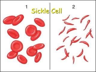
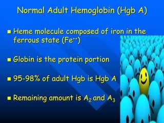
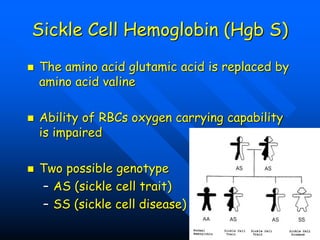
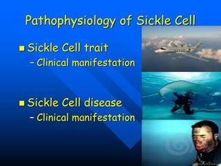
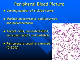
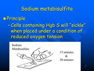
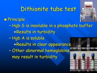
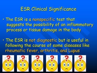
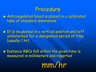
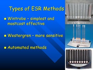
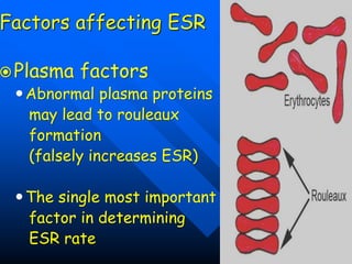
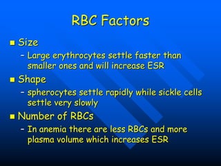
Sickle Cell
1
Genetics Review
Chromosomes
Genes
Loci
Genotype
2
Normal Adult Hemoglobin (Hgb A)
 Heme molecule composed of iron in the
ferrous state (Fe++)
 Globin is the protein portion
 95-98% of adult Hgb is Hgb A
 Remaining amount is A2 and A3
3
Sickle Cell Hemoglobin (Hgb S)
 The amino acid glutamic acid is replaced by
amino acid valine
 Ability of RBCs oxygen carrying capability
is impaired
 Two possible genotype
– AS (sickle cell trait)
– SS (sickle cell disease)
4
Pathophysiology of Sickle Cell
 Sickle Cell trait
– Clinical manifestation
 Sickle Cell disease
– Clinical manifestation
5
Peripheral Blood Picture
 Varying number of sickled forms
 Marked anisocytosis, poikilocytosis,
and polychromasia
 Target cells, nucleated RBCs,
increased WBCs and platelets
 Reticulocyte count is elevated:
(5-25%)
6
Sodium metabisulfite
 Principle
– Cells containing Hgb S will “sickle”
when placed under a condition of
reduced oxygen tension
Sodium
Metabisulfate
15 minutes
&
30 minutes
7
Dithionite tube test
 Principle
– Hgb S is insoluble in a phosphate buffer
»Results in turbidity
– Hgb A is soluble
»Results in clear appearance
– Other abnormal hemoglobins
may result in turbidity
8
ESR Clinical Significance
• The ESR is a nonspecific test that
suggests the possibility of an inflammatory
process or tissue damage in the body
• The ESR is not diagnostic but is useful in
following the course of some diseases like
rheumatic fever, arthritis, and Lupus
Principle
 Based on principle of
sedimentation where solids settle
to the bottom of a liquid
 A known quantity of
anticoagulated blood is left
undisturbed, the RBCs separate
from the plasma and fall to the
bottom of the container
Procedure
 Anticoagulated blood is placed in a calibrated
tube of standard dimensions
 It is incubated in a vertical position and left
undisturbed for a designated period of time
(usually 1 hr)
 Distance RBCs fall within the given time is
measured in millimeters and reported
mm/hr
Types of ESR Methods
 Wintrobe – simplest and
mostcost effective
 Westergren – more sensitive
 Automated methods
Factors affecting ESR
Plasma factors
Abnormal plasma proteins
may lead to rouleaux
formation
(falsely increases ESR)
The single most important
factor in determining
ESR rate
RBC Factors
 Size
– Large erythrocytes settle faster than
smaller ones and will increase ESR
 Shape
– spherocytes settle rapidly while sickle cells
settle very slowly
 Number of RBCs
– In anemia there are less RBCs and more
plasma volume which increases ESR
Technical Factors
 Tilting
 Noise and vibration
 Temperature
 Test set up within 2hrs of blood collection
 ESR tube
 Well mixed sample
 Air bubbles
 Accurately timed

methods used for testing sickling test .ppt

  • 1.
  • 2.
  • 3.
    Normal Adult Hemoglobin(Hgb A)  Heme molecule composed of iron in the ferrous state (Fe++)  Globin is the protein portion  95-98% of adult Hgb is Hgb A  Remaining amount is A2 and A3 3
  • 4.
    Sickle Cell Hemoglobin(Hgb S)  The amino acid glutamic acid is replaced by amino acid valine  Ability of RBCs oxygen carrying capability is impaired  Two possible genotype – AS (sickle cell trait) – SS (sickle cell disease) 4
  • 5.
    Pathophysiology of SickleCell  Sickle Cell trait – Clinical manifestation  Sickle Cell disease – Clinical manifestation 5
  • 6.
    Peripheral Blood Picture Varying number of sickled forms  Marked anisocytosis, poikilocytosis, and polychromasia  Target cells, nucleated RBCs, increased WBCs and platelets  Reticulocyte count is elevated: (5-25%) 6
  • 7.
    Sodium metabisulfite  Principle –Cells containing Hgb S will “sickle” when placed under a condition of reduced oxygen tension Sodium Metabisulfate 15 minutes & 30 minutes 7
  • 8.
    Dithionite tube test Principle – Hgb S is insoluble in a phosphate buffer »Results in turbidity – Hgb A is soluble »Results in clear appearance – Other abnormal hemoglobins may result in turbidity 8
  • 9.
    ESR Clinical Significance •The ESR is a nonspecific test that suggests the possibility of an inflammatory process or tissue damage in the body • The ESR is not diagnostic but is useful in following the course of some diseases like rheumatic fever, arthritis, and Lupus
  • 10.
    Principle  Based onprinciple of sedimentation where solids settle to the bottom of a liquid  A known quantity of anticoagulated blood is left undisturbed, the RBCs separate from the plasma and fall to the bottom of the container
  • 11.
    Procedure  Anticoagulated bloodis placed in a calibrated tube of standard dimensions  It is incubated in a vertical position and left undisturbed for a designated period of time (usually 1 hr)  Distance RBCs fall within the given time is measured in millimeters and reported mm/hr
  • 12.
    Types of ESRMethods  Wintrobe – simplest and mostcost effective  Westergren – more sensitive  Automated methods
  • 13.
    Factors affecting ESR Plasmafactors Abnormal plasma proteins may lead to rouleaux formation (falsely increases ESR) The single most important factor in determining ESR rate
  • 14.
    RBC Factors  Size –Large erythrocytes settle faster than smaller ones and will increase ESR  Shape – spherocytes settle rapidly while sickle cells settle very slowly  Number of RBCs – In anemia there are less RBCs and more plasma volume which increases ESR
  • 15.
    Technical Factors  Tilting Noise and vibration  Temperature  Test set up within 2hrs of blood collection  ESR tube  Well mixed sample  Air bubbles  Accurately timed