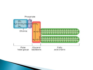
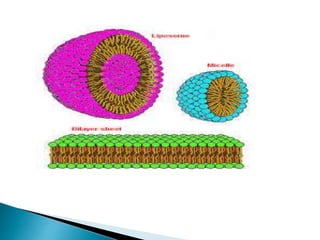
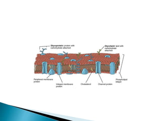
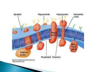
Bio-membrane
Bibhudatta Mohanty
B.Tech Biotechnology
VIT University
 Introduction-
 Phospholipids Associate Non-covalently to
Form the Basic bi-layer Structure of Bio-
membranes.
 Bio membranes are large flexible sheets that
serve as the boundaries of cells and their
intracellular organelles and form the outer
surfaces of some viruses.
 Unlike the proteins, nucleic acids, and
polysaccharides, membranes are assembled
by the non-covalent association of their
component building block
 The primary building blocks of all bio-membranes
are phospholipids, whose physical properties are
responsible for the formation of the sheet like
structure of membranes.
 Phospholipids consist of two long-chain, non-polar
fatty acyl groups linked (usually by an ester bond)
to small, highly polar groups, including a
phosphate.
 In most phospholipids found in membranes, the
phosphate group is esterified to a hydroxyl group
on another hydrophilic compound.
 In phosphatidylcholine, for example, choline is
attached to the phosphate.
 The negative charge on the phosphate as well as
the charged or polar groups esterified to it can
interact strongly with water .
 The phosphate and its associated esterified group,
the “head” group of a phospholipid, is hydrophilic,
whereas the fatty acyl chains, the “tails,” are
hydrophobic.
 The amphipathic nature of phospholipids, which
governs their interactions, is critical to the
structure of bio-membranes.
 When a suspension of phospholipids is
mechanically dispersed in aqueous solution, the
phospholipids aggregate into one of three forms:
spherical Micelles and Liposomes and sheetlike,
two-molecule-thick phospholipid bi-layers.
 The type of structure formed by a pure
phospholipid or a mixture of phospholipids
depends on several factors, including the length of
the fatty acyl chains, their degree of saturation,
and temperature
 In all three structures, the hydrophobic effect
causes the fatty acyl chains to aggregate and
exclude water molecules from the “core.”
 Under suitable conditions, phospholipids of the
composition present in cells spontaneously form
symmetric phospholipid bi-layers .
 Each phospholipid layer in this lamellar structure
is called a leaflet .
 The fatty acyl chains in each leaflet minimize
contact with water by aligning themselves tightly
together in the center of the bilayer, forming a
hydrophobic core that is about 3 nm thick .
 The close packing of these nonpolar tails is
stabilized by the hydrophobic effect and van der
Waals interactions between them. Ionic and
hydrogen bonds stabilize the interaction of the
phospholipid polar head groups with one another
and with water.
 Because of their hydrophobic core, bilayers are
virtually impermeable to salts, sugars, and most
other small hydrophilic molecules .
 The phospholipid bilayer is the basic structural unit
of nearly all biological membranes; thus, although
they contain other molecules (e.g., cholesterol,
glycolipids, proteins), biomembranes have a
hydrophobic core that separates two aqueous
solutions and acts as a permeability barrier
 First, the hydrophobic core is an impermeable
barrier that prevents the diffusion of water-soluble
(hydrophilic) solutes across the membrane.
Importantly, this simple barrier function is
modulated by the presence of membrane proteins
that mediate the transport of specific molecules
across this otherwise impermeable bilayer.
 The second property of the bilayer is its stability.
The bilayer structure is maintained by hydrophobic
and van der Waals interactions between the lipid
chains. Even though the exterior aqueous
environment can vary widely in ionic strength and
pH, the bilayer has the strength to retain its
characteristic architecture
A typical biomembrane is assembled from –
1) phosphoglycerides
2)Sphingolipids
3)steroids
All three classes of lipids are amphipathic
molecules having a polar (hydrophilic) head group
and hydrophobic tail.
 I. Phosphoglycerides, the most abundant class of
lipids in most membranes, are derivatives of
glycerol 3-phosphate.
 A typical phosphoglyceride molecule consists of a
hydrophobic tail composed of two fatty acyl chains
esterified to the two hydroxyl groups in glycerol
phosphate and a polar head group attached to the
phosphate group.
 The two fatty acyl chains may differ in the number
of carbons that they contain (commonly 16 or 18)
and their degree of saturation (0, 1, or 2 double
bonds). A phosphogyceride is classified according
to the nature of its head group.
 Phosphatidylcholines, the most abundant
phospholipids in the plasma membrane.
 All of these compounds are derived from
sphingosine, an amino alcohol with a long
hydrocarbon chain, and contain a long-chain fatty
acid attached to the sphingosine amino group.
 In sphingomyelin, the most abundant sphingolipid,
phosphocholine is attached to the terminal
hydroxyl group of sphingosine . Thus
sphingomyelin is a phospholipid, and its overall
structure is quite similar to that of
phosphatidylcholine.
 Other sphingolipids are amphipathic glycolipids
whose polar head groups are sugars.
Glucosylcerebroside, the simplest
glycosphingolipid, contains a single glucose unit
attached to sphingosine. In the complex
glycosphingolipids called gangliosides, one or two
branched sugar chains containing sialic acid
groups are attached to sphingosine. Glycolipids
constitute 2– 10 percent of the total lipid in plasma
membranes; they are most abundant in nervous
tissue.
 The basic structure of steroids is a four-ring
hydrocarbon.
 Cholesterol, the major steroidal constituent of
animal tissues, has a hydroxyl substituent on one
ring . Although cholesterol is almost entirely
hydrocarbon in composition, it is amphipathic
because its hydroxyl group can interact with water.
 Cholesterol is especially abundant in the plasma
membranes of mammalian cells but is absent from
most prokaryotic cells.
 As much as 30–50 percent of the lipids in plant
plasma membranes consist of certain steroids
 Membrane proteins are defined by their location
within or at the surface of a phospholipid bilayer.
Although every biological membrane has the
same basic bilayer structure, the proteins
associated with a particular membrane are
responsible for its distinctive activities .
 The density and complement of proteins
associated with biomembranes vary, depending
on cell type and subcellular location. For
example, the inner mitochondrial membrane is
76 percent protein; the myelin membrane, only
18 percent.
Biomembranes: Protein Components and Basic Functions
 The lipid bilayer presents a unique two-
dimensional hydrophobic environment for
membrane proteins.
 Some proteins are buried within the lipid-rich
bilayer; other proteins are associated with the
exoplasmic or cytosolic leaflet of the bilayer.
 Protein domains on the extracellular surface of
the plasma membrane generally bind to other
molecules, including external signaling proteins,
ions, and small metabolites (e.g., glucose, fatty
acids), and to adhesion molecules on other cells
or in the external environment
 Domains within the plasma membrane, particularly
those that form channels and pores, move
molecules in and out of cells.
 Domains lying along the cytosolic face of the
plasma membrane have a wide range of functions,
from anchoring cytoskeletal proteins to the
membrane to triggering intracellular signaling
pathways .
 In many cases, the function of a membrane
protein and the topology of its polypeptide chain in
the membrane can be predicted on the basis of its
homology with another, well characterized protein
 Membrane proteins can be classified into three
categories
 1) integral,
 2)lipid-anchored,
 3) peripheral
 on the basis of the nature of the membrane–
protein interactions
 Integral membrane proteins, also called
transmembrane proteins, span a phospholipid bilayer
and are built of three segments.
 The cytosolic and exoplasmic domains have
hydrophilic exterior surfaces that interact with the
aqueous solutions on the cytosolic and exoplasmic
faces of the membrane.
 These domains resemble other water-soluble proteins
in their amino acid composition and structure. In
contrast, the 3-nm-thick membrane-spanning domain
contains many hydrophobic amino acids whose side
chains protrude outward and interact with the
hydrocarbon core of the phospholipid bilayer
 In all transmembrane proteins examined to date,
the membrane-spanning domains consist of one
or more helices or of multiple strands. In addition,
most transmembrane proteins are glycosylated
with a complex branched sugar group attached to
one or several amino acid side chains.
 Invariably these sugar chains are localized to the
exoplasmic domains.
 Lipid-anchored membrane proteins are bound
covalently to one or more lipid molecules .
 The hydrophobic carbon chain of the attached
lipid is embedded in one leaflet of the membrane
and anchors the protein to the membrane.
 The polypeptide chain itself does not enter the
phospholipid bilayer
 Peripheral membrane proteins do not interact with
the hydrophobic core of the phospholipid bilayer.
Instead they are usually bound to the membrane
indirectly by interactions with integral membrane
proteins or directly by interactions with lipid head
groups. Peripheral proteins are localized to either
the cytosolic or the exoplasmic face of the plasma
membrane.
 The most common type of IMP is
the transmembrane protein (TM), which
spans the entire biological
membrane. Single-pass membrane
proteins cross the membrane only once,
while multi-pass membrane proteins
weave in and out, crossing several times.
 Single pass TM proteins can be categorized as Type I,
which are positioned such that their carboxyl-terminus
is towards the cytosol, or Type II, which have their
amino-terminus towards the cytosol.
 Type III proteins have multiple transmembrane
domains in a single polypeptide, while type IV consists
of several different polypeptides assembled together in
a channel through the membrane.
 Type V proteins are anchored to the lipid bilayer
through covalently linked lipids. Finally Type VI
proteins have both a transmembrane domains and lipid
anchors
 Example of single pass membrane protein is
Glycophorin A
 Example of multi pass membrane protein is
Bacteriorhodopsin.
Glycophorin A
Biomembrane basic
Biomembrane basic

Biomembrane basic

  • 1.
  • 2.
     Introduction-  PhospholipidsAssociate Non-covalently to Form the Basic bi-layer Structure of Bio- membranes.  Bio membranes are large flexible sheets that serve as the boundaries of cells and their intracellular organelles and form the outer surfaces of some viruses.  Unlike the proteins, nucleic acids, and polysaccharides, membranes are assembled by the non-covalent association of their component building block
  • 3.
     The primarybuilding blocks of all bio-membranes are phospholipids, whose physical properties are responsible for the formation of the sheet like structure of membranes.  Phospholipids consist of two long-chain, non-polar fatty acyl groups linked (usually by an ester bond) to small, highly polar groups, including a phosphate.
  • 5.
     In mostphospholipids found in membranes, the phosphate group is esterified to a hydroxyl group on another hydrophilic compound.  In phosphatidylcholine, for example, choline is attached to the phosphate.  The negative charge on the phosphate as well as the charged or polar groups esterified to it can interact strongly with water .  The phosphate and its associated esterified group, the “head” group of a phospholipid, is hydrophilic, whereas the fatty acyl chains, the “tails,” are hydrophobic.
  • 6.
     The amphipathicnature of phospholipids, which governs their interactions, is critical to the structure of bio-membranes.  When a suspension of phospholipids is mechanically dispersed in aqueous solution, the phospholipids aggregate into one of three forms: spherical Micelles and Liposomes and sheetlike, two-molecule-thick phospholipid bi-layers.  The type of structure formed by a pure phospholipid or a mixture of phospholipids depends on several factors, including the length of the fatty acyl chains, their degree of saturation, and temperature
  • 8.
     In allthree structures, the hydrophobic effect causes the fatty acyl chains to aggregate and exclude water molecules from the “core.”  Under suitable conditions, phospholipids of the composition present in cells spontaneously form symmetric phospholipid bi-layers .  Each phospholipid layer in this lamellar structure is called a leaflet .
  • 9.
     The fattyacyl chains in each leaflet minimize contact with water by aligning themselves tightly together in the center of the bilayer, forming a hydrophobic core that is about 3 nm thick .  The close packing of these nonpolar tails is stabilized by the hydrophobic effect and van der Waals interactions between them. Ionic and hydrogen bonds stabilize the interaction of the phospholipid polar head groups with one another and with water.
  • 10.
     Because oftheir hydrophobic core, bilayers are virtually impermeable to salts, sugars, and most other small hydrophilic molecules .  The phospholipid bilayer is the basic structural unit of nearly all biological membranes; thus, although they contain other molecules (e.g., cholesterol, glycolipids, proteins), biomembranes have a hydrophobic core that separates two aqueous solutions and acts as a permeability barrier
  • 11.
     First, thehydrophobic core is an impermeable barrier that prevents the diffusion of water-soluble (hydrophilic) solutes across the membrane. Importantly, this simple barrier function is modulated by the presence of membrane proteins that mediate the transport of specific molecules across this otherwise impermeable bilayer.  The second property of the bilayer is its stability. The bilayer structure is maintained by hydrophobic and van der Waals interactions between the lipid chains. Even though the exterior aqueous environment can vary widely in ionic strength and pH, the bilayer has the strength to retain its characteristic architecture
  • 12.
    A typical biomembraneis assembled from – 1) phosphoglycerides 2)Sphingolipids 3)steroids All three classes of lipids are amphipathic molecules having a polar (hydrophilic) head group and hydrophobic tail.
  • 13.
     I. Phosphoglycerides,the most abundant class of lipids in most membranes, are derivatives of glycerol 3-phosphate.  A typical phosphoglyceride molecule consists of a hydrophobic tail composed of two fatty acyl chains esterified to the two hydroxyl groups in glycerol phosphate and a polar head group attached to the phosphate group.  The two fatty acyl chains may differ in the number of carbons that they contain (commonly 16 or 18) and their degree of saturation (0, 1, or 2 double bonds). A phosphogyceride is classified according to the nature of its head group.  Phosphatidylcholines, the most abundant phospholipids in the plasma membrane.
  • 15.
     All ofthese compounds are derived from sphingosine, an amino alcohol with a long hydrocarbon chain, and contain a long-chain fatty acid attached to the sphingosine amino group.  In sphingomyelin, the most abundant sphingolipid, phosphocholine is attached to the terminal hydroxyl group of sphingosine . Thus sphingomyelin is a phospholipid, and its overall structure is quite similar to that of phosphatidylcholine.
  • 16.
     Other sphingolipidsare amphipathic glycolipids whose polar head groups are sugars. Glucosylcerebroside, the simplest glycosphingolipid, contains a single glucose unit attached to sphingosine. In the complex glycosphingolipids called gangliosides, one or two branched sugar chains containing sialic acid groups are attached to sphingosine. Glycolipids constitute 2– 10 percent of the total lipid in plasma membranes; they are most abundant in nervous tissue.
  • 18.
     The basicstructure of steroids is a four-ring hydrocarbon.  Cholesterol, the major steroidal constituent of animal tissues, has a hydroxyl substituent on one ring . Although cholesterol is almost entirely hydrocarbon in composition, it is amphipathic because its hydroxyl group can interact with water.  Cholesterol is especially abundant in the plasma membranes of mammalian cells but is absent from most prokaryotic cells.  As much as 30–50 percent of the lipids in plant plasma membranes consist of certain steroids
  • 20.
     Membrane proteinsare defined by their location within or at the surface of a phospholipid bilayer. Although every biological membrane has the same basic bilayer structure, the proteins associated with a particular membrane are responsible for its distinctive activities .  The density and complement of proteins associated with biomembranes vary, depending on cell type and subcellular location. For example, the inner mitochondrial membrane is 76 percent protein; the myelin membrane, only 18 percent. Biomembranes: Protein Components and Basic Functions
  • 21.
     The lipidbilayer presents a unique two- dimensional hydrophobic environment for membrane proteins.  Some proteins are buried within the lipid-rich bilayer; other proteins are associated with the exoplasmic or cytosolic leaflet of the bilayer.  Protein domains on the extracellular surface of the plasma membrane generally bind to other molecules, including external signaling proteins, ions, and small metabolites (e.g., glucose, fatty acids), and to adhesion molecules on other cells or in the external environment
  • 22.
     Domains withinthe plasma membrane, particularly those that form channels and pores, move molecules in and out of cells.  Domains lying along the cytosolic face of the plasma membrane have a wide range of functions, from anchoring cytoskeletal proteins to the membrane to triggering intracellular signaling pathways .  In many cases, the function of a membrane protein and the topology of its polypeptide chain in the membrane can be predicted on the basis of its homology with another, well characterized protein
  • 24.
     Membrane proteinscan be classified into three categories  1) integral,  2)lipid-anchored,  3) peripheral  on the basis of the nature of the membrane– protein interactions
  • 25.
     Integral membraneproteins, also called transmembrane proteins, span a phospholipid bilayer and are built of three segments.  The cytosolic and exoplasmic domains have hydrophilic exterior surfaces that interact with the aqueous solutions on the cytosolic and exoplasmic faces of the membrane.  These domains resemble other water-soluble proteins in their amino acid composition and structure. In contrast, the 3-nm-thick membrane-spanning domain contains many hydrophobic amino acids whose side chains protrude outward and interact with the hydrocarbon core of the phospholipid bilayer
  • 27.
     In alltransmembrane proteins examined to date, the membrane-spanning domains consist of one or more helices or of multiple strands. In addition, most transmembrane proteins are glycosylated with a complex branched sugar group attached to one or several amino acid side chains.  Invariably these sugar chains are localized to the exoplasmic domains.
  • 28.
     Lipid-anchored membraneproteins are bound covalently to one or more lipid molecules .  The hydrophobic carbon chain of the attached lipid is embedded in one leaflet of the membrane and anchors the protein to the membrane.  The polypeptide chain itself does not enter the phospholipid bilayer
  • 30.
     Peripheral membraneproteins do not interact with the hydrophobic core of the phospholipid bilayer. Instead they are usually bound to the membrane indirectly by interactions with integral membrane proteins or directly by interactions with lipid head groups. Peripheral proteins are localized to either the cytosolic or the exoplasmic face of the plasma membrane.
  • 31.
     The mostcommon type of IMP is the transmembrane protein (TM), which spans the entire biological membrane. Single-pass membrane proteins cross the membrane only once, while multi-pass membrane proteins weave in and out, crossing several times.
  • 32.
     Single passTM proteins can be categorized as Type I, which are positioned such that their carboxyl-terminus is towards the cytosol, or Type II, which have their amino-terminus towards the cytosol.  Type III proteins have multiple transmembrane domains in a single polypeptide, while type IV consists of several different polypeptides assembled together in a channel through the membrane.  Type V proteins are anchored to the lipid bilayer through covalently linked lipids. Finally Type VI proteins have both a transmembrane domains and lipid anchors
  • 33.
     Example ofsingle pass membrane protein is Glycophorin A  Example of multi pass membrane protein is Bacteriorhodopsin.
  • 34.