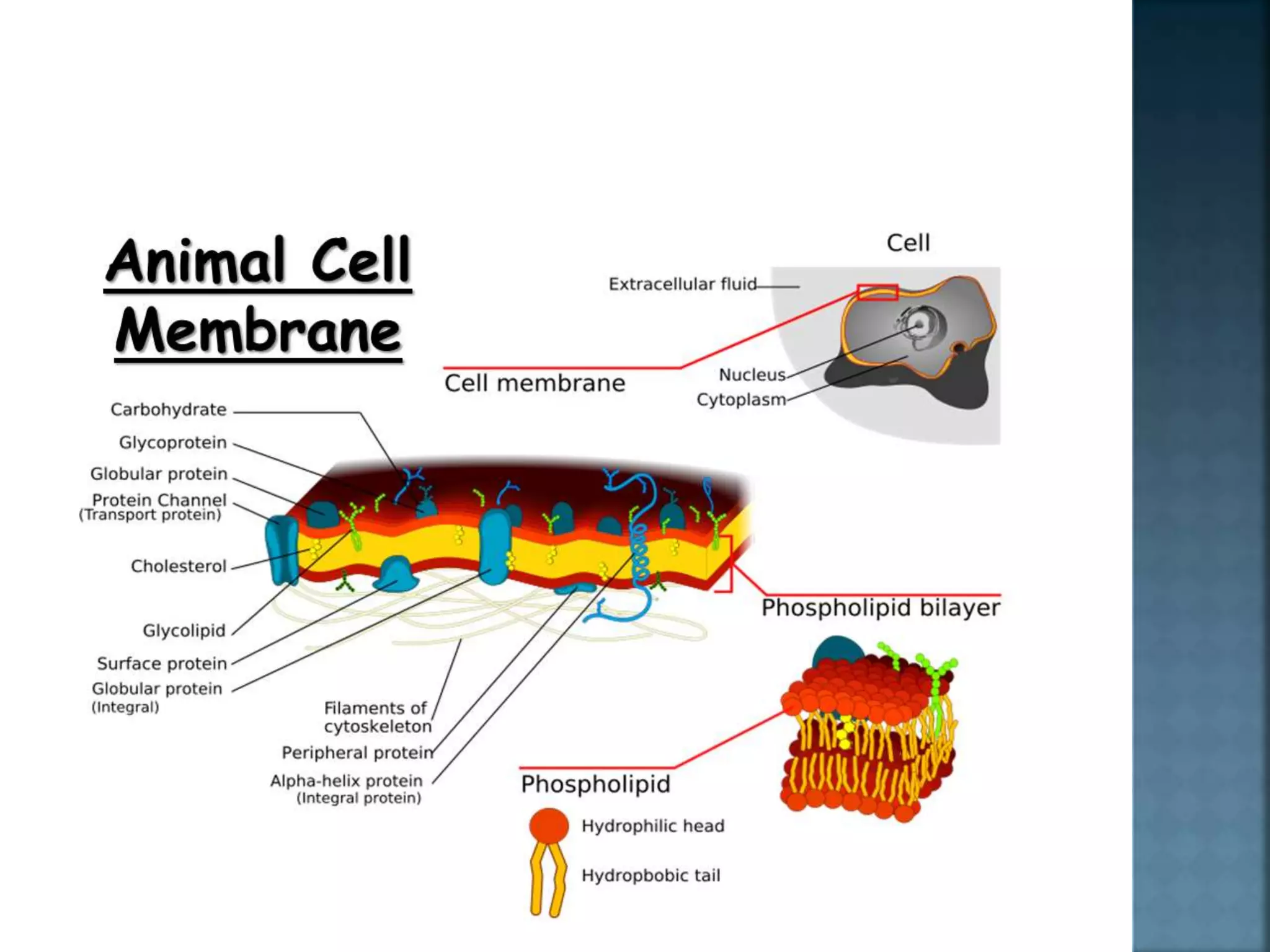
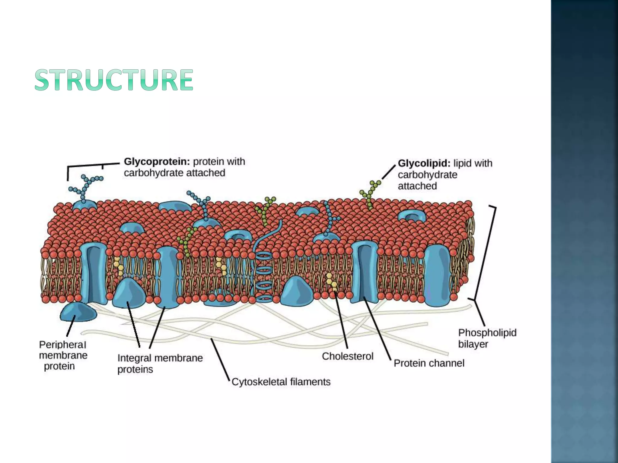
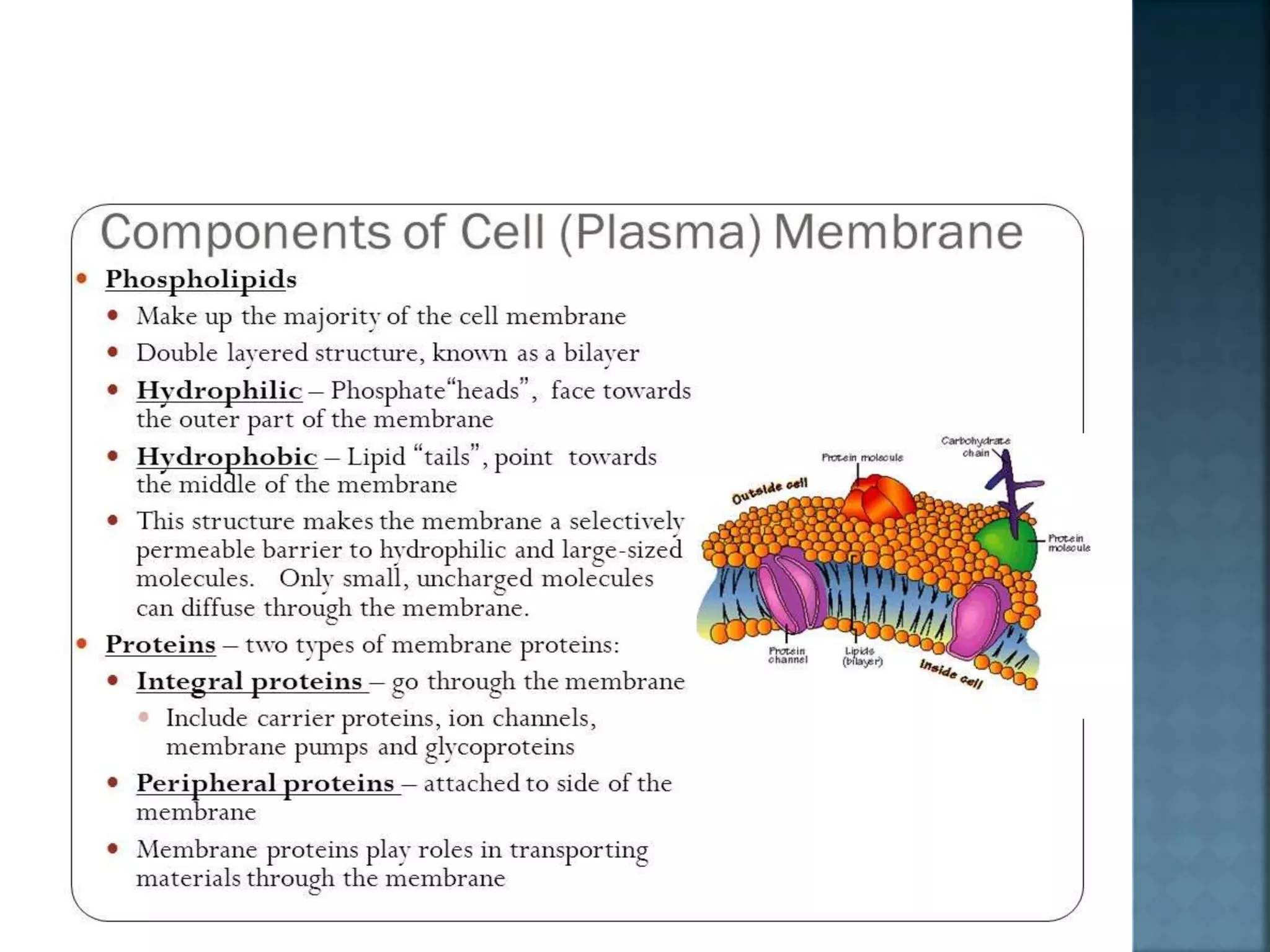
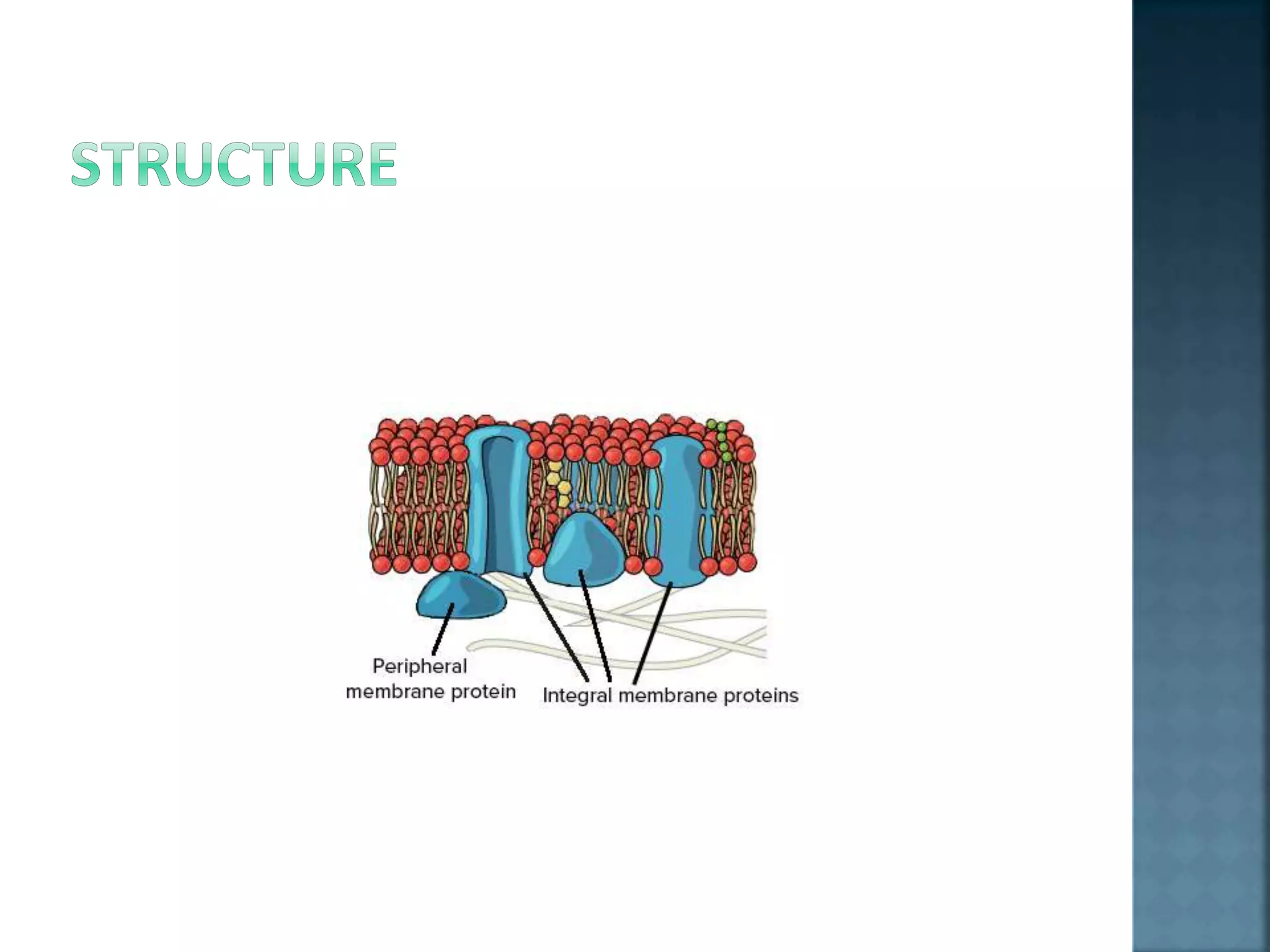
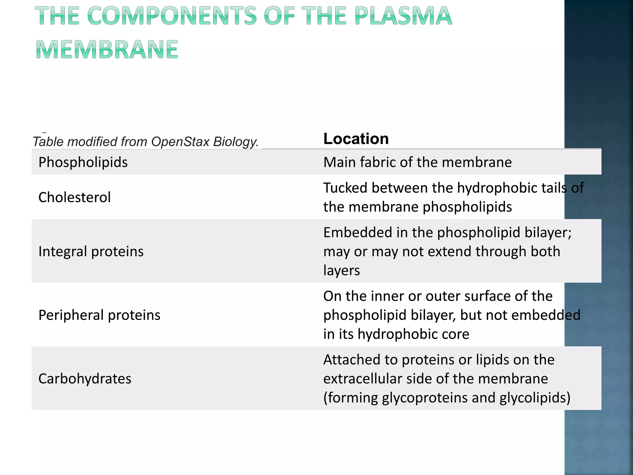
Presented by
Muhammad Akmal BHIKH
 Structure of plasma membrane was proposed as
fluid mosaic model, was first proposed by
S.J.Singer and Garth L. Nicolson in 1972 to explain
the structure of the plasma membrane.
 The fluid mosaic model explains various
observations regarding the structure of
functional cell membranes. According to this
model, there is a lipid bilayer in which the protein
molecules are embedded.
 Each cell of your body is encased in a tiny bubble of
membrane.
 This membrane has about the consistency of...salad
oil.
 The plasma membrane not only defines the borders of
the cell, but also allows the cell to interact with its
environment in a controlled way.
 Cells must be able to exclude, take in, and excrete
various substances, all in specific amounts. In addition,
they must able to communicate with other cells,
identifying themselves and sharing information.
 To perform these roles, the plasma membrane needs lipids,
which make a semi-permeable barrier between the cell and
its environment.
 It also needs proteins, which are involved in cross-
membrane transport and cell communication, and
carbohydrates (sugars and sugar chains), which decorate
both the proteins and lipids and help cells recognize each
other.
 Here, we’ll take a closer look at the different components
of the plasma membrane, examining their roles, their
diversity, and how they work together to make a flexible,
sensitive, and secure boundary around the cell.
 The currently accepted model for the structure of the
plasma membrane, called the fluid mosaic model, was
first proposed in 1972.
 This model has evolved over time, but it still provides
a good basic description of the structure and behavior
of membranes in many cells.
 According to the fluid mosaic model, the plasma
membrane is a mosaic of components—primarily,
phospholipids, cholesterol, and proteins—that move
freely and fluidly in the plane of the membrane.
 In other words, a diagram of the membrane is
just a snapshot of a dynamic process in which
phospholipids and proteins are continually
sliding past one another.
 Interestingly enough, this fluidity means that if
you insert a very fine needle into a cell, the
membrane will simply part to flow around the
needle; once the needle is removed, the
membrane will flow back together seamlessly.
 The principal components of the plasma membrane are lipids
(phospholipids and cholesterol), proteins, and carbohydrate
groups that are attached to some of the lipids and proteins.
 A phospholipid is a lipid made of glycerol, two fatty acid tails, and
a phosphate-linked head group. Biological membranes usually
involve two layers of phospholipids with their tails pointing
inward, an arrangement called a phospholipid bilayer.
 Cholesterol, another lipid composed of four fused carbon rings, is
found alongside phospholipids in the core of the membrane.
 Membrane proteins may extend partway into the plasma
membrane, cross the membrane entirely, or be loosely attached
to its inside or outside face.
 Carbohydrate groups are present only on the outer surface of the
plasma membrane and are attached to proteins,
forming glycoproteins, or lipids, forming glycolipids.
 The proportions of proteins, lipids, and carbohydrates in the
plasma membrane vary between different types of cells. For a
typical human cell, however, proteins account for about 50
percent of the composition by mass, lipids (of all types) account
for about 40 percent, and the remaining 10 percent comes from
carbohydrates.
 Phospholipids, arranged in a bilayer, make up the basic fabric of the plasma
membrane. They are well-suited for this role because they are amphipathic,
meaning that they have both hydrophilic and hydrophobic regions.
 The hydrophilic, or “water-loving,” portion of a phospholipid is its head, which contains a
negatively charged phosphate group as well as an additional small group (of varying identity,
“R” in the diagram at left), which may also or be charged or polar.
 The hydrophilic heads of phospholipids in a membrane bilayer face outward, contacting the
aqueous (watery) fluid both inside and outside the cell. Since water is a polar molecule, it
readily forms electrostatic (charge-based) interactions with the phospholipid heads.
o The hydrophobic, or “water-fearing,” part of a phospholipid consists of its long, nonpolar fatty
acid tails. The fatty acid tails can easily interact with other nonpolar molecules, but they
interact poorly with water.
 phospholipids aren’t just well-suited to form a
membrane bilayer.
 Instead, this is something they’ll do spontaneously
under the right conditions.
 In water or aqueous solution, phospholipids tend to
arrange themselves with their hydrophobic tails facing
each other and their hydrophilic heads facing out.
 If the phospholipids have small tails, they may form
a micelle (a small, single-layered sphere), while if they
have bulkier tails, they may form a liposome (a hollow
droplet of bilayer membrane).
 Proteins are the second major component of plasma membranes.
There are two main categories of membrane proteins: integral
and peripheral.
 Integral membrane proteins are, as their name suggests,
integrated into the membrane.
 they have at least one hydrophobic region that anchors them to
the hydrophobic core of the phospholipid bilayer.
 Some stick only partway into the membrane, while others
stretch from one side of the membrane to the other and are
exposed on either side^11start superscript, 1, end superscript.
 Proteins that extend all the way across the membrane are
called transmembrane proteins.
 The portions of an integral membrane protein found inside the
membrane are hydrophobic.
 while those that are exposed to the cytoplasm or extracellular
fluid tend to be hydrophilic.
 Transmembrane proteins may cross the membrane just once, or
may have as many as twelve different membrane-spanning
sections.
 A typical membrane-spanning segment consists of 20-25
hydrophobic amino acids arranged in an alpha helix, although not
all transmembrane proteins fit this model.
 Some integral membrane proteins form a channel that allows
ions or other small molecules to pass, as shown below.
 Peripheral membrane proteins are found on
the outside and inside surfaces of membranes,
attached either to integral proteins or to
phospholipids.
 Unlike integral membrane proteins, peripheral
membrane proteins do not stick into the
hydrophobic core of the membrane, and they
tend to be more loosely attached.
 Carbohydrates are the third major component of plasma
membranes. In general, they are found on the outside surface of
cells and are bound either to proteins (forming glycoproteins) or
to lipids (forming glycolipids).
 These carbohydrate chains may consist of 2-60 monosaccharide
units and can be either straight or branched.
 Along with membrane proteins, these carbohydrates form
distinctive cellular markers, sort of like molecular ID badges, that
allow cells to recognize each other.
 These markers are very important in the immune system,
allowing immune cells to differentiate between body cells, which
they shouldn’t attack, and foreign cells or tissues, which they
should.
 The structure of the fatty acid tails of the
phospholipids is important in determining the
properties of the membrane, and in particular, how
fluid it is.
 Saturated fatty acids have no double bonds (are
saturated with hydrogens), so they are relatively
straight. Unsaturated fatty acids, on the other hand,
contain one or more double bonds, often resulting in a
bend or kink. (You can see an example of a bent,
unsaturated tail in the diagram of phospholipid
structure that appears earlier in this article.) The
saturated and unsaturated fatty acid tails of
phospholipids behave differently as temperature
drops:
 At cooler temperatures, the straight tails of
saturated fatty acids can pack tightly together,
making a dense and fairly rigid membrane.
 Phospholipids with unsaturated fatty acid tails
cannot pack together as tightly because of the
bent structure of the tails. Because of this, a
membrane containing unsaturated phospholipids
will stay fluid at lower temperatures than a
membrane made of saturated ones.
 In addition to phospholipids, animals have an
additional membrane component that helps to
maintain fluidity. Cholesterol, another type of
lipid that is embedded among the phospholipids
of the membrane, helps to minimize the effects of
temperature on fluidity.
 At low temperatures, cholesterol increases fluidity by
keeping phospholipids from packing tightly together.
 while at high temperatures, it actually reduces
fluidity^{3,4}3,4start superscript, 3, comma, 4, end
superscript. In this way, cholesterol expands the range of
temperatures at which a membrane maintains a functional,
healthy fluidity.
Component Location
Phospholipids Main fabric of the membrane
Cholesterol
Tucked between the hydrophobic tails of
the membrane phospholipids
Integral proteins
Embedded in the phospholipid bilayer;
may or may not extend through both
layers
Peripheral proteins
On the inner or outer surface of the
phospholipid bilayer, but not embedded
in its hydrophobic core
Carbohydrates
Attached to proteins or lipids on the
extracellular side of the membrane
(forming glycoproteins and glycolipids)
Table modified from OpenStax Biology.Component Location
Phospholipids Main fabric of the membrane
Cholesterol
Tucked between the hydrophobic tails of
the membrane phospholipids
Integral proteins
Embedded in the phospholipid bilayer;
may or may not extend through both
layers
Peripheral proteins
On the inner or outer surface of the
phospholipid bilayer, but not embedded
in its hydrophobic core
Carbohydrates
Attached to proteins or lipids on the
extracellular side of the membrane
(forming glycoproteins and glycolipids)
Table modified from OpenStax Biology.
THANKS

Structure, composition of plasma membrane by muhammad akmal bhikh

  • 1.
  • 2.
     Structure ofplasma membrane was proposed as fluid mosaic model, was first proposed by S.J.Singer and Garth L. Nicolson in 1972 to explain the structure of the plasma membrane.  The fluid mosaic model explains various observations regarding the structure of functional cell membranes. According to this model, there is a lipid bilayer in which the protein molecules are embedded.
  • 3.
     Each cellof your body is encased in a tiny bubble of membrane.  This membrane has about the consistency of...salad oil.  The plasma membrane not only defines the borders of the cell, but also allows the cell to interact with its environment in a controlled way.  Cells must be able to exclude, take in, and excrete various substances, all in specific amounts. In addition, they must able to communicate with other cells, identifying themselves and sharing information.
  • 4.
     To performthese roles, the plasma membrane needs lipids, which make a semi-permeable barrier between the cell and its environment.  It also needs proteins, which are involved in cross- membrane transport and cell communication, and carbohydrates (sugars and sugar chains), which decorate both the proteins and lipids and help cells recognize each other.  Here, we’ll take a closer look at the different components of the plasma membrane, examining their roles, their diversity, and how they work together to make a flexible, sensitive, and secure boundary around the cell.
  • 5.
     The currentlyaccepted model for the structure of the plasma membrane, called the fluid mosaic model, was first proposed in 1972.  This model has evolved over time, but it still provides a good basic description of the structure and behavior of membranes in many cells.  According to the fluid mosaic model, the plasma membrane is a mosaic of components—primarily, phospholipids, cholesterol, and proteins—that move freely and fluidly in the plane of the membrane.
  • 7.
     In otherwords, a diagram of the membrane is just a snapshot of a dynamic process in which phospholipids and proteins are continually sliding past one another.  Interestingly enough, this fluidity means that if you insert a very fine needle into a cell, the membrane will simply part to flow around the needle; once the needle is removed, the membrane will flow back together seamlessly.
  • 9.
     The principalcomponents of the plasma membrane are lipids (phospholipids and cholesterol), proteins, and carbohydrate groups that are attached to some of the lipids and proteins.  A phospholipid is a lipid made of glycerol, two fatty acid tails, and a phosphate-linked head group. Biological membranes usually involve two layers of phospholipids with their tails pointing inward, an arrangement called a phospholipid bilayer.  Cholesterol, another lipid composed of four fused carbon rings, is found alongside phospholipids in the core of the membrane.
  • 10.
     Membrane proteinsmay extend partway into the plasma membrane, cross the membrane entirely, or be loosely attached to its inside or outside face.  Carbohydrate groups are present only on the outer surface of the plasma membrane and are attached to proteins, forming glycoproteins, or lipids, forming glycolipids.  The proportions of proteins, lipids, and carbohydrates in the plasma membrane vary between different types of cells. For a typical human cell, however, proteins account for about 50 percent of the composition by mass, lipids (of all types) account for about 40 percent, and the remaining 10 percent comes from carbohydrates.
  • 12.
     Phospholipids, arrangedin a bilayer, make up the basic fabric of the plasma membrane. They are well-suited for this role because they are amphipathic, meaning that they have both hydrophilic and hydrophobic regions.
  • 13.
     The hydrophilic,or “water-loving,” portion of a phospholipid is its head, which contains a negatively charged phosphate group as well as an additional small group (of varying identity, “R” in the diagram at left), which may also or be charged or polar.  The hydrophilic heads of phospholipids in a membrane bilayer face outward, contacting the aqueous (watery) fluid both inside and outside the cell. Since water is a polar molecule, it readily forms electrostatic (charge-based) interactions with the phospholipid heads. o The hydrophobic, or “water-fearing,” part of a phospholipid consists of its long, nonpolar fatty acid tails. The fatty acid tails can easily interact with other nonpolar molecules, but they interact poorly with water.
  • 15.
     phospholipids aren’tjust well-suited to form a membrane bilayer.  Instead, this is something they’ll do spontaneously under the right conditions.  In water or aqueous solution, phospholipids tend to arrange themselves with their hydrophobic tails facing each other and their hydrophilic heads facing out.  If the phospholipids have small tails, they may form a micelle (a small, single-layered sphere), while if they have bulkier tails, they may form a liposome (a hollow droplet of bilayer membrane).
  • 16.
     Proteins arethe second major component of plasma membranes. There are two main categories of membrane proteins: integral and peripheral.  Integral membrane proteins are, as their name suggests, integrated into the membrane.  they have at least one hydrophobic region that anchors them to the hydrophobic core of the phospholipid bilayer.  Some stick only partway into the membrane, while others stretch from one side of the membrane to the other and are exposed on either side^11start superscript, 1, end superscript.  Proteins that extend all the way across the membrane are called transmembrane proteins.
  • 18.
     The portionsof an integral membrane protein found inside the membrane are hydrophobic.  while those that are exposed to the cytoplasm or extracellular fluid tend to be hydrophilic.  Transmembrane proteins may cross the membrane just once, or may have as many as twelve different membrane-spanning sections.  A typical membrane-spanning segment consists of 20-25 hydrophobic amino acids arranged in an alpha helix, although not all transmembrane proteins fit this model.  Some integral membrane proteins form a channel that allows ions or other small molecules to pass, as shown below.
  • 20.
     Peripheral membraneproteins are found on the outside and inside surfaces of membranes, attached either to integral proteins or to phospholipids.  Unlike integral membrane proteins, peripheral membrane proteins do not stick into the hydrophobic core of the membrane, and they tend to be more loosely attached.
  • 21.
     Carbohydrates arethe third major component of plasma membranes. In general, they are found on the outside surface of cells and are bound either to proteins (forming glycoproteins) or to lipids (forming glycolipids).  These carbohydrate chains may consist of 2-60 monosaccharide units and can be either straight or branched.  Along with membrane proteins, these carbohydrates form distinctive cellular markers, sort of like molecular ID badges, that allow cells to recognize each other.  These markers are very important in the immune system, allowing immune cells to differentiate between body cells, which they shouldn’t attack, and foreign cells or tissues, which they should.
  • 22.
     The structureof the fatty acid tails of the phospholipids is important in determining the properties of the membrane, and in particular, how fluid it is.  Saturated fatty acids have no double bonds (are saturated with hydrogens), so they are relatively straight. Unsaturated fatty acids, on the other hand, contain one or more double bonds, often resulting in a bend or kink. (You can see an example of a bent, unsaturated tail in the diagram of phospholipid structure that appears earlier in this article.) The saturated and unsaturated fatty acid tails of phospholipids behave differently as temperature drops:
  • 23.
     At coolertemperatures, the straight tails of saturated fatty acids can pack tightly together, making a dense and fairly rigid membrane.  Phospholipids with unsaturated fatty acid tails cannot pack together as tightly because of the bent structure of the tails. Because of this, a membrane containing unsaturated phospholipids will stay fluid at lower temperatures than a membrane made of saturated ones.
  • 24.
     In additionto phospholipids, animals have an additional membrane component that helps to maintain fluidity. Cholesterol, another type of lipid that is embedded among the phospholipids of the membrane, helps to minimize the effects of temperature on fluidity.
  • 25.
     At lowtemperatures, cholesterol increases fluidity by keeping phospholipids from packing tightly together.  while at high temperatures, it actually reduces fluidity^{3,4}3,4start superscript, 3, comma, 4, end superscript. In this way, cholesterol expands the range of temperatures at which a membrane maintains a functional, healthy fluidity.
  • 26.
    Component Location Phospholipids Mainfabric of the membrane Cholesterol Tucked between the hydrophobic tails of the membrane phospholipids Integral proteins Embedded in the phospholipid bilayer; may or may not extend through both layers Peripheral proteins On the inner or outer surface of the phospholipid bilayer, but not embedded in its hydrophobic core Carbohydrates Attached to proteins or lipids on the extracellular side of the membrane (forming glycoproteins and glycolipids) Table modified from OpenStax Biology.Component Location Phospholipids Main fabric of the membrane Cholesterol Tucked between the hydrophobic tails of the membrane phospholipids Integral proteins Embedded in the phospholipid bilayer; may or may not extend through both layers Peripheral proteins On the inner or outer surface of the phospholipid bilayer, but not embedded in its hydrophobic core Carbohydrates Attached to proteins or lipids on the extracellular side of the membrane (forming glycoproteins and glycolipids) Table modified from OpenStax Biology.
  • 27.