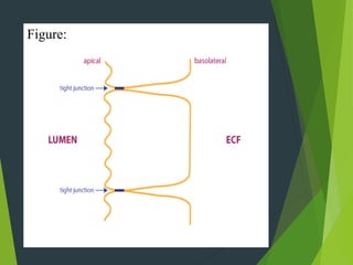
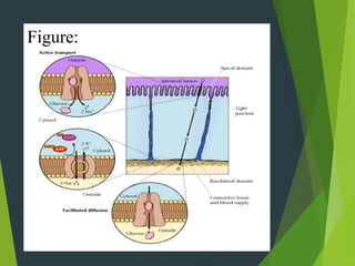
SHAH ABDUL LATIF UNIVERSITY
KHAIRPUR
By: Abdul-Rahman Shaikh , Aamir , Sadam & Muhammad-bux
( BS P-III & Previous) BIOCHEMISTRY
Transpotation through Epithelial
cells:
Epithelia form linings
throughout the body. In the
small intestine, for instance, the
simple columnar epithelium
forms a barrier that separates the
lumen from the internal
environment of the body
(note that the internal
environment in which body cells
exist is the extracellular fluid or
ECF).
The epithelium forms a barrier
because cells are linked by tight
junctions, which prevent many
substances from diffusing
between adjacent cells.
For a substance to cross the
epithelium, it must be transported
across the cell's plasma
membranes by membrane
transporters.
The apical plasma membrane
faces the lumen. In the drawing
at the right, the apical plasma
membrane is drawn as a wavy
line, because intestinal epithelial
Figure:
cells have a high degree of apical
plasma membrane folding to
increase the surface area
available for membrane transport
(these apical plasma membrane
folds are known as microvilli).
The basolateral plasma
membrane faces the ECF.
Epithelial cells are able to
transport substances in one
direction across the epithelium
because different sets of
transporters are localized in
either the apical or basolateral
membranes.
Absorption is the means whereby
nutrients such as glucose are
taken into the body to nourish
cells.
Glucose is transported across the
apical plasma membrane of the
intestine by the sodium-glucose
cotransporter (purple).
Because transport of Na+ and
glucose is coupled, we need to
add the free energy inherent in
Na+ transport to the free energy
inherent in glucose transport to
get the overall free energy for
the process. Just after a meal,
there will be abundant glucose in
the lumen of the intestine,
favoring absorption. Towards the
end of the absorptive phase of a
meal, however, the cotransporter
is still able to move glucose into
the cell (uphill against its
concentration gradient) because
of the strong Na+ concentration
gradient. The Na+ gradient is
established by the Na+/K+-
ATPase (red), which is located on
the basolateral membrane. The
activity of the cotransporter
increases the glucose
concentration inside the cells,
allowing glucose to be
transported into the ECF via the
glucose transporter (blue).
Figure:
Facilitated diffusion of glucose
into the ECF is a passive process,
since glucose flows down its
concentration gradient.
A therapy that takes advantage of
this absorption mechanism is
oral rehydration therapy.
Oral rehydration therapy is a life
saving treatment for people with
severe diarrhea due to intestinal
infections such as cholera. Fluid
loss from the gastrointestinal
tract depletes the ECF. The goal
in oral rehydration is to expand
the ECF. This requires getting
Na+ absorbed into the body,
since the amount of Na+ in the
ECF is the major determinant of
ECF volume. Because both Na+
and glucose need to bind to the
sodium-glucose cotransporter
for transport to occur, glucose is
included in oral rehydration
solutions to help speed
absorption of Na+.
Figure:
About 1500 ml of fluid per day is
moved from the extracellular
fluid into the lumen of the small
intestine in order to provide
lubrication that can protect the
epithelium and help with
intestinal motility. The
mechanism for fluid secretion is
that solutes are moved across the
epithelium, which then draw
water into the lumen by osmosis.
The rate-limiting and regulated
step in intestinal secretion is the
movement of Cl- ions across the
apical plasma membrane.
The important proteins involved
in secretion are diagrammed in
the figure. First, Cl- is
transported into the epithelial cell
by a cotransporter expressed on
Figure:
the basolateral membrane. As
with the previous example, the
Na+ gradient, established by the
Na+/K+-ATPase, provides the
energy to power transport of ions
into the cell (this cotransporter
moves 2 Cl-, one K+, and one
Na+ ion with each round of
transport). Cl- flows down its
concentration gradient into the
lumen via the Cl- channel CFTR
(green) located on the apical
plasma membrane. (Not shown is
that Na+ also flows into the
lumen, by a passive mechanism).
The CFTR protein is a member
of the ATP-binding cassette
(ABC) protein family. CFTR is
an atypical ABC protein; like
other members of the ABC
protein family, it binds ATP, but
in this case ATP binding is used
to open an ion channel.
Importantly, the CFTR protein
also has a regulatory domain that
is phosphorylated by protein
kinase A (PKA), also known as
cAMP-dependent kinase.
Intestinal secretion is turned on
when a regulatory molecule
binds a G-protein coupled
receptor, causing the alpha
subunit of the G-protein to
activate the enzyme adenylyl
cyclase. Adenylyl cyclase
produces the second messenger
cAMP, which activates PKA to
phosphorylate CFTR.
Figure:
With few exceptions, all the
internal and external body
surfaces of animals, such as the
skin, stomach, and intestines, are
covered with a layer of epithelial
cells called an epithelium. Many
epithelial cells transport ions or
small molecules from one side to
the other of the epithelium.
Those lining the stomach, for
instance, secrete hydrochloric
acid into the stomach lumen,
which after a meal becomes pH
1, while those lining the small
intestine transport products of
digestion (e.g., glucose and
amino acids) into the blood. All
epithelial cells in a sheet are
interconnected by several
types of specialized regions of the
plasma membrane called cell
junctions. These impart strength
and rigidity to the tissue and
prevent water-soluble material on
one side of the sheet (as in the
intestinal lumen) from moving
across to the other side. In this
section we first describe the
polarized nature of epithelia and
how different combinations of
membrane proteins enable
epithelial cells to carry out their
transport or secretory functions.
Then we discuss the structure
and function of the junctions that
interconnect epithelial cells.
Figure:
“Life laughs at you
when you are
unhappy... Life smiles
at you when you are
happy... Life salutes
you when you make
others happy…”
Biomembrane and cell signalling

Biomembrane and cell signalling

  • 1.
    SHAH ABDUL LATIFUNIVERSITY KHAIRPUR
  • 2.
    By: Abdul-Rahman Shaikh, Aamir , Sadam & Muhammad-bux ( BS P-III & Previous) BIOCHEMISTRY
  • 3.
    Transpotation through Epithelial cells: Epitheliaform linings throughout the body. In the small intestine, for instance, the simple columnar epithelium forms a barrier that separates the lumen from the internal environment of the body
  • 4.
    (note that theinternal environment in which body cells exist is the extracellular fluid or ECF). The epithelium forms a barrier because cells are linked by tight junctions, which prevent many substances from diffusing between adjacent cells.
  • 5.
    For a substanceto cross the epithelium, it must be transported across the cell's plasma membranes by membrane transporters. The apical plasma membrane faces the lumen. In the drawing at the right, the apical plasma membrane is drawn as a wavy line, because intestinal epithelial
  • 6.
  • 7.
    cells have ahigh degree of apical plasma membrane folding to increase the surface area available for membrane transport (these apical plasma membrane folds are known as microvilli). The basolateral plasma membrane faces the ECF. Epithelial cells are able to transport substances in one
  • 8.
    direction across theepithelium because different sets of transporters are localized in either the apical or basolateral membranes. Absorption is the means whereby nutrients such as glucose are taken into the body to nourish cells.
  • 9.
    Glucose is transportedacross the apical plasma membrane of the intestine by the sodium-glucose cotransporter (purple). Because transport of Na+ and glucose is coupled, we need to add the free energy inherent in Na+ transport to the free energy inherent in glucose transport to get the overall free energy for
  • 10.
    the process. Justafter a meal, there will be abundant glucose in the lumen of the intestine, favoring absorption. Towards the end of the absorptive phase of a meal, however, the cotransporter is still able to move glucose into the cell (uphill against its concentration gradient) because of the strong Na+ concentration
  • 11.
    gradient. The Na+gradient is established by the Na+/K+- ATPase (red), which is located on the basolateral membrane. The activity of the cotransporter increases the glucose concentration inside the cells, allowing glucose to be transported into the ECF via the glucose transporter (blue).
  • 12.
  • 13.
    Facilitated diffusion ofglucose into the ECF is a passive process, since glucose flows down its concentration gradient. A therapy that takes advantage of this absorption mechanism is oral rehydration therapy. Oral rehydration therapy is a life saving treatment for people with severe diarrhea due to intestinal
  • 14.
    infections such ascholera. Fluid loss from the gastrointestinal tract depletes the ECF. The goal in oral rehydration is to expand the ECF. This requires getting Na+ absorbed into the body, since the amount of Na+ in the ECF is the major determinant of ECF volume. Because both Na+ and glucose need to bind to the
  • 15.
    sodium-glucose cotransporter for transportto occur, glucose is included in oral rehydration solutions to help speed absorption of Na+.
  • 16.
  • 17.
    About 1500 mlof fluid per day is moved from the extracellular fluid into the lumen of the small intestine in order to provide lubrication that can protect the epithelium and help with intestinal motility. The mechanism for fluid secretion is that solutes are moved across the epithelium, which then draw
  • 18.
    water into thelumen by osmosis. The rate-limiting and regulated step in intestinal secretion is the movement of Cl- ions across the apical plasma membrane. The important proteins involved in secretion are diagrammed in the figure. First, Cl- is transported into the epithelial cell by a cotransporter expressed on
  • 19.
  • 20.
    the basolateral membrane.As with the previous example, the Na+ gradient, established by the Na+/K+-ATPase, provides the energy to power transport of ions into the cell (this cotransporter moves 2 Cl-, one K+, and one Na+ ion with each round of transport). Cl- flows down its concentration gradient into the
  • 21.
    lumen via theCl- channel CFTR (green) located on the apical plasma membrane. (Not shown is that Na+ also flows into the lumen, by a passive mechanism). The CFTR protein is a member of the ATP-binding cassette (ABC) protein family. CFTR is an atypical ABC protein; like other members of the ABC
  • 22.
    protein family, itbinds ATP, but in this case ATP binding is used to open an ion channel. Importantly, the CFTR protein also has a regulatory domain that is phosphorylated by protein kinase A (PKA), also known as cAMP-dependent kinase. Intestinal secretion is turned on when a regulatory molecule binds a G-protein coupled
  • 23.
    receptor, causing thealpha subunit of the G-protein to activate the enzyme adenylyl cyclase. Adenylyl cyclase produces the second messenger cAMP, which activates PKA to phosphorylate CFTR.
  • 24.
  • 25.
    With few exceptions,all the internal and external body surfaces of animals, such as the skin, stomach, and intestines, are covered with a layer of epithelial cells called an epithelium. Many epithelial cells transport ions or small molecules from one side to the other of the epithelium.
  • 26.
    Those lining thestomach, for instance, secrete hydrochloric acid into the stomach lumen, which after a meal becomes pH 1, while those lining the small intestine transport products of digestion (e.g., glucose and amino acids) into the blood. All epithelial cells in a sheet are interconnected by several
  • 27.
    types of specializedregions of the plasma membrane called cell junctions. These impart strength and rigidity to the tissue and prevent water-soluble material on one side of the sheet (as in the intestinal lumen) from moving across to the other side. In this section we first describe the polarized nature of epithelia and
  • 28.
    how different combinationsof membrane proteins enable epithelial cells to carry out their transport or secretory functions. Then we discuss the structure and function of the junctions that interconnect epithelial cells.
  • 29.
  • 30.
    “Life laughs atyou when you are unhappy... Life smiles at you when you are happy... Life salutes you when you make others happy…”