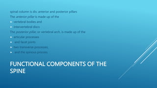
SPINE
STRUCTURE AND FUNCTION OF THE
SPINE
Structure
 Spinal column consists of 33 vertebrae
 (7 cervical, 12 thoracic, 5 lumbar, 5 fused sacral, and 3 or 4 coccygeal)
and their respective intervertebral discs.
FUNCTIONAL COMPONENTS OF THE
SPINE
spinal column is div. anterior and posterior pillars
The anterior pillar is made up of the
 vertebral bodies and
 intervertebral discs
The posterior pillar, or vertebral arch, is made up of the
 articular processes
 and facet joints
 two transverse processes,
 and the spinous process
MOTIONS OF THE SPINAL COLUMN
 Motion of the spinal column is described both globally and at the
functional unit or motion segment.
The functional unit
is comprised of two vertebrae and the joints in between(typically, two
zygapophyseal facet joints and one intervertebral disc).
THE SIX DEGREES OF MOTION
Flexion/Extension
 With flexion, the anterior portion of the bodies approximate and the
spinous processes separate;
 with extension, the anterior portion of the bodies separate and the
spinous processes approximate.
SIDE BENDING.
With side bending, the lateral edges of the vertebral bodies approximate
on the side toward which the spine is bending and separate the opposite
side.
ROTATION
 Motion in the transverse plane results in rotation.
 Rotation to the right results in relative movement of the body of the
superior vertebrae to the right and its spinous process to the left; the
opposite occurs with rotation to the left.
Anterior/posterior shear.
Forward or backward shear (translation) occurs when the body of the superior
vertebra translates forward or backward on the vertebra below.
 Lateral shear. Lateral shear (translation) occurs when the body of the
superior vertebra translates sideways on the vertebra below.
 Compression/distraction. Separation or approximation occurs with a
longitudinal force, either away from or toward the vertebral bodies
ARTHROKINEMATICS OF THE
ZYGAPOPHYSEAL (FACET) JOINTS
 Coupled motions typically occur at a segmental level when a person
side bends or rotates their spine.
Coupled motion is
 defined as “consistent association of one motion about an axis with
another motion around a different axis”and varies depending on the
region, the spinal posture, the orientation of the facets, and factors such
as extensibility of the soft tissues.
 When motions of side bending and rotation are coupled, foraminal
opening is dictated by the side bending component.
STRUCTURE AND FUNCTION OF
INTERVERTEBRAL DISCS
 The intervertebral disc, consisting of the annulus fibrosus and nucleus
pulposus, is one component of a three-joint complex between two
adjacent vertebrae
INTERVERTEBRAL FORAMINA
 The intervertebral foramina are between each vertebral segment in the posterior pillar.
Their anterior boundary is the
 intervertebral disc;
the posterior boundary is the
facet joint;
superior and inferior boundaries are the
 pedicles of the superior and inferior vertebrae of the spinal segment.
The mixed spinal nerve exits the spinal canal via the foramen along with blood vessels and recurrent
meningeal or sinuvertebral nerves.
 The size of the intervertebral foramina is affected by spinal motion, being larger with forward bending
and contralateral side bending and smaller with extension and ipsilateral side bending.
CURVES OF THE SPINE
 two primary, or posterior, curves, so named because they are present in
the infant and the convexity is posterior
 and two compensatory,or anterior, curves, so named because they
develop as the infant learns to lift the head and eventually stand, and
the convexity is anterior
CURVES OF THE SPINE
Posterior curves are in the thoracic and sacral regions.
 Kyphosis is a term used to denote a posterior curve.
 Kyphotic posture refers to an excessive posterior curvature of the thoracic spine.
Anterior curves are in the cervical and lumbar regions.
Lordosis is a term also used to denote an anterior curve, although some sources reserve
the term lordosis to denote abnormal conditions such as those that occur with a sway
Back.
■ The curves and flexibility in the spinal column are important for withstanding the
effects of gravity and other external forces.
■ The structure of the bones, joints, muscles, and inert tissues of the lower extremities
are designed for weight bearing; they support and balance the trunk in the upright
posture.
GRAVITY
 Ankle. For the ankle, the gravity line is anterior to the joint, so it tends to
rotate the tibia forward about the ankle. Stability is provided by the
plantarflexor muscles, primarily the soleus muscle.
 Knee. The normal gravity line is anterior to the knee joint, which tends to
keep the knee in extension. Stability is provided by the anterior cruciate
ligament, posterior capsule (locking mechanism of the knee), and tension in
the muscles posterior to the knee (the gastrocnemius and hamstring
muscles). The soleus provides active stability by pulling posteriorly on the
tibia. With the knees fully extended, no muscle support is required at that
joint to maintain an uprightposture; however, if the knees flex slightly, the
gravity line shifts posterior to the joint, and the quadriceps femoris muscle
must contract to prevent the knee from buckling.
 Hip. The gravity line at the hip varies with the swaying of the body. When the line
passes through the hip joint, there is equilibrium, and no external support is
necessary. When the gravitational line shifts posterior to the joint, some posterior
rotation of the pelvis occurs, but is controlled by tension in the hip flexor muscles
(primarily the iliopsoas). During relaxed standing, the iliofemoral ligament provides
passive stability to the joint, and no muscle tension is necessary. When the
gravitational line shifts anteriorly, stability is provided by active support of the hip
extensor muscles.
 Trunk. Normally, the gravity line in the trunk goes through the bodies of the lumbar
and cervical vertebrae, and the curves are balanced. Some activity in the muscles of
the trunk and pelvis helps maintain the balance. (This is described in greater detail in
the following sections.) As the trunk shifts, contralateral muscles contract and
function as guy wires. Extreme or sustained deviations are supported by inert
structures.
 Head. The center of gravity of the head falls anterior to the atlanto-
occipital joints. The posterior cervical muscles contract to keep the head
head balanced
STABILIZING FEATURES OF MUSCLES
CONTROLLING THE SPINE
Global Muscles
Characteristics
 Superficial: farther from axis of motion
 ■ Cross multiple vertebral segments
 ■ Produce motion and provide large guy wire function
 ■ Compressive loading with strong contractions
 Lumbar region
 Rectus abdominis External and internal obliques
 ■ Quadratus lumborum (lateral portion)
 ■ Erector spinae
 ■ Iliopsoas
CERVICAL REGION
 Sternocleidomastoid
 Scalene
 Levator scapulae
 Upper trapezius
 Erector spinae
DEEP SEGMENTAL MUSCLES
Deep
closer to axis of motion
 Attach to each vertebral segment
 ■ Control segmental motion; segmental guy wire function
 ■ Greater percentage of type I muscle fibers for muscular endurance
Lumber region
 transversus abdominis
 Multifidus
 Quadratus lumborum (deep portion)
Cervical
 Deep rotators
 Rectus capitis anterior and lateralis
 ■ Longus colli
BACK MUSCLES CAN BE DIVIDED INTO FOUR
FUNCTIONAL GROUPS: FLEXORS, EXTENSORS,
LATERAL FLEXORS AND ROTATORS
 Extensors, arranged in three layers
Most superficial is the strong Erector Spinae or sacrospinalis muscle
Middle layer is the multifidus. The fibers of the multifidus are centered on
each of the lumbar spinous processes.
 Third layer is made up of small muscles arranged from level to level,
which not only have an extension function but are also rotators and
lateral flexors.
FLEXORS
 intrinsic group (psoas major, psoas minor and iliacus)
 extrinsic group (abdominal wall muscles)
LATERAL FLEXORS AND ROTATORS
 internal and external oblique,
 the intertransverse and quadratus lumborum muscles.
Remember that pure lateral flexion is brought about only by the
quadratus lumborum.
COMMON FAULTY
POSTURES:CHARACTERISTICS AND
IMPAIRMENTS
 Pelvic and Lumbar Region/Lordotic Posture
 Relaxed or Slouched Posture
 Flat Low-Back Posture
LORDOTIC POSTURE
Lordotic Posture
 Lordosis is the normal curve (anterior convexity) of cervical and lumbar
spine which is found all normal individual pathologically. it is an
exaggeration of the normal curve found in cervical and lumbar spine.
Potential Sources of Pain
• Stress to the anterior longitudinal ligament
• Narrowing of the posterior disk space and narrowing of intervertebral
foramen.
• Approximation of the articular facets. The facets may become weight
bearing which may cause syonovial irritation and joint inflammation.
COMMON CAUSE OF EXCESSIVE
LUMBAR LORDOSIS
 Weakness of abdominals muscle
 Tightness or contracture of hip flexor (iliopsoas)
 Congenital problems such as bilateral congenital dislocation of hip
 Pregnancy
 High heel shoes / foot wears
 Spondylolisthesis
 Anterior tilt of pelvis as a result of weak extensor of hip and Abdominals
 Tightness or shortening of cervical extensor
TREATMENT FOR EXCESSIVE LUMBAR
LORDOSIS
 Mobilization of the lumbar spine.
 Anterior stretching of the lumbar spine
 Strengthening of the abdominals, glutei and hamstring.
 Training in grade correction of pelvic tilt has to be emphasized active
backward or posterior pelvic tilting by contracting abdominals and
glutei in supine is initiated.
 Toe touching in long sitting or forward bending sitting exercise
 Spinal extension or hyper extension should be strictly avoided.
 Treat the cause of increase lumbar lordosis.
KYPHOTIC POSTURE / ROUND BACK
 It is a faulty posture in which lumbar spine and cervical spine get hyper
extended while thoracic spine get flexed and head become slightly
forward.
 Potential Sources of Pain
 * Stressed to the posterior longitudinal ligament.
 * Fatigue of the thoracic erector spinae and rhomboid muscle.
 * Thoracic outlet syndromes.
 * Cervical posture syndromes
COMMON CAUSE OF KYPHOSIS
 Shortening or tightness of extensors of cervical spine and lumbar spine
and flexor of hip joint.
 Weakness of neck flexors,upper back extensors (erector spinae) and
Hamstring muscle.
 Bony anomaly generally in anterior tilt of pelvis, abdominals get
elongated but in this posture excessive flexion of thoracic spine offsets
the effect of anterior pelvic tilt.
 Ankylosing spondylitis.
 Other congenital anomalies.
TREATMENT OF KYPHOSIS
 Relaxation
 Postural belt
 Repeated stretching session
 Posture of head, neck and shoulder
 Mobilization of the whole spine
 Resistive exercise for longitudinal and transverse back muscle
 Controlled pelvic tilt
SCOLIOTIC POSTURE
 A lateral curvature of spine which exceeds 10 degree bending of the
vertebral from the normal is tended as scoliosis column to one side
combined with rotation of the vertebral bodies towards the convexity
and the spinous process towards the concavity
CLASSIFICATION
I. Non structural Scoliosis (Postural)
II. Transient Structural Scoliosis
III. Structural Scoliosis
1:Non structural Scoliosis
• Postural Scoliosis
• Compensatory Scoliosis
2: Transient Structural Scoliosis
• Sciatic Scoliosis
• Hysterical Scoliosis
• Inflamatory Scoliosis
 III. Structural Scoliosis
Structural scoliosis – Neuromuscular disease,osteopathic disorder, and
idiopathic disorder
 • Idiopathic Scoliosis
Old classification
Infantile Onset < 3 yrs Age
Juvenile Onset 3-10 yrs Age
Adolescent Onset > 10 yrs Age
 New Classification
Early onset Onset < 8 yrs Age – Late onset Onset > 8 yrs Age
POTENTIAL SOURCE OF PAIN
 Muscle fatigue and ligamentous strain on the side of convexity.Nerve
root irritation on the side of concavity.
Non structural – Leg length discrepancy,either structural or functional,
muscle guarding or spasm a painful stimuli in the back or neck, and
habitual or asymmetric posture.
PHYSIOLOGICAL EFFECTS OF SCOLIOSIS
• Mid-back pain
• lower back pain,
• neck pain, headaches,
• premature disc and joint degeneration
• Decreased pulmonary function
CLINICAL EVALUATION
• Plumb line - On posterior aspect, line drawn from occiput should
normally align with gluteal cleft
SCOLIOMETRY
ADAM’S FORWARD BEND TEST
TREATMENT OF SCOLIOSIS
 Active Correction with postural adaptation
 Passive Correction by Hanging
 Educate the patient by active effort
 Relaxation technique
 Repeated sessions of maintenance
 General free mobility exercises
 Deep breathing
 Balance Exercises
 Traction
TREATMENT
 Nonoperative treatment • Observation • Orthotics – braces • Traction and Casting
 • Exercises maintain muscle tone but no effect on the curve • If curve between 20
degree & 30 is progressing, bracing done TREATMENT
 Orthotics • Hibbs and Risser – Turnbuckle cast • Milwaukee brace ( CTLSO )•
Thoracolumbosacral othosis (TLSO’s) Milwaukee brace .
Contra indictions for orthosis
• Curve > 40 ° • Extreme thoracic kyphosis • Mature adolescent ; girls 2 yrs post
menarchal • High thoracic or cervicothoracic curve.
Halo traction device • Spinal skeletal traction & fixation device • Halo traction device
attached to skull & is connected to a plaster body cast by a steel frame
STREACHING
 Daily application of longitudinal & lateral traction forces mobilize the
spine gradually
• Patient in lying position, head end attached with 10 pounds weight pulls
proximally
• Pelvic girdle & traction straps with 20 to 30 pounds weight pull distally
Stretching.
CCP
 Curve correcting pad Milwaukee brace
SWAY BACK POSTURE/SLOUCHED
It is faulty posture in which head becomes slightly forward there is
extension of cervical spine, flexion of thoracic and loss of lordosis of
lumbar spine extension of hip and knee joint during standing are also the
feature of sway back posture pelvis rotates posteriorly.
* In this there is increased pelvic inclination up to 40.
When standing for prolonged period the person usually assumes an
asymmetric stance.
* In which most of the weight is borne on one lower extremity with
periodic shifting of weight to the opposite extremity.
POTENTIAL SOURCE OF PAIN
 Stress to iliofemoral ligament, the anterior longitudinal ligament of
lower lumbar spine and posterior longitudinal ligament of upper
lumbar and thoracic spine.
 Narrowing of intervertebral foramen in lower lumbar spine that may
compress the blood vessel dura & nerve root. Approximation of
articular facets in to lower lumbar spine
COMMON CAUSE OF SWAY BACK
1)Tightness of hamstring and abdominal muscle.
2)Weakness of one joint iliopsoas
3)Bony anomaly
TREATMENT OF SWAY BACK
• Stretching of hamstring and abdominal muscle
• Relaxation of the body
• Strengthening of Iliopsoas
• Maintain position of head is backward, extension of thoracic Spine
• Maintain normal lordosis of lumbar spine
• Always standing in erect position
FLAT BACK POSTURE SWAY BACK
POSTURE
 Flat Back Posture –
Flat back is faulty posture in which whole lumbar and thoracic spine gets
flattened. Although the cause and symptom of both flat back and sway
back are common but can be differentiated by excessive flexion and back
ward deviation of the upper thoracic spine in sway back posture while in
flat back posture spine become almost straight. It is reverse a lumbar
lordosis. There is flattening of normal lumbar lordosis
POTENTIAL SOURCE OF PAIN
 Lack of the normal physiologic lumbar curve which reduces the shock
absorbing effect of lumbar region and predisposes the person to injury.
• Stress to the posterior longitudinal ligament.
• Increase of the posterior disk space which allow the nucleus pulposus to
imbibe extra fluid and under certain circumstance may protrude
posteriorly when the person attempts extension.
COMMON CAUSE OF FLAT BACK
 1) Tight trunk flexor (rectus abdominis and intercostal) and hip
extensor muscle.
 2) Stretched and weak lumbar extensor and possibly hip flexor muscle.
TREATMENT OF FLAT BACK
• Increase lumbar lordosis which results in forward tilting of pelvis.
• Maintance of arch by active holding and also passive support in sitting
are effective in maintaining lordosis.
• Mobility and strengthening exercise of lumbar extensor are important.
• Stretching of trunk flexor and hip extensor muscle
MANAGEMENT OF
IMPAIRED POSTURE
 Structural and Functional Impairments
 • Pain from mechanical stress to sensitive structures and from muscle tension
 • Impaired mobility from muscle, joint, or fascial restrictions
 • Impaired muscle performance associated with an imbalance in muscle length and
strength between antagonistic muscle groups
 • Impaired muscle performance associated with poor muscular endurance
 • Insufficient postural control of scapular and trunk stabilizing muscles
 • Decreased cardiopulmonary endurance
 • Altered kinesthetic sense of posture associated with poor neuromuscular control
and prolonged faulty postural habits
 • Lack of knowledge of healthy spinal control and mechanics
PLAN OF CARE INTERVENTION
 1. Develop awareness and control of spinal posture
 2. Educate the patient about the relationship between
 faulty posture and symptoms
 3. Increase mobility in restricting muscles, joints, fascia
 4. Develop neuromuscular control, strength, and
 endurance in postural and extremity muscles
 5. Teach safe body mechanics
 6. Ergonomic assessment of home, work, recreational
 environments
 7. Stress management/relaxation
 8. Identify safe aerobic activities
 9. Promote healthy exercise habits for self-maintenance
INTERVENTION
 1. Kinesthetic training; cervical and scapular motions, pelvic tilts, control of neutral spine. Utilize
procedures to develop and reinforce control of posture when sitting, standing, walking, and
performing targeted functional activities
 2. Practice positions and movements to experience control of symptoms with various postures
 3. Manual stretching and joint mobilization/manipulation; teach self-stretching
 4. Stabilization exercises; progress repetitions and challenge with extremity motions; progress to
dynamic trunk strengthening exercises
 5. Functional exercises to prepare for safe mechanics(squatting, lunges, reaching, pushing/pulling,
lifting and turning loads with stable spine)
 6. Adapt work, home, recreational environment
 7. Relaxation exercises and postural stress relief
 8. Implement and progress an aerobic exercise program
 9. Integration of a fitness program, regular exercise, and safe body mechanics into daily life
SPINAL PATHOLOGIES AND IMPAIRED SPINAL
FUNCTION
 ■ Postural stress, strain
 ■ Abnormal posture
 ■ Muscle strain, tear, contusion
 ■ Acute low back or cervical pain
 ■ Degenerative disc disease (DDD), disc herniation
 ■ Degenerative joint disease (DJD), spondylosis
 ■ Rheumatoid arthritis
 ■ Radiculopathy, nerve root lesions, sciatica
 ■ Spinal stenosis
 ■ Segmental instability
 ■ Spondylolistheses
 ■ Sprains, strains
 ■ Laminectomy
 ■ Anterior cervical disc fusion (ACDF)
 ■ Transforaminal lumbar interbody fusion (TLIF)
 Compression fracture
 ■ Spondylosis with myelopathy
 ■ Intervertebral disc disorders
PATHOLOGY OF THE INTERVERTEBRAL
DISC
 Herniation: a general term used when there is any change in the shape
of the annulus that causes it to bulge beyond its normal perimeter.
 Protrusion: nuclear material is contained by the outer layers of the
annulus and supporting ligamentous structures.
 Prolapse: frank rupture of the nuclear material into the vertebral
canal.
 Extrusion: extension of nuclear material beyond the confines of the
posterior longitudinal ligament or above and below the disc space, as
detected on magnetic resonance imaging (MRI),but still in contact with
the disc.
 Free sequestration: the extruded nucleus has separated from the
disc and moved away from the prolapsed
COMMON IMPAIRMENTS RELATED TO DISC
PROTRUSIONS IN THE LUMBAR SPINE
 Pain, muscle-guarding
 ■ Flexed posture and deviation away from (usually) the symptomatic side
 ■ Neurological symptoms in dermatome and possibly myotome of affected
nerve roots
 ■ Increased symptoms (peripheralization) with sitting, prolonged flexed
postures, transition from sit to stand, coughing, straining
 ■ Limited nerve mobility, such as straight-leg raising (usually between 30°
and 60°)
 ■ Peripheralization of symptoms with repeated forward bending (spinal
flexion) test
PATHOLOGY OF THE ZYGAPOPHYSEAL
(FACET) JOINTS
 Facet joints are synovial articulations that are enclosed in a capsule and
supported by ligaments
 locked-back mechanism
 Pain: When acute, there is pain and muscle guarding with all motions; pain when
subacute and chronic is related to periods of immobility or excessive activity.
 ■ Impaired mobility: Usually hypomobility and decreased joint play in affected joints;
there may be hypermobility or instability during early stages.
 ■ Impaired posture.
 ■ Impaired spinal extension: Extension may cause or increase neurological
symptoms due to foraminal stenosis; therefore, may be unable to sustain or perform
repetitive extension activities without exacerbating symptoms.
 ■ Any functional activity that requires flexibility or prolonged repetition of trunk
motions, such as repetitive lifting and carrying of heavy objects, may exacerbate
symptoms in the arthritic spine
SPONDYLOSIS, OSTEOARTHRITIS, AND
DEGENERATIVE
JOINT DISEASE
 Spondylosis and OA are synonymous terms. This pathology may also be
referred to as DJD. Osteoarthritis involves degeneration of the IV disc as
well as the facet joints. Usually, ther is a history of faulty posture,
prolonged immobilization after injury, or severe or repetitive trauma.
DJD
 osteophyte formation with spurring and lipping along the joint margins
and vertebral bodies. Progressive hypomobility with boney stenosis
results. The encroachment of osteophytes on the spinal canal and
intervertebral foramina may cause neurological signs, especially with
spinal extension and side bending.
 The degenerating joint is vulnerable to facet impingement,sprains, and
inflammation, as is any arthritic joint.
 ■ In some patients, movement relieves the symptoms; in others,
movement irritates the joints, and painful symptoms increase.
SPONDOLYSTHESIS
 Forward translation of one vertebra on another in the sagittal plane of
the spine
 Spondylolisthesis is the slippage of one vertebral body with respect to
the adjacent vertebral body causing mechanical or radicular symptoms
or pain
MEYERDING
CLASSIFICATION
CLINICAL PRESENTATION
 Pain is exacerbated by extending at the affected segment
 Pain decreases as the patient assumes flexed posture
 Pain may be exacerbated by direct palpation of the affected segment
 Pain can sometimes improve in certain positions such as lying supine.
 Atrophy of the muscles, muscle weakness
 Tense hamstrings, hamstrings spasms
 Disturbances in coordination and balance, difficulty walking
 Rarely loss of bowel or bladder control.
CONSERVATIVE TREATMENT
 Initially resting and avoiding movements like lifting, bending, and sports.
 Analgesics and NSAIDs reduce musculoskeletal pain and have an anti-
inflammatory effect on nerve root and joint irritation.
 Epidural steroid injections can be used to relieve low back pain, lower extremity
pain related to radiculopathy and neurogenic claudication.
 A brace may be useful to decrease segmental spinal instability and pain.
 Physiotherapy focuses on relieving extension stresses from the lumbosacral
junction (hamstring and hip flexor stretching), as well as working on core
strengthening (deep abdominal muscles and lumbar multifidus strengthening).
SI JOINT
 The Sacroiliac joint (simply called the SI joint) is the joint connection
between the spine and the pelvis.
 Large diarthrodial joint made up of the sacrum and the two
innominates of the pelvis
CLINICAL PRESENTATION
 Low back pain
 Thigh pain
 Difficulty sitting in one place for too long due to pain
 Local tenderness of the posterior aspect of the sacroiliac joint (near the PSIS)
 Pain occurs when the joint is mechanically stressed eg forward bending
 Absence of neurological deficit/nerve root tension signs
 Aberrant sacroiliac movement pattern
 The joint can be hyper or hypo-mobile which can cause pain
 Pain is usually localized over the buttock
 Patients can often complain of sharp, stabbing, and/or shooting pain which extends down the posterior thigh
usually not past the knee.
 Pain can frequently mimic and be misdiagnosed as radicular pain
 Patients will frequently complain of pain while sitting down, lying on the ipsilateral side of pain, or climbing
stairs
DIAGNOSTIC PROCEDURES
 Physiotherapists use a variety of orthopaedic provocation tests.
 Gaenslen Test
 Distraction Test/compression test
 Faber test(Patrick Sign)
 Yeoman's test
 Sacral Thrust Test
 Thigh Thrust test
PHYSICAL THERAPY MANAGEMENT
 First stage of treatment is to reduce the inflammation with ice packs and
anti-inflammatory medication.
 second goal is to improve mobility using mobilizations, manipulation or
exercise therapy.
 complaints of instability, it can be useful to make use of a sacroiliac belt to
temporarily support the pelvis, together with progressive stabilization
training to increase motor control and stability.
 If there are complaints of instability, it can be useful to make use of a
sacroiliac belt to temporarily support the pelvis, together with progressive
stabilization training to increase motor control and stability.
 If the sacroiliac joint is severely inflamed, a sacroiliac belt can also be
used.
 Postural and ergonomic advice will help the patient to decrease the risk
of reinjure
 If the sacroiliac joint is severely inflamed, a sacroiliac belt can also be
used
 Postural and ergonomic advice will help the patient to decrease the risk
of reinjury.
MOTIONS AVAILABLE
 The main function of the SI joint is to provide stability and attenuate
forces to the lower extremities.
 The strong ligamentous system of the joint makes it better designed for
stability and limits the amount of motion available.
Nutation
 sacrum moves anteriorly and inferiorly, the coccyx moves posteriorly
relative to the ilium.
Counternutation
 sacrum moves up, backward,
NUTATION AND COUNTER NUTATION
ANKYLOSING SPONDYLITIS (AS)
 Ankylosing Spondylitis > previously known as Bechterew's disease ,
Bechterew syndrome , Marie Strümpell disease
 It is a form of arthritis that is long-lasting (chronic) and most often
affects the spine. It affects joints in the spine and the sacroilium in the
pelvis , causing eventual fusion of the spine.
 Complete fusion results in a complete rigidity of the spine, a condition
known as bamboo spine
 AS is a systemic rheumatic disease and is one of the seronegative
spondyloarthropathies.
 The typical patient is young, aged 18-30 Men are affected more than
women by a ratio about of 3:1
What causes ankylosing spondylitis?
The cause of ankylosing spondylitis is unknown , but a tendency to
develop the condition may be genetic .
> HLA-B27 genotype. -90% of patients
Tumor necrosis factor-alpha (TNF α) > IL-1 10%
 ■ There is a gradual loss of motion and the person will complain of
general stiffness. The patient may initially complain of bilateral pain in
his or her sacroiliac joints, thoracic spine,or shoulders. The person will
wake up early with pain and stiffness and have difficulty standing up
straight.
 ■ In advanced cases, radiographs will reveal a “bamboo”spine. This
imaging identifies where the anterior longitudinal ligament has fused
to the vertebral bodies. Decreased joint spaces may also be identified
on the film.
 PRECAUTION: Atlanto-axial subluxation is the hallmark of
 cervical spine involvement. Extreme caution should be used when
assessing and manipulating the cervical spine region to avoid causing
serious or fatal injury.
FACET JOINT IMPINGEMENT (BLOCKING,
FIXATION,EXTRAPMENT)
 With a sudden or unusual movement, the meniscoid of a facet capsule
may be extrapped, impinged (entrapped), or stressed,which causes pain
and muscle guarding. The onset is sudden and usually involves forward
bending and rotation.
■ There is loss of specific motions and attempted movement induces pain.
At rest, the individual has no pain.
■ There are no true neurological signs, but there may be referred pain in
the related dermatome.
■ Over time, stress is placed on the contralateral joint and on the disc,
leading to problems in these structures.
DIAGNOSIS
 a blood test for the HLA-B27 gene
 X-ray -which show characteristic spinal changes and sacroiliitis.
 tomography and magnetic resonance imaging of the sacroiliac joints -
but the reliability of these tests is still unclear
 Schober's test -a useful clinical measure of flexion of the lumbar spine
performed during examination.
 X-ray demonstrating in ankylosing spondylitis
 educate about the nature of the disease >baseline ROM including chest
expansion should be advised
 pain should be managed by appropriate medications, heat, massage and
gentle exercise.
 excessive physical exertion during periods of active inflammation should be
discouraged.
 proper positioning at rest is essential >the mattress should be firm,
 the patient should sleep on the back and avoid positions that encourage
flexion deformity.
 postural training emphasizes avoiding flexion, heavy lifting and prolonged
walking, standing or sitting.
SPINE 2.pptx

SPINE 2.pptx

  • 1.
  • 2.
    STRUCTURE AND FUNCTIONOF THE SPINE Structure  Spinal column consists of 33 vertebrae  (7 cervical, 12 thoracic, 5 lumbar, 5 fused sacral, and 3 or 4 coccygeal) and their respective intervertebral discs.
  • 3.
    FUNCTIONAL COMPONENTS OFTHE SPINE spinal column is div. anterior and posterior pillars The anterior pillar is made up of the  vertebral bodies and  intervertebral discs The posterior pillar, or vertebral arch, is made up of the  articular processes  and facet joints  two transverse processes,  and the spinous process
  • 5.
    MOTIONS OF THESPINAL COLUMN  Motion of the spinal column is described both globally and at the functional unit or motion segment. The functional unit is comprised of two vertebrae and the joints in between(typically, two zygapophyseal facet joints and one intervertebral disc).
  • 6.
    THE SIX DEGREESOF MOTION Flexion/Extension  With flexion, the anterior portion of the bodies approximate and the spinous processes separate;  with extension, the anterior portion of the bodies separate and the spinous processes approximate.
  • 7.
    SIDE BENDING. With sidebending, the lateral edges of the vertebral bodies approximate on the side toward which the spine is bending and separate the opposite side.
  • 8.
    ROTATION  Motion inthe transverse plane results in rotation.  Rotation to the right results in relative movement of the body of the superior vertebrae to the right and its spinous process to the left; the opposite occurs with rotation to the left. Anterior/posterior shear. Forward or backward shear (translation) occurs when the body of the superior vertebra translates forward or backward on the vertebra below.  Lateral shear. Lateral shear (translation) occurs when the body of the superior vertebra translates sideways on the vertebra below.  Compression/distraction. Separation or approximation occurs with a longitudinal force, either away from or toward the vertebral bodies
  • 9.
    ARTHROKINEMATICS OF THE ZYGAPOPHYSEAL(FACET) JOINTS  Coupled motions typically occur at a segmental level when a person side bends or rotates their spine. Coupled motion is  defined as “consistent association of one motion about an axis with another motion around a different axis”and varies depending on the region, the spinal posture, the orientation of the facets, and factors such as extensibility of the soft tissues.  When motions of side bending and rotation are coupled, foraminal opening is dictated by the side bending component.
  • 10.
    STRUCTURE AND FUNCTIONOF INTERVERTEBRAL DISCS  The intervertebral disc, consisting of the annulus fibrosus and nucleus pulposus, is one component of a three-joint complex between two adjacent vertebrae
  • 14.
    INTERVERTEBRAL FORAMINA  Theintervertebral foramina are between each vertebral segment in the posterior pillar. Their anterior boundary is the  intervertebral disc; the posterior boundary is the facet joint; superior and inferior boundaries are the  pedicles of the superior and inferior vertebrae of the spinal segment. The mixed spinal nerve exits the spinal canal via the foramen along with blood vessels and recurrent meningeal or sinuvertebral nerves.  The size of the intervertebral foramina is affected by spinal motion, being larger with forward bending and contralateral side bending and smaller with extension and ipsilateral side bending.
  • 16.
    CURVES OF THESPINE  two primary, or posterior, curves, so named because they are present in the infant and the convexity is posterior  and two compensatory,or anterior, curves, so named because they develop as the infant learns to lift the head and eventually stand, and the convexity is anterior
  • 17.
    CURVES OF THESPINE Posterior curves are in the thoracic and sacral regions.  Kyphosis is a term used to denote a posterior curve.  Kyphotic posture refers to an excessive posterior curvature of the thoracic spine. Anterior curves are in the cervical and lumbar regions. Lordosis is a term also used to denote an anterior curve, although some sources reserve the term lordosis to denote abnormal conditions such as those that occur with a sway Back. ■ The curves and flexibility in the spinal column are important for withstanding the effects of gravity and other external forces. ■ The structure of the bones, joints, muscles, and inert tissues of the lower extremities are designed for weight bearing; they support and balance the trunk in the upright posture.
  • 19.
    GRAVITY  Ankle. Forthe ankle, the gravity line is anterior to the joint, so it tends to rotate the tibia forward about the ankle. Stability is provided by the plantarflexor muscles, primarily the soleus muscle.  Knee. The normal gravity line is anterior to the knee joint, which tends to keep the knee in extension. Stability is provided by the anterior cruciate ligament, posterior capsule (locking mechanism of the knee), and tension in the muscles posterior to the knee (the gastrocnemius and hamstring muscles). The soleus provides active stability by pulling posteriorly on the tibia. With the knees fully extended, no muscle support is required at that joint to maintain an uprightposture; however, if the knees flex slightly, the gravity line shifts posterior to the joint, and the quadriceps femoris muscle must contract to prevent the knee from buckling.
  • 20.
     Hip. Thegravity line at the hip varies with the swaying of the body. When the line passes through the hip joint, there is equilibrium, and no external support is necessary. When the gravitational line shifts posterior to the joint, some posterior rotation of the pelvis occurs, but is controlled by tension in the hip flexor muscles (primarily the iliopsoas). During relaxed standing, the iliofemoral ligament provides passive stability to the joint, and no muscle tension is necessary. When the gravitational line shifts anteriorly, stability is provided by active support of the hip extensor muscles.  Trunk. Normally, the gravity line in the trunk goes through the bodies of the lumbar and cervical vertebrae, and the curves are balanced. Some activity in the muscles of the trunk and pelvis helps maintain the balance. (This is described in greater detail in the following sections.) As the trunk shifts, contralateral muscles contract and function as guy wires. Extreme or sustained deviations are supported by inert structures.
  • 21.
     Head. Thecenter of gravity of the head falls anterior to the atlanto- occipital joints. The posterior cervical muscles contract to keep the head head balanced
  • 23.
    STABILIZING FEATURES OFMUSCLES CONTROLLING THE SPINE Global Muscles Characteristics  Superficial: farther from axis of motion  ■ Cross multiple vertebral segments  ■ Produce motion and provide large guy wire function  ■ Compressive loading with strong contractions  Lumbar region  Rectus abdominis External and internal obliques  ■ Quadratus lumborum (lateral portion)  ■ Erector spinae  ■ Iliopsoas
  • 24.
    CERVICAL REGION  Sternocleidomastoid Scalene  Levator scapulae  Upper trapezius  Erector spinae
  • 25.
    DEEP SEGMENTAL MUSCLES Deep closerto axis of motion  Attach to each vertebral segment  ■ Control segmental motion; segmental guy wire function  ■ Greater percentage of type I muscle fibers for muscular endurance Lumber region  transversus abdominis  Multifidus  Quadratus lumborum (deep portion) Cervical  Deep rotators  Rectus capitis anterior and lateralis  ■ Longus colli
  • 26.
    BACK MUSCLES CANBE DIVIDED INTO FOUR FUNCTIONAL GROUPS: FLEXORS, EXTENSORS, LATERAL FLEXORS AND ROTATORS  Extensors, arranged in three layers Most superficial is the strong Erector Spinae or sacrospinalis muscle Middle layer is the multifidus. The fibers of the multifidus are centered on each of the lumbar spinous processes.  Third layer is made up of small muscles arranged from level to level, which not only have an extension function but are also rotators and lateral flexors.
  • 27.
    FLEXORS  intrinsic group(psoas major, psoas minor and iliacus)  extrinsic group (abdominal wall muscles)
  • 28.
    LATERAL FLEXORS ANDROTATORS  internal and external oblique,  the intertransverse and quadratus lumborum muscles. Remember that pure lateral flexion is brought about only by the quadratus lumborum.
  • 30.
    COMMON FAULTY POSTURES:CHARACTERISTICS AND IMPAIRMENTS Pelvic and Lumbar Region/Lordotic Posture  Relaxed or Slouched Posture  Flat Low-Back Posture
  • 32.
    LORDOTIC POSTURE Lordotic Posture Lordosis is the normal curve (anterior convexity) of cervical and lumbar spine which is found all normal individual pathologically. it is an exaggeration of the normal curve found in cervical and lumbar spine. Potential Sources of Pain • Stress to the anterior longitudinal ligament • Narrowing of the posterior disk space and narrowing of intervertebral foramen. • Approximation of the articular facets. The facets may become weight bearing which may cause syonovial irritation and joint inflammation.
  • 33.
    COMMON CAUSE OFEXCESSIVE LUMBAR LORDOSIS  Weakness of abdominals muscle  Tightness or contracture of hip flexor (iliopsoas)  Congenital problems such as bilateral congenital dislocation of hip  Pregnancy  High heel shoes / foot wears  Spondylolisthesis  Anterior tilt of pelvis as a result of weak extensor of hip and Abdominals  Tightness or shortening of cervical extensor
  • 34.
    TREATMENT FOR EXCESSIVELUMBAR LORDOSIS  Mobilization of the lumbar spine.  Anterior stretching of the lumbar spine  Strengthening of the abdominals, glutei and hamstring.  Training in grade correction of pelvic tilt has to be emphasized active backward or posterior pelvic tilting by contracting abdominals and glutei in supine is initiated.  Toe touching in long sitting or forward bending sitting exercise  Spinal extension or hyper extension should be strictly avoided.  Treat the cause of increase lumbar lordosis.
  • 35.
    KYPHOTIC POSTURE /ROUND BACK  It is a faulty posture in which lumbar spine and cervical spine get hyper extended while thoracic spine get flexed and head become slightly forward.  Potential Sources of Pain  * Stressed to the posterior longitudinal ligament.  * Fatigue of the thoracic erector spinae and rhomboid muscle.  * Thoracic outlet syndromes.  * Cervical posture syndromes
  • 37.
    COMMON CAUSE OFKYPHOSIS  Shortening or tightness of extensors of cervical spine and lumbar spine and flexor of hip joint.  Weakness of neck flexors,upper back extensors (erector spinae) and Hamstring muscle.  Bony anomaly generally in anterior tilt of pelvis, abdominals get elongated but in this posture excessive flexion of thoracic spine offsets the effect of anterior pelvic tilt.  Ankylosing spondylitis.  Other congenital anomalies.
  • 38.
    TREATMENT OF KYPHOSIS Relaxation  Postural belt  Repeated stretching session  Posture of head, neck and shoulder  Mobilization of the whole spine  Resistive exercise for longitudinal and transverse back muscle  Controlled pelvic tilt
  • 39.
    SCOLIOTIC POSTURE  Alateral curvature of spine which exceeds 10 degree bending of the vertebral from the normal is tended as scoliosis column to one side combined with rotation of the vertebral bodies towards the convexity and the spinous process towards the concavity
  • 40.
    CLASSIFICATION I. Non structuralScoliosis (Postural) II. Transient Structural Scoliosis III. Structural Scoliosis
  • 41.
    1:Non structural Scoliosis •Postural Scoliosis • Compensatory Scoliosis 2: Transient Structural Scoliosis • Sciatic Scoliosis • Hysterical Scoliosis • Inflamatory Scoliosis
  • 42.
     III. StructuralScoliosis Structural scoliosis – Neuromuscular disease,osteopathic disorder, and idiopathic disorder  • Idiopathic Scoliosis Old classification Infantile Onset < 3 yrs Age Juvenile Onset 3-10 yrs Age Adolescent Onset > 10 yrs Age  New Classification Early onset Onset < 8 yrs Age – Late onset Onset > 8 yrs Age
  • 43.
    POTENTIAL SOURCE OFPAIN  Muscle fatigue and ligamentous strain on the side of convexity.Nerve root irritation on the side of concavity. Non structural – Leg length discrepancy,either structural or functional, muscle guarding or spasm a painful stimuli in the back or neck, and habitual or asymmetric posture.
  • 44.
    PHYSIOLOGICAL EFFECTS OFSCOLIOSIS • Mid-back pain • lower back pain, • neck pain, headaches, • premature disc and joint degeneration • Decreased pulmonary function
  • 45.
    CLINICAL EVALUATION • Plumbline - On posterior aspect, line drawn from occiput should normally align with gluteal cleft SCOLIOMETRY ADAM’S FORWARD BEND TEST
  • 46.
    TREATMENT OF SCOLIOSIS Active Correction with postural adaptation  Passive Correction by Hanging  Educate the patient by active effort  Relaxation technique  Repeated sessions of maintenance  General free mobility exercises  Deep breathing  Balance Exercises  Traction
  • 47.
    TREATMENT  Nonoperative treatment• Observation • Orthotics – braces • Traction and Casting  • Exercises maintain muscle tone but no effect on the curve • If curve between 20 degree & 30 is progressing, bracing done TREATMENT  Orthotics • Hibbs and Risser – Turnbuckle cast • Milwaukee brace ( CTLSO )• Thoracolumbosacral othosis (TLSO’s) Milwaukee brace . Contra indictions for orthosis • Curve > 40 ° • Extreme thoracic kyphosis • Mature adolescent ; girls 2 yrs post menarchal • High thoracic or cervicothoracic curve. Halo traction device • Spinal skeletal traction & fixation device • Halo traction device attached to skull & is connected to a plaster body cast by a steel frame
  • 48.
    STREACHING  Daily applicationof longitudinal & lateral traction forces mobilize the spine gradually • Patient in lying position, head end attached with 10 pounds weight pulls proximally • Pelvic girdle & traction straps with 20 to 30 pounds weight pull distally Stretching.
  • 51.
    CCP  Curve correctingpad Milwaukee brace
  • 52.
    SWAY BACK POSTURE/SLOUCHED Itis faulty posture in which head becomes slightly forward there is extension of cervical spine, flexion of thoracic and loss of lordosis of lumbar spine extension of hip and knee joint during standing are also the feature of sway back posture pelvis rotates posteriorly. * In this there is increased pelvic inclination up to 40. When standing for prolonged period the person usually assumes an asymmetric stance. * In which most of the weight is borne on one lower extremity with periodic shifting of weight to the opposite extremity.
  • 53.
    POTENTIAL SOURCE OFPAIN  Stress to iliofemoral ligament, the anterior longitudinal ligament of lower lumbar spine and posterior longitudinal ligament of upper lumbar and thoracic spine.  Narrowing of intervertebral foramen in lower lumbar spine that may compress the blood vessel dura & nerve root. Approximation of articular facets in to lower lumbar spine
  • 54.
    COMMON CAUSE OFSWAY BACK 1)Tightness of hamstring and abdominal muscle. 2)Weakness of one joint iliopsoas 3)Bony anomaly
  • 55.
    TREATMENT OF SWAYBACK • Stretching of hamstring and abdominal muscle • Relaxation of the body • Strengthening of Iliopsoas • Maintain position of head is backward, extension of thoracic Spine • Maintain normal lordosis of lumbar spine • Always standing in erect position
  • 56.
    FLAT BACK POSTURESWAY BACK POSTURE  Flat Back Posture – Flat back is faulty posture in which whole lumbar and thoracic spine gets flattened. Although the cause and symptom of both flat back and sway back are common but can be differentiated by excessive flexion and back ward deviation of the upper thoracic spine in sway back posture while in flat back posture spine become almost straight. It is reverse a lumbar lordosis. There is flattening of normal lumbar lordosis
  • 57.
    POTENTIAL SOURCE OFPAIN  Lack of the normal physiologic lumbar curve which reduces the shock absorbing effect of lumbar region and predisposes the person to injury. • Stress to the posterior longitudinal ligament. • Increase of the posterior disk space which allow the nucleus pulposus to imbibe extra fluid and under certain circumstance may protrude posteriorly when the person attempts extension.
  • 58.
    COMMON CAUSE OFFLAT BACK  1) Tight trunk flexor (rectus abdominis and intercostal) and hip extensor muscle.  2) Stretched and weak lumbar extensor and possibly hip flexor muscle.
  • 59.
    TREATMENT OF FLATBACK • Increase lumbar lordosis which results in forward tilting of pelvis. • Maintance of arch by active holding and also passive support in sitting are effective in maintaining lordosis. • Mobility and strengthening exercise of lumbar extensor are important. • Stretching of trunk flexor and hip extensor muscle
  • 60.
    MANAGEMENT OF IMPAIRED POSTURE Structural and Functional Impairments  • Pain from mechanical stress to sensitive structures and from muscle tension  • Impaired mobility from muscle, joint, or fascial restrictions  • Impaired muscle performance associated with an imbalance in muscle length and strength between antagonistic muscle groups  • Impaired muscle performance associated with poor muscular endurance  • Insufficient postural control of scapular and trunk stabilizing muscles  • Decreased cardiopulmonary endurance  • Altered kinesthetic sense of posture associated with poor neuromuscular control and prolonged faulty postural habits  • Lack of knowledge of healthy spinal control and mechanics
  • 61.
    PLAN OF CAREINTERVENTION  1. Develop awareness and control of spinal posture  2. Educate the patient about the relationship between  faulty posture and symptoms  3. Increase mobility in restricting muscles, joints, fascia  4. Develop neuromuscular control, strength, and  endurance in postural and extremity muscles  5. Teach safe body mechanics  6. Ergonomic assessment of home, work, recreational  environments  7. Stress management/relaxation  8. Identify safe aerobic activities  9. Promote healthy exercise habits for self-maintenance
  • 62.
    INTERVENTION  1. Kinesthetictraining; cervical and scapular motions, pelvic tilts, control of neutral spine. Utilize procedures to develop and reinforce control of posture when sitting, standing, walking, and performing targeted functional activities  2. Practice positions and movements to experience control of symptoms with various postures  3. Manual stretching and joint mobilization/manipulation; teach self-stretching  4. Stabilization exercises; progress repetitions and challenge with extremity motions; progress to dynamic trunk strengthening exercises  5. Functional exercises to prepare for safe mechanics(squatting, lunges, reaching, pushing/pulling, lifting and turning loads with stable spine)  6. Adapt work, home, recreational environment  7. Relaxation exercises and postural stress relief  8. Implement and progress an aerobic exercise program  9. Integration of a fitness program, regular exercise, and safe body mechanics into daily life
  • 63.
    SPINAL PATHOLOGIES ANDIMPAIRED SPINAL FUNCTION  ■ Postural stress, strain  ■ Abnormal posture  ■ Muscle strain, tear, contusion  ■ Acute low back or cervical pain  ■ Degenerative disc disease (DDD), disc herniation  ■ Degenerative joint disease (DJD), spondylosis  ■ Rheumatoid arthritis  ■ Radiculopathy, nerve root lesions, sciatica  ■ Spinal stenosis
  • 64.
     ■ Segmentalinstability  ■ Spondylolistheses  ■ Sprains, strains  ■ Laminectomy  ■ Anterior cervical disc fusion (ACDF)  ■ Transforaminal lumbar interbody fusion (TLIF)  Compression fracture  ■ Spondylosis with myelopathy  ■ Intervertebral disc disorders
  • 65.
    PATHOLOGY OF THEINTERVERTEBRAL DISC  Herniation: a general term used when there is any change in the shape of the annulus that causes it to bulge beyond its normal perimeter.  Protrusion: nuclear material is contained by the outer layers of the annulus and supporting ligamentous structures.  Prolapse: frank rupture of the nuclear material into the vertebral canal.
  • 66.
     Extrusion: extensionof nuclear material beyond the confines of the posterior longitudinal ligament or above and below the disc space, as detected on magnetic resonance imaging (MRI),but still in contact with the disc.  Free sequestration: the extruded nucleus has separated from the disc and moved away from the prolapsed
  • 68.
    COMMON IMPAIRMENTS RELATEDTO DISC PROTRUSIONS IN THE LUMBAR SPINE  Pain, muscle-guarding  ■ Flexed posture and deviation away from (usually) the symptomatic side  ■ Neurological symptoms in dermatome and possibly myotome of affected nerve roots  ■ Increased symptoms (peripheralization) with sitting, prolonged flexed postures, transition from sit to stand, coughing, straining  ■ Limited nerve mobility, such as straight-leg raising (usually between 30° and 60°)  ■ Peripheralization of symptoms with repeated forward bending (spinal flexion) test
  • 69.
    PATHOLOGY OF THEZYGAPOPHYSEAL (FACET) JOINTS  Facet joints are synovial articulations that are enclosed in a capsule and supported by ligaments  locked-back mechanism
  • 70.
     Pain: Whenacute, there is pain and muscle guarding with all motions; pain when subacute and chronic is related to periods of immobility or excessive activity.  ■ Impaired mobility: Usually hypomobility and decreased joint play in affected joints; there may be hypermobility or instability during early stages.  ■ Impaired posture.  ■ Impaired spinal extension: Extension may cause or increase neurological symptoms due to foraminal stenosis; therefore, may be unable to sustain or perform repetitive extension activities without exacerbating symptoms.  ■ Any functional activity that requires flexibility or prolonged repetition of trunk motions, such as repetitive lifting and carrying of heavy objects, may exacerbate symptoms in the arthritic spine
  • 71.
    SPONDYLOSIS, OSTEOARTHRITIS, AND DEGENERATIVE JOINTDISEASE  Spondylosis and OA are synonymous terms. This pathology may also be referred to as DJD. Osteoarthritis involves degeneration of the IV disc as well as the facet joints. Usually, ther is a history of faulty posture, prolonged immobilization after injury, or severe or repetitive trauma.
  • 72.
    DJD  osteophyte formationwith spurring and lipping along the joint margins and vertebral bodies. Progressive hypomobility with boney stenosis results. The encroachment of osteophytes on the spinal canal and intervertebral foramina may cause neurological signs, especially with spinal extension and side bending.  The degenerating joint is vulnerable to facet impingement,sprains, and inflammation, as is any arthritic joint.  ■ In some patients, movement relieves the symptoms; in others, movement irritates the joints, and painful symptoms increase.
  • 73.
    SPONDOLYSTHESIS  Forward translationof one vertebra on another in the sagittal plane of the spine  Spondylolisthesis is the slippage of one vertebral body with respect to the adjacent vertebral body causing mechanical or radicular symptoms or pain
  • 74.
  • 75.
    CLINICAL PRESENTATION  Painis exacerbated by extending at the affected segment  Pain decreases as the patient assumes flexed posture  Pain may be exacerbated by direct palpation of the affected segment  Pain can sometimes improve in certain positions such as lying supine.  Atrophy of the muscles, muscle weakness  Tense hamstrings, hamstrings spasms  Disturbances in coordination and balance, difficulty walking  Rarely loss of bowel or bladder control.
  • 76.
    CONSERVATIVE TREATMENT  Initiallyresting and avoiding movements like lifting, bending, and sports.  Analgesics and NSAIDs reduce musculoskeletal pain and have an anti- inflammatory effect on nerve root and joint irritation.  Epidural steroid injections can be used to relieve low back pain, lower extremity pain related to radiculopathy and neurogenic claudication.  A brace may be useful to decrease segmental spinal instability and pain.  Physiotherapy focuses on relieving extension stresses from the lumbosacral junction (hamstring and hip flexor stretching), as well as working on core strengthening (deep abdominal muscles and lumbar multifidus strengthening).
  • 77.
  • 78.
     The Sacroiliacjoint (simply called the SI joint) is the joint connection between the spine and the pelvis.  Large diarthrodial joint made up of the sacrum and the two innominates of the pelvis
  • 79.
    CLINICAL PRESENTATION  Lowback pain  Thigh pain  Difficulty sitting in one place for too long due to pain  Local tenderness of the posterior aspect of the sacroiliac joint (near the PSIS)  Pain occurs when the joint is mechanically stressed eg forward bending  Absence of neurological deficit/nerve root tension signs  Aberrant sacroiliac movement pattern  The joint can be hyper or hypo-mobile which can cause pain  Pain is usually localized over the buttock  Patients can often complain of sharp, stabbing, and/or shooting pain which extends down the posterior thigh usually not past the knee.  Pain can frequently mimic and be misdiagnosed as radicular pain  Patients will frequently complain of pain while sitting down, lying on the ipsilateral side of pain, or climbing stairs
  • 80.
    DIAGNOSTIC PROCEDURES  Physiotherapistsuse a variety of orthopaedic provocation tests.  Gaenslen Test  Distraction Test/compression test  Faber test(Patrick Sign)  Yeoman's test  Sacral Thrust Test  Thigh Thrust test
  • 81.
    PHYSICAL THERAPY MANAGEMENT First stage of treatment is to reduce the inflammation with ice packs and anti-inflammatory medication.  second goal is to improve mobility using mobilizations, manipulation or exercise therapy.  complaints of instability, it can be useful to make use of a sacroiliac belt to temporarily support the pelvis, together with progressive stabilization training to increase motor control and stability.  If there are complaints of instability, it can be useful to make use of a sacroiliac belt to temporarily support the pelvis, together with progressive stabilization training to increase motor control and stability.
  • 82.
     If thesacroiliac joint is severely inflamed, a sacroiliac belt can also be used.  Postural and ergonomic advice will help the patient to decrease the risk of reinjure  If the sacroiliac joint is severely inflamed, a sacroiliac belt can also be used  Postural and ergonomic advice will help the patient to decrease the risk of reinjury.
  • 83.
    MOTIONS AVAILABLE  Themain function of the SI joint is to provide stability and attenuate forces to the lower extremities.  The strong ligamentous system of the joint makes it better designed for stability and limits the amount of motion available. Nutation  sacrum moves anteriorly and inferiorly, the coccyx moves posteriorly relative to the ilium. Counternutation  sacrum moves up, backward,
  • 84.
  • 85.
    ANKYLOSING SPONDYLITIS (AS) Ankylosing Spondylitis > previously known as Bechterew's disease , Bechterew syndrome , Marie Strümpell disease  It is a form of arthritis that is long-lasting (chronic) and most often affects the spine. It affects joints in the spine and the sacroilium in the pelvis , causing eventual fusion of the spine.  Complete fusion results in a complete rigidity of the spine, a condition known as bamboo spine  AS is a systemic rheumatic disease and is one of the seronegative spondyloarthropathies.
  • 86.
     The typicalpatient is young, aged 18-30 Men are affected more than women by a ratio about of 3:1 What causes ankylosing spondylitis? The cause of ankylosing spondylitis is unknown , but a tendency to develop the condition may be genetic . > HLA-B27 genotype. -90% of patients Tumor necrosis factor-alpha (TNF α) > IL-1 10%
  • 87.
     ■ Thereis a gradual loss of motion and the person will complain of general stiffness. The patient may initially complain of bilateral pain in his or her sacroiliac joints, thoracic spine,or shoulders. The person will wake up early with pain and stiffness and have difficulty standing up straight.  ■ In advanced cases, radiographs will reveal a “bamboo”spine. This imaging identifies where the anterior longitudinal ligament has fused to the vertebral bodies. Decreased joint spaces may also be identified on the film.
  • 88.
     PRECAUTION: Atlanto-axialsubluxation is the hallmark of  cervical spine involvement. Extreme caution should be used when assessing and manipulating the cervical spine region to avoid causing serious or fatal injury.
  • 89.
    FACET JOINT IMPINGEMENT(BLOCKING, FIXATION,EXTRAPMENT)  With a sudden or unusual movement, the meniscoid of a facet capsule may be extrapped, impinged (entrapped), or stressed,which causes pain and muscle guarding. The onset is sudden and usually involves forward bending and rotation.
  • 90.
    ■ There isloss of specific motions and attempted movement induces pain. At rest, the individual has no pain. ■ There are no true neurological signs, but there may be referred pain in the related dermatome. ■ Over time, stress is placed on the contralateral joint and on the disc, leading to problems in these structures.
  • 91.
    DIAGNOSIS  a bloodtest for the HLA-B27 gene  X-ray -which show characteristic spinal changes and sacroiliitis.  tomography and magnetic resonance imaging of the sacroiliac joints - but the reliability of these tests is still unclear  Schober's test -a useful clinical measure of flexion of the lumbar spine performed during examination.  X-ray demonstrating in ankylosing spondylitis
  • 92.
     educate aboutthe nature of the disease >baseline ROM including chest expansion should be advised  pain should be managed by appropriate medications, heat, massage and gentle exercise.  excessive physical exertion during periods of active inflammation should be discouraged.  proper positioning at rest is essential >the mattress should be firm,  the patient should sleep on the back and avoid positions that encourage flexion deformity.  postural training emphasizes avoiding flexion, heavy lifting and prolonged walking, standing or sitting.