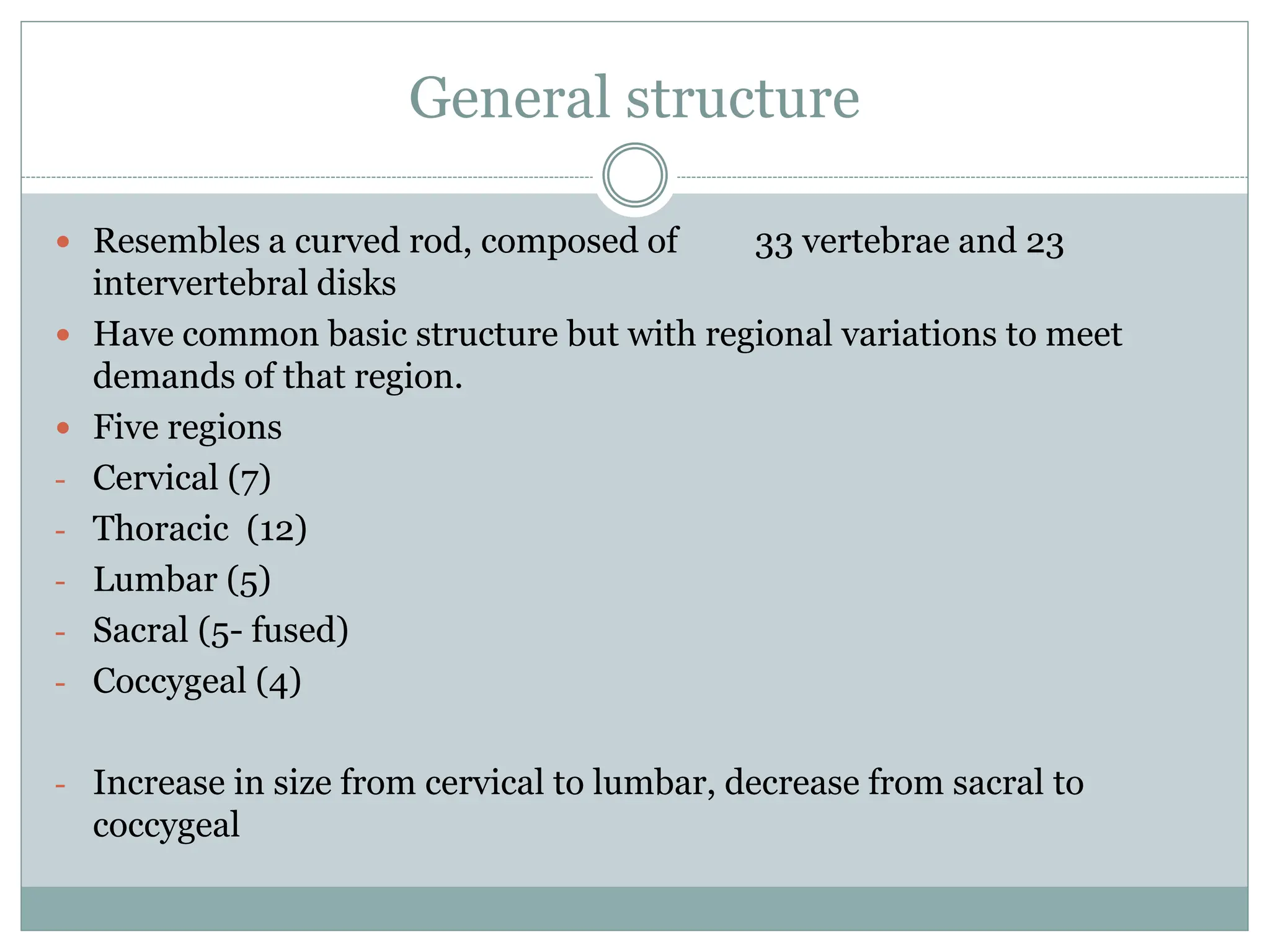
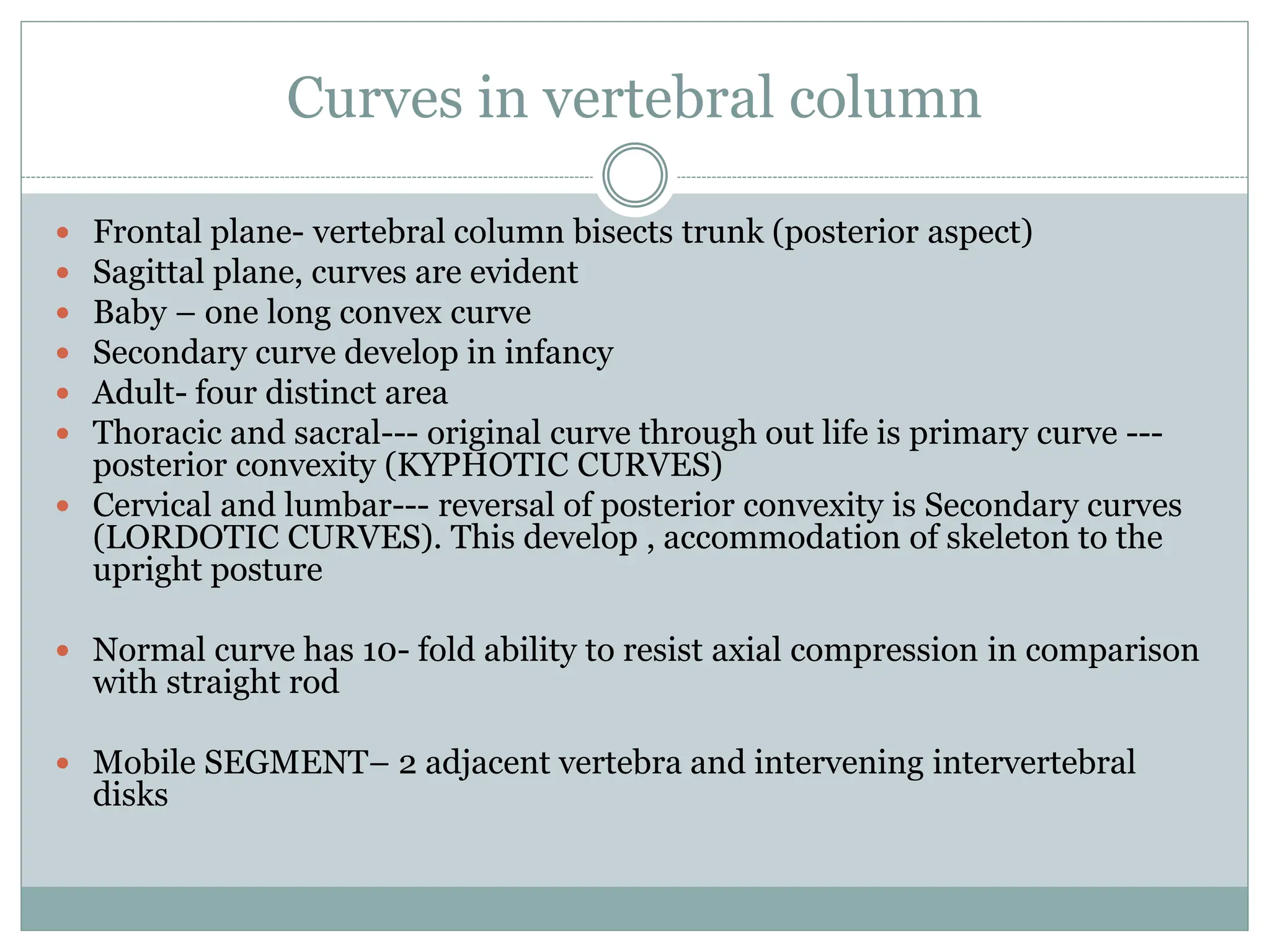
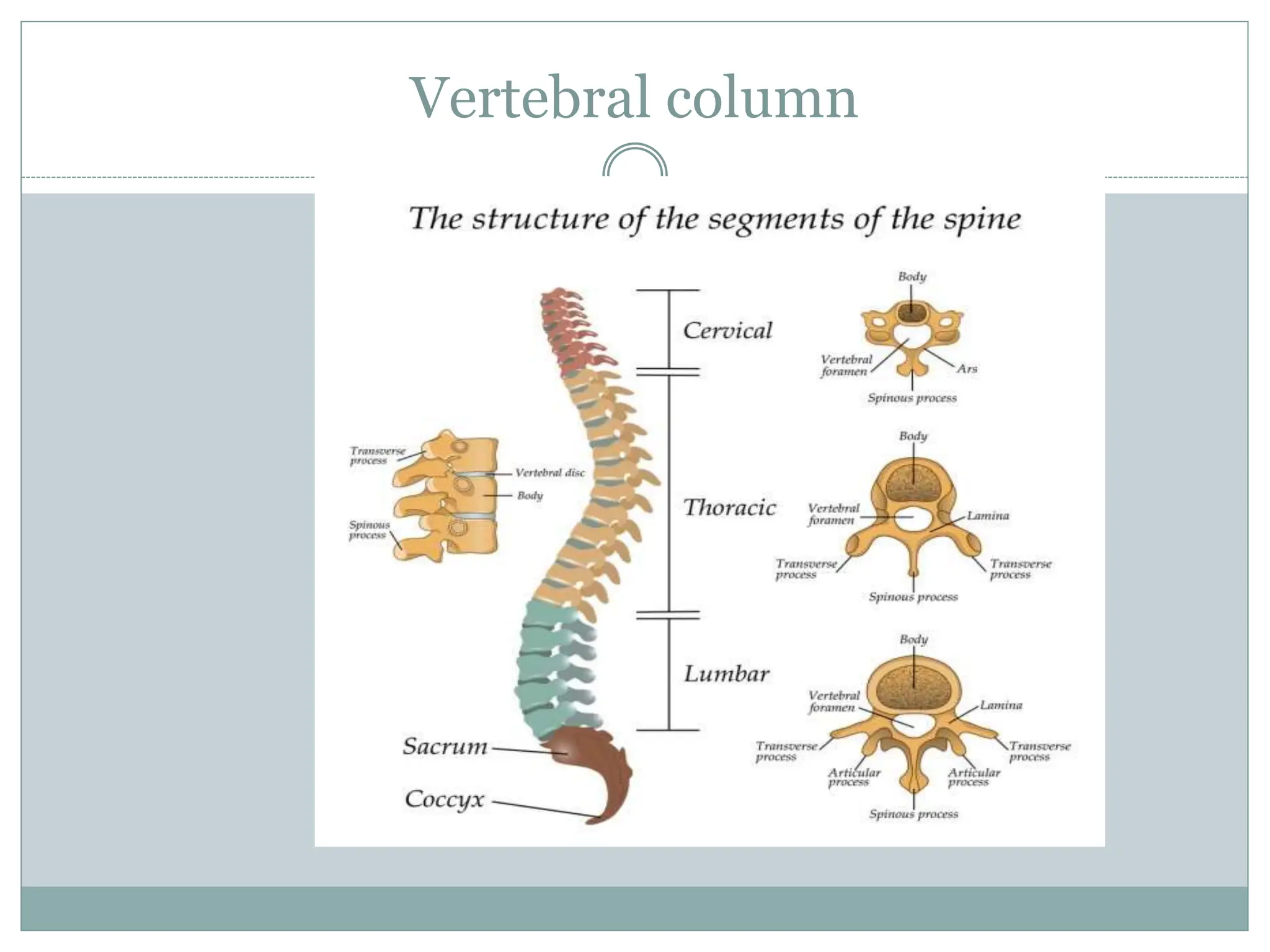
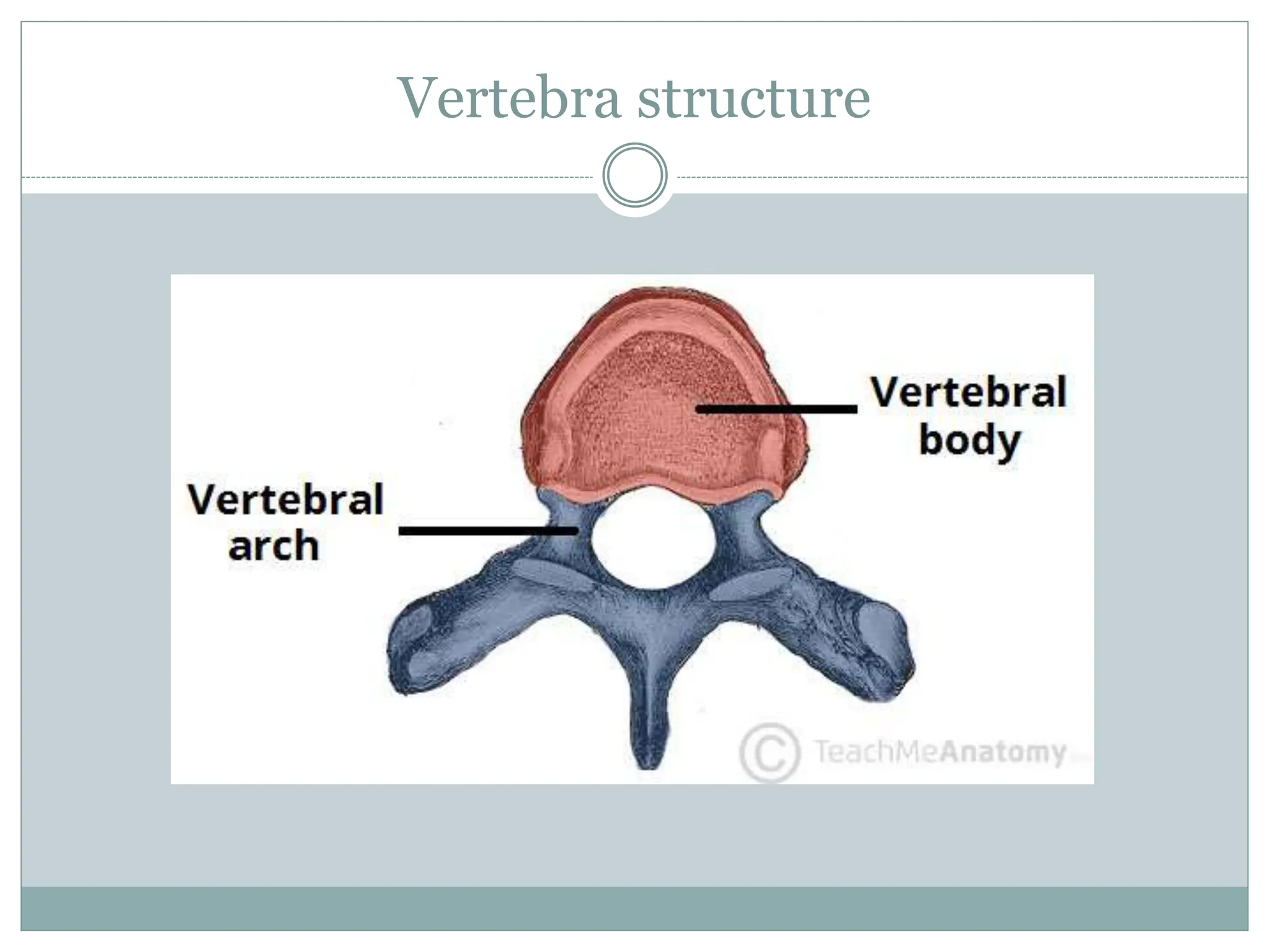
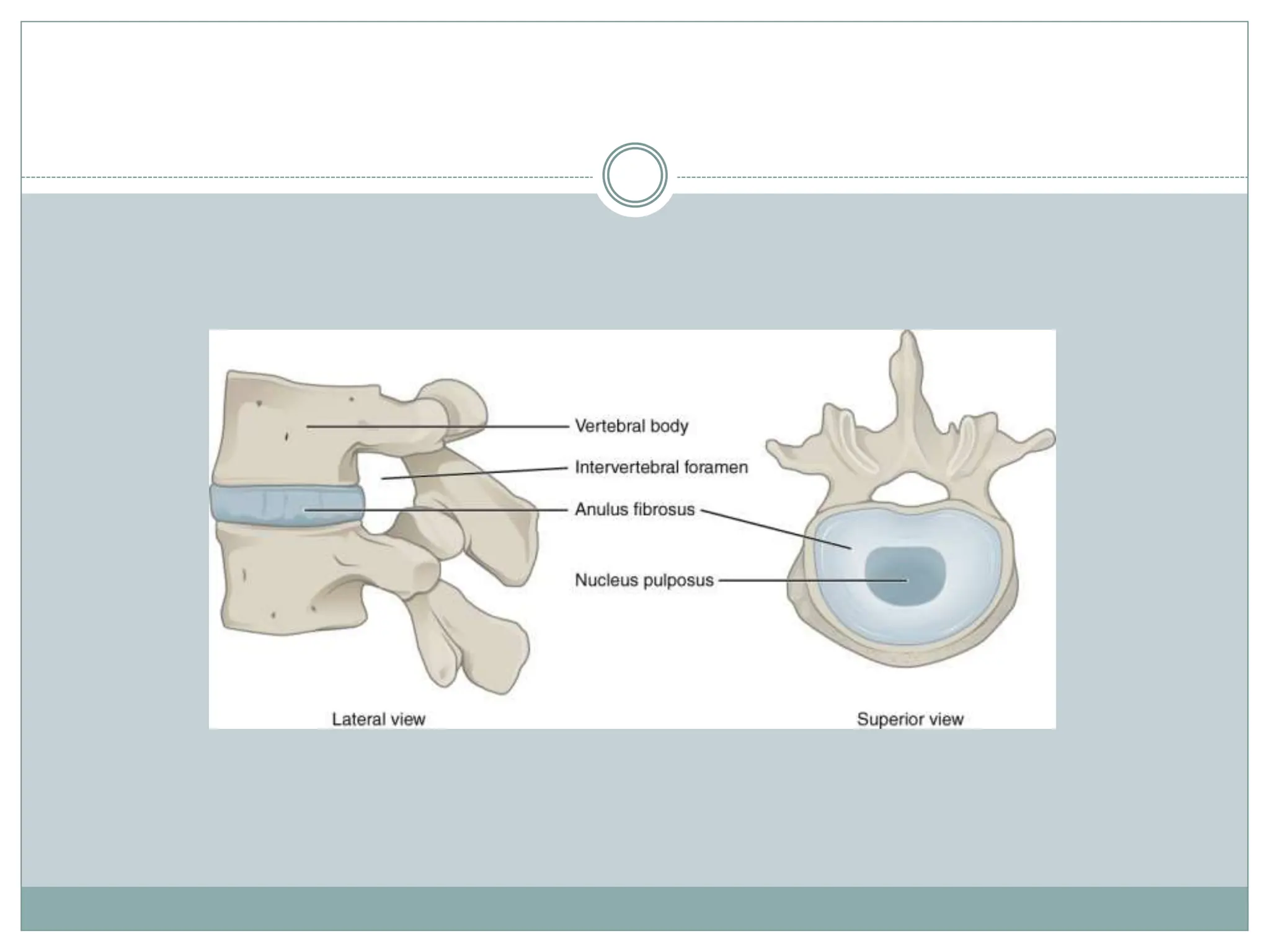
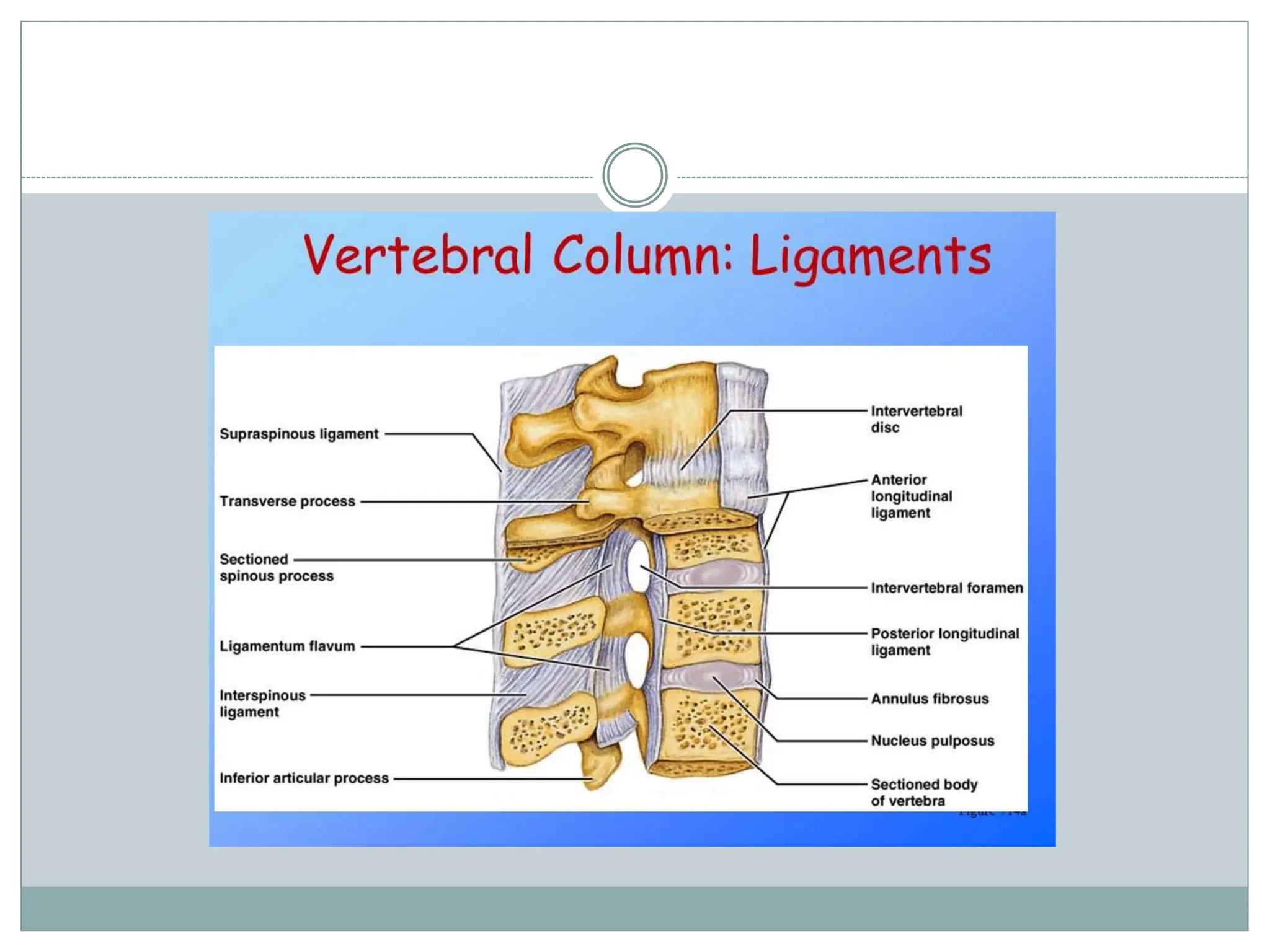
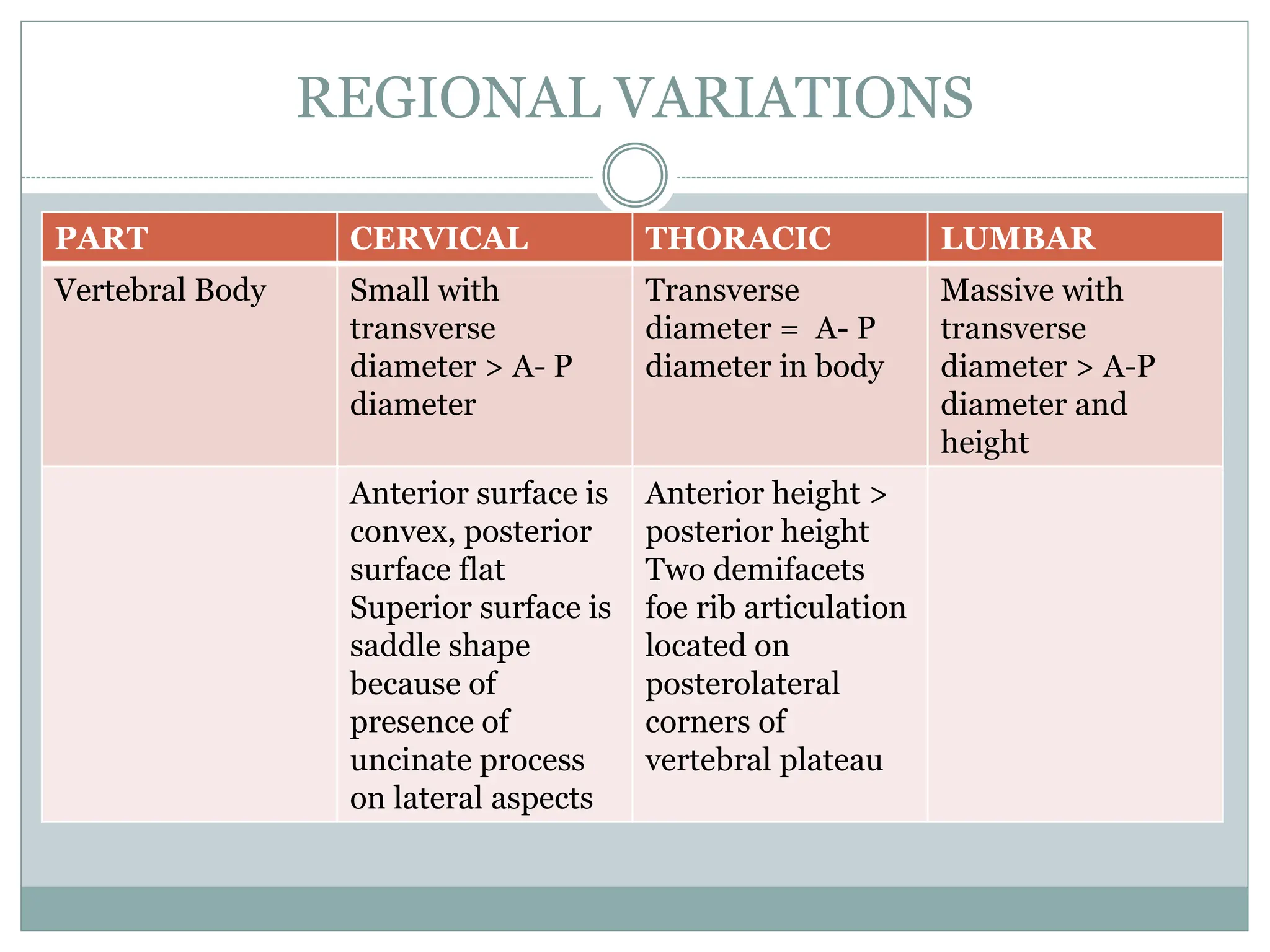
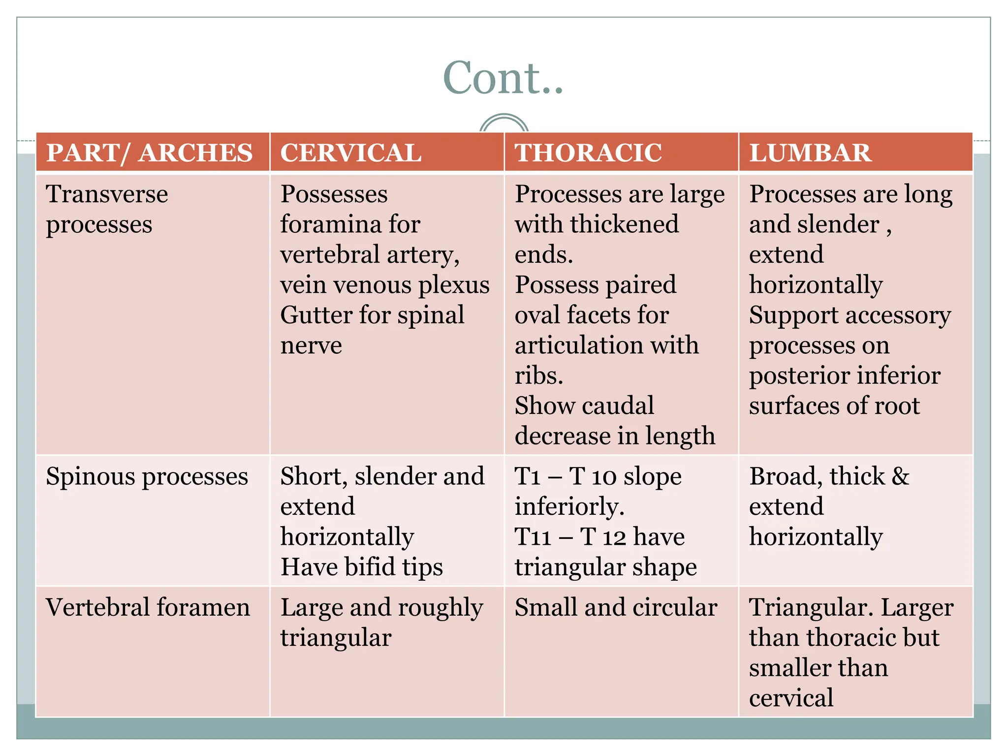
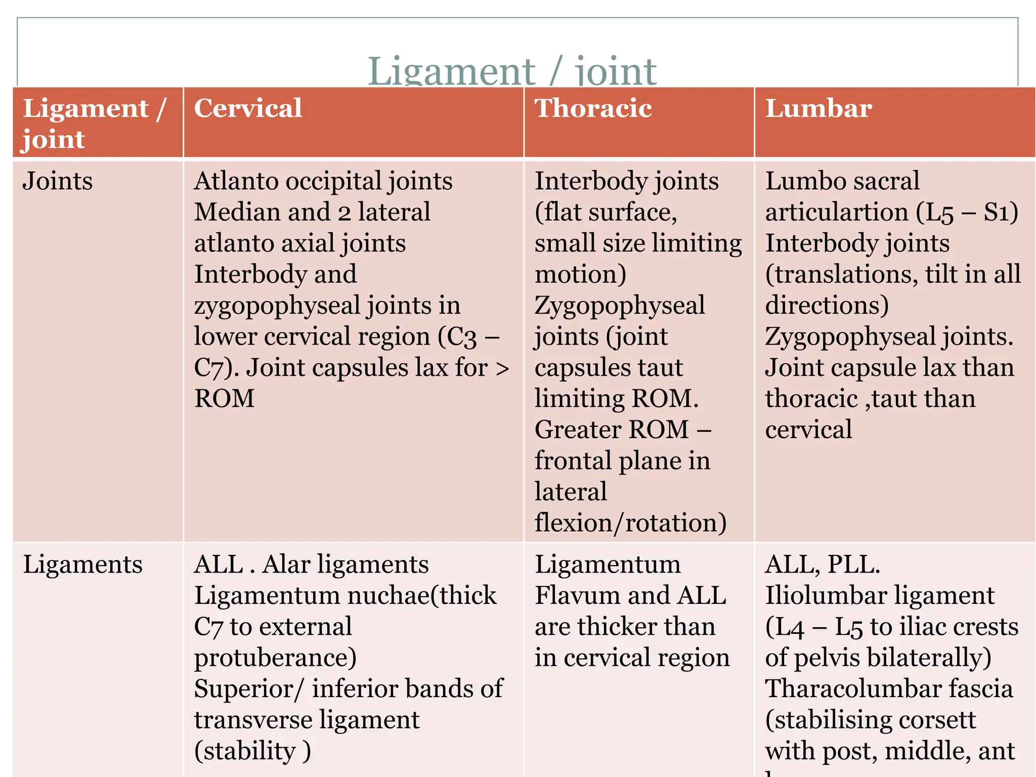
 S T R U C T U R E & F U N C T I O N
 R E G I O N A L S T R U C T U R E & F U N C T I O N
 M U S C L E S O F V E R T E B R A L C O L U M N
 E F F E C T S O F A G I N G & A P P L I E D A S P E C T S
Vertebral column
Vertebral column
 Amazing complex structure
 Meet contradictory demands of mobility and stability
of trunk
 Protection for spinal cord
 Pelvic attachment to vertebral column is crucial and
dealt with vertebral column
General structure
 Resembles a curved rod, composed of 33 vertebrae and 23
intervertebral disks
 Have common basic structure but with regional variations to meet
demands of that region.
 Five regions
- Cervical (7)
- Thoracic (12)
- Lumbar (5)
- Sacral (5- fused)
- Coccygeal (4)
- Increase in size from cervical to lumbar, decrease from sacral to
coccygeal
Curves in vertebral column
 Frontal plane- vertebral column bisects trunk (posterior aspect)
 Sagittal plane, curves are evident
 Baby – one long convex curve
 Secondary curve develop in infancy
 Adult- four distinct area
 Thoracic and sacral--- original curve through out life is primary curve ---
posterior convexity (KYPHOTIC CURVES)
 Cervical and lumbar--- reversal of posterior convexity is Secondary curves
(LORDOTIC CURVES). This develop , accommodation of skeleton to the
upright posture
 Normal curve has 10- fold ability to resist axial compression in comparison
with straight rod
 Mobile SEGMENT– 2 adjacent vertebra and intervening intervertebral
disks
Vertebral column
Typical vertebra
 2 parts
 - anterior (cylindrical shaped vertebral body)
 - posterior (irregularly shaped vertebral / neural arch)
 VERTEBRAL BODY
 - weight bearing structure of spinal column
 Block like shape with flat superior and inferior surfaces
 Not a solid block of bone, but a shell of cortical bone
surrounding a cancellous cavity (dynamic loading)
 - cortical shell reinforced by trabeculae in cancellous bone
providing resistance to compressive forces
Vertebra … cont
 NEURAL ARCH
1. Pedicles – connection between posterior elements and
vertebral bodies. Transmit tension and bending forces.
Short, stout pillars with thick walls. Increase in size from
cervical to lumbar (greater force in lumbar)
2. Posterior elements –
a) laminae (roof to neural arch. pars intercularis- force
transfers occur. Subjected to bending forces. Most developed in
lumber region
b) articular process- 2 superior and 2 inferior facets for
articulation from cranial and caudal vertebra
c) spinous process and d) transverse process sites for muscle
attachments, increasing lever arm
Cont..
 Intervertebral Disk
- To separate two vertebral bodies
- To transmit load from one vertebral body to the
next
- 20 – 30% 0f length of vertebral column
- Thickness 3 mm in – cervical and 9 mm in lumbar
region
- Greater functional needs for mobility found in
cervical and lumbar regions and for stability in
thoracic region
Cont..
- It comprises of 3 parts:
Nucleus Pulposus ( gelatinous mass in the center. 70
– 90% of water with collagen,
glycosaminoglycans,…)
Annulus Fibrosus ( fibrous outer ring. Composition
made such it resist greater proportions of tensile
forces. Attached to cartilagenous end plates by
SHARPEY FIBRES)
Vertebral End Plate (layers of cartilage . 0.6 to 1 mm
thick. Both hyaline and fibrocartilage are present)
Vertebra structure
 Innervation & Nutrition
Innervated in outer 1/3 rd to ½ of fibres of annulus
fibrosus
Innervation by branches from vertebral and
sinuvertebral nerves
Metaphyseal arteries
Articulations
 2 types
 1. cartilagenous joints of symphysis type between
vertebral bodies including interposed disks
(interbody joints). Gliding, distraction,
compression and rotation movements
 2. Diarthrodial or synovial joints between
zygapophyseal facets of one vertebra above and other
below (zygopophyseal joints). Protect
articular surfaces that exposed during flexion
and extension of the vertebral column
Ligaments
 Anterior longitudinal ligament (ALL)– increase in
thickness and width from lower thoracic vertebra to L5/S1.
associated with interbody joints in anterior/lateral surfaces.
C2 to occiput. To intervertebral disks
 posterior longitudinal ligaments (PLL)- posterior vertebral
bodies.. Vertebral end plates. C2 to sacrum
 Ligamentum flavum – thick, elastic ligament. Strong in
lower thoracic and week in midthoracic level. Even in
neutral this ligament inn constant tension. Compressive to
disks, such gives support for spine. Ligament not buckle.
Cont..
 Interspinous- Spinous process of adjacent vertebra.
Possible source of low back pain. First to be damaged in
excessive flexion.
 Supraspinous- ligamentum nuchae in cervical. Tips of
spinous process of C7 to L3/L4
 intertransverse ligaments - transverse process and deep
muscles of the back. Stressed and compressed during
lateral bending
 Zygopophyseal Joint Capsules----- assist ligament in
limiting motion, providing stability to vertebral column.
vulnerable to hyperflexion mainly in lumbar region
FUNCTION – Kinematics
 Coupling
 Flexion
 Extension
 Lateral flexion
Coupling
 Motions in vertebral column occur independently of each
other , but at level motion they coupled
 Coupling- consistent association of one motion about an
axis, another motion around different axis.
 More predominant- lateral flexion and rotation. They do
not exhibit as individual motion
 Coupling pattern depends on spinal posture, curves,
fluidity, elasticity, muscles
 Motion in interbody and zygopophyseal --- amount of
motion determined by size of disks, direction by
orientation of facets
 Gliding motion occur at interbody and zygopophyseal
joints
Flexion
 Vertebral flexion– anterior tilting and gliding of
superior vertebra.
 Widening of intervertebral foramen and separation
of spinous process
 Anterior portion of annulus fibrosus is compressed
and bulges anteriorly, posterior portion stretched
and resist separation of vertebral bodies
Extension
 Vertebral extension – posterior tilting and gliding of
superior vertebra
 Narrowing of intervertebral foramen
 Spinous process move close together
 Limited by size of disks, bony process.
 The only ligament limiting is ALL
Lateral Flexion
 Lateral flexion- - superior vertebra laterally tilts,
rotates and translates over adjacent vertebra below
 Annulus fibrosus compressed on concavity of the
curve, stretched on convexity of the curve
 Direction of rotation accomplishing lateral flexion
differs from region to region because of orientation
of facets
Kinetics
 Axial compression
 Bending
 Torsion
 Shear
Axial compression
 Force acting through long axis of spine at right angles to
the disks
 Occurs as a result of force of gravity, ground reaction
forces, and forces by ligaments and muscular
contractions
 Compressive load transfer from superior to inferior end
plate
 Intervertebral disks exhibit creep
 Zygopophyseal joints carry 0 – 30% of compression load
 Spinous processes share load when spine in
hyperextension
 Nucleus pulposus acts as ball of fluid that can be
deformed by a compression force and exhibits swelling
pressure
Cont..
 Annulus fibrosus and end plates provide sufficient resistance
to swelling pressure and maintain equilibrium
 Disks and trabecular bone undergo greater amount
deformation in axial compression
 End plates undergo least amount of deformation , and will be
first to fail
 Intervertebral disks exhibit creep producing diurnal changes
in disk composition and function
 Expressed fluid is absorbed through microscopic pores in
cartilagenous end plates
 Recovery fluid returns disk in unloading hence person getting
up from bed in morning looks taller
 Running is a form of dynamic loading increases disk height
 Height variations- 19 mm with loss of 1.5 mm
Bending
 Causes both compression and tension in spine
 Forward FLEXION – anterior structures compression,
posterior structures tension. Resistance offered by tensile
forces in collagen fibres, ligaments, hence providing stability
 Creep occurs in sustained loading, fully flexed postures, fully
extended postures. Resulting deformation curve places
vertebral structures at injury
 EXTENSION – posterior structures unloaded/compressed.
Anterior structures in tension. Resistance by annulus fibrosus,
passive tension in ALL
 LATERAL BENDING – ipsilateral side of disk is compressed,
other side stretched in Right and left. annulus fibrosus and
intertransverse ligament provide stability during and ressit
extreme motion
Torsion
 During axial rotation as a part of coupled motions in
supine
 Torsional stiffness low from T1 – T6 , increase from
T7 to T8 ---- L3/L4
 When disk subjected to torsion, half annulus
fibrosus, resist clock wise directions, others resist
counterclockwise direction.
 Risk of rupture increase when torsion, heavy
compression and bending combined
Shear
 Act on mid plane of disk and cause each vertebra to
undergo translation (move anteriorly, posteriorly,
sidewards in relation to inferior vertebra)
 When load is sustained, disk exhibit creep
REGIONAL VARIATIONS
PART CERVICAL THORACIC LUMBAR
Vertebral Body Small with
transverse
diameter > A- P
diameter
Transverse
diameter = A- P
diameter in body
Massive with
transverse
diameter > A-P
diameter and
height
Anterior surface is
convex, posterior
surface flat
Superior surface is
saddle shape
because of
presence of
uncinate process
on lateral aspects
Anterior height >
posterior height
Two demifacets
foe rib articulation
located on
posterolateral
corners of
vertebral plateau
Cont
PART /ARCHES CERVICAL THORACIC LUMBAR
Pedicles Project
posterolaterally
Variable in shape
and orientation
Short & thick
Laminae Project
posteromedially.
Thin and slightly
curved
Short, thick and
broad
Short & broad
Superior
zygopophyseal
facets
Face superiorly
and medially
Thin, flat. Face
posteriorly,
superiorly and
laterally
Vertical & concave.
Face
posteromedially
Support mamillary
processes
Inferior
zygopophyseal
facets
Face anteriorly
and laterally
Face anteriorly,
superiorly and
medially
Cont..
PART/ ARCHES CERVICAL THORACIC LUMBAR
Transverse
processes
Possesses
foramina for
vertebral artery,
vein venous plexus
Gutter for spinal
nerve
Processes are large
with thickened
ends.
Possess paired
oval facets for
articulation with
ribs.
Show caudal
decrease in length
Processes are long
and slender ,
extend
horizontally
Support accessory
processes on
posterior inferior
surfaces of root
Spinous processes Short, slender and
extend
horizontally
Have bifid tips
T1 – T 10 slope
inferiorly.
T11 – T 12 have
triangular shape
Broad, thick &
extend
horizontally
Vertebral foramen Large and roughly
triangular
Small and circular Triangular. Larger
than thoracic but
smaller than
cervical
Ligament / joint
Ligament /
joint
Cervical Thoracic Lumbar
Joints Atlanto occipital joints
Median and 2 lateral
atlanto axial joints
Interbody and
zygopophyseal joints in
lower cervical region (C3 –
C7). Joint capsules lax for >
ROM
Interbody joints
(flat surface,
small size limiting
motion)
Zygopophyseal
joints (joint
capsules taut
limiting ROM.
Greater ROM –
frontal plane in
lateral
flexion/rotation)
Lumbo sacral
articulartion (L5 – S1)
Interbody joints
(translations, tilt in all
directions)
Zygopophyseal joints.
Joint capsule lax than
thoracic ,taut than
cervical
Ligaments ALL . Alar ligaments
Ligamentum nuchae(thick
C7 to external
protuberance)
Superior/ inferior bands of
transverse ligament
(stability )
Ligamentum
Flavum and ALL
are thicker than
in cervical region
ALL, PLL.
Iliolumbar ligament
(L4 – L5 to iliac crests
of pelvis bilaterally)
Tharacolumbar fascia
(stabilising corsett
with post, middle, ant

Vertebral column... and Biomechanics.pptx

  • 1.
     S TR U C T U R E & F U N C T I O N  R E G I O N A L S T R U C T U R E & F U N C T I O N  M U S C L E S O F V E R T E B R A L C O L U M N  E F F E C T S O F A G I N G & A P P L I E D A S P E C T S Vertebral column
  • 2.
    Vertebral column  Amazingcomplex structure  Meet contradictory demands of mobility and stability of trunk  Protection for spinal cord  Pelvic attachment to vertebral column is crucial and dealt with vertebral column
  • 3.
    General structure  Resemblesa curved rod, composed of 33 vertebrae and 23 intervertebral disks  Have common basic structure but with regional variations to meet demands of that region.  Five regions - Cervical (7) - Thoracic (12) - Lumbar (5) - Sacral (5- fused) - Coccygeal (4) - Increase in size from cervical to lumbar, decrease from sacral to coccygeal
  • 4.
    Curves in vertebralcolumn  Frontal plane- vertebral column bisects trunk (posterior aspect)  Sagittal plane, curves are evident  Baby – one long convex curve  Secondary curve develop in infancy  Adult- four distinct area  Thoracic and sacral--- original curve through out life is primary curve --- posterior convexity (KYPHOTIC CURVES)  Cervical and lumbar--- reversal of posterior convexity is Secondary curves (LORDOTIC CURVES). This develop , accommodation of skeleton to the upright posture  Normal curve has 10- fold ability to resist axial compression in comparison with straight rod  Mobile SEGMENT– 2 adjacent vertebra and intervening intervertebral disks
  • 5.
  • 6.
    Typical vertebra  2parts  - anterior (cylindrical shaped vertebral body)  - posterior (irregularly shaped vertebral / neural arch)  VERTEBRAL BODY  - weight bearing structure of spinal column  Block like shape with flat superior and inferior surfaces  Not a solid block of bone, but a shell of cortical bone surrounding a cancellous cavity (dynamic loading)  - cortical shell reinforced by trabeculae in cancellous bone providing resistance to compressive forces
  • 7.
    Vertebra … cont NEURAL ARCH 1. Pedicles – connection between posterior elements and vertebral bodies. Transmit tension and bending forces. Short, stout pillars with thick walls. Increase in size from cervical to lumbar (greater force in lumbar) 2. Posterior elements – a) laminae (roof to neural arch. pars intercularis- force transfers occur. Subjected to bending forces. Most developed in lumber region b) articular process- 2 superior and 2 inferior facets for articulation from cranial and caudal vertebra c) spinous process and d) transverse process sites for muscle attachments, increasing lever arm
  • 8.
    Cont..  Intervertebral Disk -To separate two vertebral bodies - To transmit load from one vertebral body to the next - 20 – 30% 0f length of vertebral column - Thickness 3 mm in – cervical and 9 mm in lumbar region - Greater functional needs for mobility found in cervical and lumbar regions and for stability in thoracic region
  • 9.
    Cont.. - It comprisesof 3 parts: Nucleus Pulposus ( gelatinous mass in the center. 70 – 90% of water with collagen, glycosaminoglycans,…) Annulus Fibrosus ( fibrous outer ring. Composition made such it resist greater proportions of tensile forces. Attached to cartilagenous end plates by SHARPEY FIBRES) Vertebral End Plate (layers of cartilage . 0.6 to 1 mm thick. Both hyaline and fibrocartilage are present)
  • 10.
  • 11.
     Innervation &Nutrition Innervated in outer 1/3 rd to ½ of fibres of annulus fibrosus Innervation by branches from vertebral and sinuvertebral nerves Metaphyseal arteries
  • 12.
    Articulations  2 types 1. cartilagenous joints of symphysis type between vertebral bodies including interposed disks (interbody joints). Gliding, distraction, compression and rotation movements  2. Diarthrodial or synovial joints between zygapophyseal facets of one vertebra above and other below (zygopophyseal joints). Protect articular surfaces that exposed during flexion and extension of the vertebral column
  • 14.
    Ligaments  Anterior longitudinalligament (ALL)– increase in thickness and width from lower thoracic vertebra to L5/S1. associated with interbody joints in anterior/lateral surfaces. C2 to occiput. To intervertebral disks  posterior longitudinal ligaments (PLL)- posterior vertebral bodies.. Vertebral end plates. C2 to sacrum  Ligamentum flavum – thick, elastic ligament. Strong in lower thoracic and week in midthoracic level. Even in neutral this ligament inn constant tension. Compressive to disks, such gives support for spine. Ligament not buckle.
  • 15.
    Cont..  Interspinous- Spinousprocess of adjacent vertebra. Possible source of low back pain. First to be damaged in excessive flexion.  Supraspinous- ligamentum nuchae in cervical. Tips of spinous process of C7 to L3/L4  intertransverse ligaments - transverse process and deep muscles of the back. Stressed and compressed during lateral bending  Zygopophyseal Joint Capsules----- assist ligament in limiting motion, providing stability to vertebral column. vulnerable to hyperflexion mainly in lumbar region
  • 17.
    FUNCTION – Kinematics Coupling  Flexion  Extension  Lateral flexion
  • 18.
    Coupling  Motions invertebral column occur independently of each other , but at level motion they coupled  Coupling- consistent association of one motion about an axis, another motion around different axis.  More predominant- lateral flexion and rotation. They do not exhibit as individual motion  Coupling pattern depends on spinal posture, curves, fluidity, elasticity, muscles  Motion in interbody and zygopophyseal --- amount of motion determined by size of disks, direction by orientation of facets  Gliding motion occur at interbody and zygopophyseal joints
  • 19.
    Flexion  Vertebral flexion–anterior tilting and gliding of superior vertebra.  Widening of intervertebral foramen and separation of spinous process  Anterior portion of annulus fibrosus is compressed and bulges anteriorly, posterior portion stretched and resist separation of vertebral bodies
  • 20.
    Extension  Vertebral extension– posterior tilting and gliding of superior vertebra  Narrowing of intervertebral foramen  Spinous process move close together  Limited by size of disks, bony process.  The only ligament limiting is ALL
  • 21.
    Lateral Flexion  Lateralflexion- - superior vertebra laterally tilts, rotates and translates over adjacent vertebra below  Annulus fibrosus compressed on concavity of the curve, stretched on convexity of the curve  Direction of rotation accomplishing lateral flexion differs from region to region because of orientation of facets
  • 22.
    Kinetics  Axial compression Bending  Torsion  Shear
  • 23.
    Axial compression  Forceacting through long axis of spine at right angles to the disks  Occurs as a result of force of gravity, ground reaction forces, and forces by ligaments and muscular contractions  Compressive load transfer from superior to inferior end plate  Intervertebral disks exhibit creep  Zygopophyseal joints carry 0 – 30% of compression load  Spinous processes share load when spine in hyperextension  Nucleus pulposus acts as ball of fluid that can be deformed by a compression force and exhibits swelling pressure
  • 24.
    Cont..  Annulus fibrosusand end plates provide sufficient resistance to swelling pressure and maintain equilibrium  Disks and trabecular bone undergo greater amount deformation in axial compression  End plates undergo least amount of deformation , and will be first to fail  Intervertebral disks exhibit creep producing diurnal changes in disk composition and function  Expressed fluid is absorbed through microscopic pores in cartilagenous end plates  Recovery fluid returns disk in unloading hence person getting up from bed in morning looks taller  Running is a form of dynamic loading increases disk height  Height variations- 19 mm with loss of 1.5 mm
  • 25.
    Bending  Causes bothcompression and tension in spine  Forward FLEXION – anterior structures compression, posterior structures tension. Resistance offered by tensile forces in collagen fibres, ligaments, hence providing stability  Creep occurs in sustained loading, fully flexed postures, fully extended postures. Resulting deformation curve places vertebral structures at injury  EXTENSION – posterior structures unloaded/compressed. Anterior structures in tension. Resistance by annulus fibrosus, passive tension in ALL  LATERAL BENDING – ipsilateral side of disk is compressed, other side stretched in Right and left. annulus fibrosus and intertransverse ligament provide stability during and ressit extreme motion
  • 26.
    Torsion  During axialrotation as a part of coupled motions in supine  Torsional stiffness low from T1 – T6 , increase from T7 to T8 ---- L3/L4  When disk subjected to torsion, half annulus fibrosus, resist clock wise directions, others resist counterclockwise direction.  Risk of rupture increase when torsion, heavy compression and bending combined
  • 27.
    Shear  Act onmid plane of disk and cause each vertebra to undergo translation (move anteriorly, posteriorly, sidewards in relation to inferior vertebra)  When load is sustained, disk exhibit creep
  • 28.
    REGIONAL VARIATIONS PART CERVICALTHORACIC LUMBAR Vertebral Body Small with transverse diameter > A- P diameter Transverse diameter = A- P diameter in body Massive with transverse diameter > A-P diameter and height Anterior surface is convex, posterior surface flat Superior surface is saddle shape because of presence of uncinate process on lateral aspects Anterior height > posterior height Two demifacets foe rib articulation located on posterolateral corners of vertebral plateau
  • 29.
    Cont PART /ARCHES CERVICALTHORACIC LUMBAR Pedicles Project posterolaterally Variable in shape and orientation Short & thick Laminae Project posteromedially. Thin and slightly curved Short, thick and broad Short & broad Superior zygopophyseal facets Face superiorly and medially Thin, flat. Face posteriorly, superiorly and laterally Vertical & concave. Face posteromedially Support mamillary processes Inferior zygopophyseal facets Face anteriorly and laterally Face anteriorly, superiorly and medially
  • 30.
    Cont.. PART/ ARCHES CERVICALTHORACIC LUMBAR Transverse processes Possesses foramina for vertebral artery, vein venous plexus Gutter for spinal nerve Processes are large with thickened ends. Possess paired oval facets for articulation with ribs. Show caudal decrease in length Processes are long and slender , extend horizontally Support accessory processes on posterior inferior surfaces of root Spinous processes Short, slender and extend horizontally Have bifid tips T1 – T 10 slope inferiorly. T11 – T 12 have triangular shape Broad, thick & extend horizontally Vertebral foramen Large and roughly triangular Small and circular Triangular. Larger than thoracic but smaller than cervical
  • 31.
    Ligament / joint Ligament/ joint Cervical Thoracic Lumbar Joints Atlanto occipital joints Median and 2 lateral atlanto axial joints Interbody and zygopophyseal joints in lower cervical region (C3 – C7). Joint capsules lax for > ROM Interbody joints (flat surface, small size limiting motion) Zygopophyseal joints (joint capsules taut limiting ROM. Greater ROM – frontal plane in lateral flexion/rotation) Lumbo sacral articulartion (L5 – S1) Interbody joints (translations, tilt in all directions) Zygopophyseal joints. Joint capsule lax than thoracic ,taut than cervical Ligaments ALL . Alar ligaments Ligamentum nuchae(thick C7 to external protuberance) Superior/ inferior bands of transverse ligament (stability ) Ligamentum Flavum and ALL are thicker than in cervical region ALL, PLL. Iliolumbar ligament (L4 – L5 to iliac crests of pelvis bilaterally) Tharacolumbar fascia (stabilising corsett with post, middle, ant