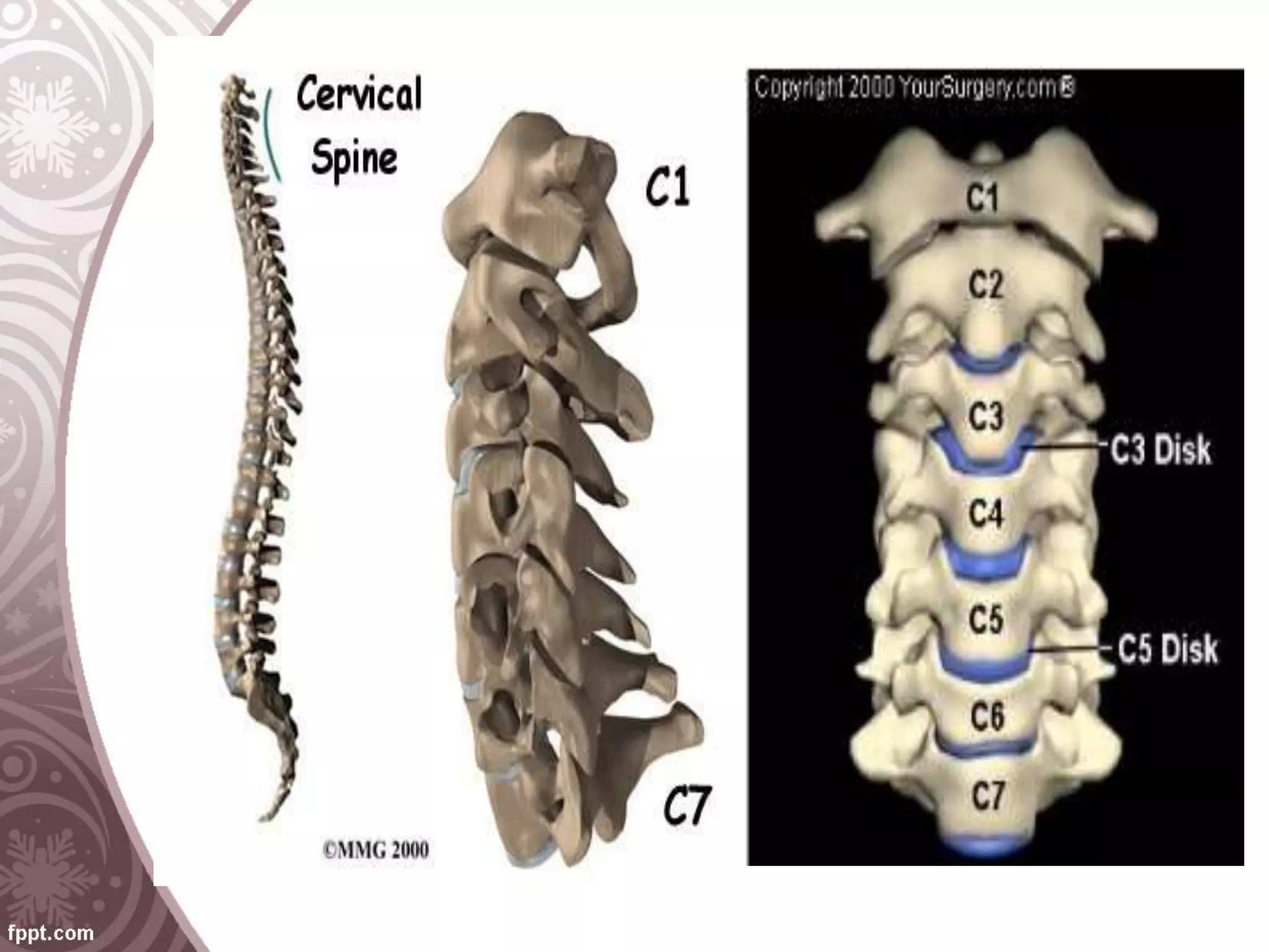
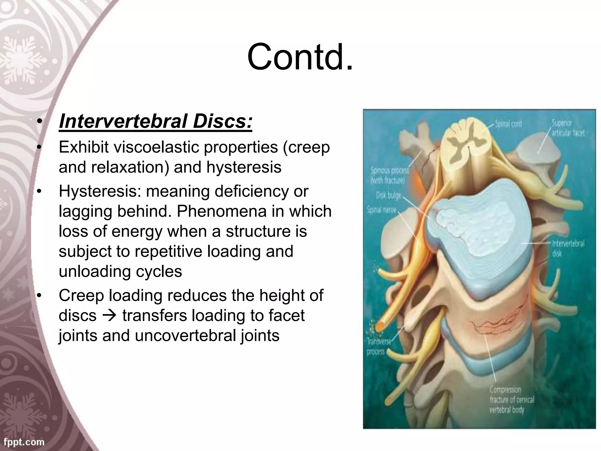
BIOMECHANICS OF
THE CERVICAL SPINE
Chapter # 11
Dr.Muhammad Tahir Hafeez
Objectives
• Introduction to anatomy and biomechanics including:
• Anatomy of Osseous structures, intervertebral discs
• Mechanical properties of Vertebrae, Discs, Ligaments,
Muscles and Neural elements
• Describe the importance of Kinematics including:
• ROM, surface joint motion, Spinal stability
• Introduction to Applied Biomechanics: Decompression,
Arthrodesis, Cervical spine fixation
• Biomechanics of Cervical Trauma: airbag injuries and
whiplash syndrome
• Conclusion and summary
Introduction
• Sir Frank Holdsworth (1963)
describe two-column model of the
spine
• Denis (1983) refined the
principles of spinal stability and
introduce three-column model
Component Anatomy And
Biomechanics
• Anatomy:
• Cervical spine supports the skull
• Acts as a shock absorber for the brain
• Protects brainstem, spinal cord and various
neurovascular structures
• Biomechanical function is to facilitate the transfer of
weight and bending movements of head
• Neuromuscular control combined with articulations of
cervical spine, allows wide range of physiologic motion
Cont.
• Spine consists of 33 vertebrae divided into five regions:
• Cervical (7)
• Thoracic (12)
• Lumbar (5)
• Sacral (5 fused segments)
• Coccygeal (approx. 4)
• Two most cranial vertebrae C1 (atlas) and C2 (axis) =
atypical
• Functional part of the cervical spine = atlanto-occipital joint
• C3  C7 = typical vertebrae
• Cervical + Lumbar = convex anteriorly (lordotic)
• Thoracic + Sacral = convex posteriorly (kyphotic)
Contd.
• Motion segment/Functional spinal unit = biomechanical
building block of spinal column
• Motion segment = 2 adjacent vertebrae, intervertebral
disc, ligaments b/w vertebrae
• Ligaments: ant. & post. Longitudinal, intertransverse,
interspinous, supraspinous and facet capsular
• Collagenous tissues exhibit both viscoelastic and
anisotropic behavior
Contd.
• Viscoelastic: properties are rate-dependent
(time-dependent) behaviors under loading 
present in both bone and soft tissues 
mechanical strength increases with increased
rates of loading
• Anisotropy: alteration in mechanical properties
 seen when bone is loaded along diff. axes 
occurs as a result of dissimilar crystalline
microstructure of bone
Osseous Structures
• Occiput C1-C2: constitutes upper cervical spine  C/S
flexion: 40%  C/S rotation: 60%
• Primary motion: Flexion and extension (much of sagittal
ROM)
• Primarily responsible for rotation
• Occipitocervical joint: extension is limited by bony anatomy
 Flexion is limited by post. Ligamentous structures
• Alar ligaments: attach dens to occiput to prevent excessive
rotation
• Act to limit motion during side bending
• Facet joint: resist most of shear forces (approx. 16%)
• Bipeds use head rotation to look around
Intervertebral Discs
• Contribute up to one-third of height of the vertebral column
• Activities such as running, jumping apply  short-duration, high-
amplitude loads
• Normal physical activity and upright stance  result in long-
duration, low-magnitude loads
• Nucleus pulposus is centrally located within disc  consist of 90%
water
• Rest of nucleus pulposus consist of PGs and collagen (type II)
• Type II collagen fibrils able to absorb compressive forces
• Annulus fibrosus is outer portion of disc  water content is less
approx. 78%
• Type-II 60% and Type-I 40% in annulus
Mechanical Properties
• Stress: load per unit area of a perpendicularly
applied load
• Strain: change in length per unit of original
length, usually expressed as percentage
Contd.
• Vertebrae:
• Cortical bone is stiffer than cancellous bone  withstand greater
stresses before failure
• When strain exceeds 2% of original length  cortical bone fractures
• Cancellous bone withstand greater strain before fracturing
• Vertebral compression strength increases from upper cervical to
lower lumbar
• Development of osteoporosis  abnormal bone loss ensues  risk
of fracture  minor trauma, falls or “silent” vertebral compression
• 25% decrease in osseous tissue  results in 50% decrease in
strength in vertebrae
• Cortical shell of vertebrae  responsible for 10% of strength during
compression
Contd.
• Intervertebral Discs:
• Exhibit viscoelastic properties (creep
and relaxation) and hysteresis
• Hysteresis: meaning deficiency or
lagging behind. Phenomena in which
loss of energy when a structure is
subject to repetitive loading and
unloading cycles
• Creep loading reduces the height of
discs  transfers loading to facet
joints and uncovertebral joints
Contd.
• Ligaments:
• Stability depends primarily on soft tissue components, especially in
cervical spine
• Ligament strength and extensibility help maintain stability
• All ligaments have high collagen content except for ligamentum
flavum (having large % of elastin)
• Elastic properties also assist in limiting the inward buckling of
ligaments during extension  potentially compress neural elements
Contd.
• Muscle:
• Muscular strength and control help maintain head and neck balance
• It also reduces stress on bone
• In flexion: tensile stress to posterior  compression to anterior
• Play a critical role in basic postural homeostasis
Contd.
• Neural Elements:
• Spinal cord injuries also result from  sudden flexion-extension
movements
• Neurologic injuries may result from  anterior posterior
compression of cord  more common in stenotic canal
• Flexion motion result in injuries  when spinal cord contacts with
osteophytes
• Extension result in  pincer-like compression of cord b/w
osteophytes (ant.) and ligamentum flavum (post.)
Kinematics
• Study of the motion of rigid bodies without taking relevant forces
• It describes the physiologic and pathologic motions occur in
various spinal units
• Motion segment/functional spinal unit is the unit of study in
kinematics
• Degree of Freedom: motion in which a rigid body can either
translate back and forth along straight line or rotate around
particular axis
• Each vertebral body can translate or rotate in each of three
orthogonal planes  for total of six degrees of freedom
Range Of Motion
• C1-C2 Axial Rotation range:
• Active  27° to 49° (mean = 39°)
• Passive  29° to 46° (mean = 41°)
• They account for approx. 50% of total cervical rotation
• C1-C2 Flexion and Extension:
• Flexion  5° to 20°
• Extension  Active (12°) to passive (15°)
• Subaxial cervical spine: 90° rotation takes place in C3-C7
• ROM of Flexion and Extension  64° (24° extension, 40° flexion)
Surface Joint Motion
• Instant Center Technique (Reuleaux): analyze the motion
b/w joint surfaces of two adjacent vertebrae
• Used to analyze surface motion of cervical spine  in flexion-
extension & lat. Flexion
• IC of flexion-extension  located in ant. part of lower vertebrae in
each motion segment
• IC indicates tangential motion (gliding)  takes place b/w facet
joints in flex. & ext.
• Size of intervertebral foramina increases with flex. and decreases
with ext.
• Foam collars place in slight ext.  aggravate symptoms  turning
collar around puts neck in flex.  increase size of intervertebral
foramina  relieve pressure on an inflamed nerve root
• Pathologic process (disc degeneration,lig. Impairment) can displace
IC  jamming (compression) and distraction of facet joint surfaces
 instead of gliding
Coupled Motion Of Cervical spine
• Atlantoaxial Segment: this area is extremely mobile
• IC of both rotation and flexion-extension lie in the center of dens
itself
• C1-2 joint most stable in neutral position
• C1 rises on C2  when head rotates away from midline
• Subaxial Spine: coupling patterns in lower c/s spine ;
• In lateral bending (left)  spinous process move to right
• In lateral bending (right)  spinous process move to left
• Facet joints and uncovertebral joints  major contributors to
coupled motion in lower c/s spine
Abnormal Kinematics
• Refers to “excessive motion” within functional spinal units
• May also refers to “atypical patterns of motion” such as abnormal
coupling, paradoxical motion
• Paradoxical Motion: when overall pattern of motion of one aspect is
in one direction and local pattern in opposite
• Example: Paradoxical flexion  seen when flexion occurs at single
functional spinal unit  although spine is extended as whole
• These can create a pattern of movement known as “instability”
Spinal Stability
• What is stability? How it is determined?
• describe the term clinically
• “ability of the spine under physiologic loads to
maintain its pattern of displacement, so there is no
neurologic deficit, no major deformity and no
incapacitating pain”
• Ligamentous stability plays a large part in the stability of spine
• Kinematic instability: "focuses on quantity of motion
(too much or too little) or the quality of motion
present (alterations), or both”
• Component instability: “addresses the clinical
biomechanical role of various anatomic components
of functional spinal unit”
Conceptual Types of Instability
Kinematic
instability
Component
instability
Combined
stability
• Motion
increased
• Instantaneous
axes of rotation
altered
• Coupling
characteristics
changed
• Paradoxical
motion present
• Trauma
• Tumor
• Surgery
• Degenerative
changes
• Developmental
changes
• Kinematic
• Component
Occipitoatlantoaxial Complex
• Transverse ligament  of atlas allows dens to rotate  limits its ant.
translation  non-elastic  doesn’t permit more than 2 – 3mm of ant.
translation
• Ant. displacement of C1 on C2  3 – 5mm  indicates rupture of
transverse lig.
• Displacements of 5 – 10mm  indicates accessory ligament damage
• Displacements > 10mm  occurs with rupture of all ligaments
• Ant. Translations or displacements of C1 on C2 assessed
radiographically by measuring distance from ant ring of atlas to back
of dens
• Steel’s rule of thirds (1968): guide to amount of Atlantoaxial
displacement can occur before spinal cord compression ensues
Subaxial Cervical Spine
• Nuchal ligament: plays major role in proprioception and correct
functioning of erector spinae muscles
• Motion segment considered unstable when  all anterior or all
posterior elements are destroyed or are unable to function
• Guidelines for the determination of clinical instability in lower
cervical spine have been provided in form of a scoring system
checklist
• Total of 5 or more = clinically unstable
Checklist for diagnosis of clinical instability in middle and lower cervical
spine
Element Point Value
Ant. element destroyed/ unable o function 2
Post. element destroyed/ unable to function 2
Positive stretch test 2
Radiographic criteria:
Flexion and Extension radiographs
Or
Resting radiographs
4
Narrow spinal canal 1
Disc narrowing 1
Spinal cord damage 2
Nerve root damage 1
Dangerous loading anticipated 1
Applied biomechanics
• Understanding of biomechanical principles is an important aspect
bcoz structure and function of spinal column is frequently altered
during surgery
• Knowledge benefits patient care and is valuable in planning and
executing treatment
Decompression
• To decompress spinal cord  Cervical laminectomy is performed
• Causes of compression: stenotic process  resulting neurologic
symptoms (radiculopathy/myelopathy)
• Posterior decompressive procedures  full/partial facetectomies for
visualization or decompression of nerve root pathology  commonly
performed
• Herkowitz, 1988: development of postlaminectomy kyphosis develop
in children  17% to 25% of adults
• Lonstein, 1977: children who undergo laminectomies for spinal cord
tumors  spinal deformity occur in up to 50% of children
Cont.
• Primary cause of postlaminectomy deformity  resection of one or
more spinous processes  post. Ligamentous structures
(ligamentum flavum/ interspinous / supraspinous ligaments
• Removal of ligaments and spinous process  causes tensile forces
to become unbalanced and extra stress on facet
• Cervical subluxations and dislocations  narrow spinal canal 
cause neurologic impairment
• Adequate reduction and realignment of vertebrae  followed by
stabilization  decompress neural elements  without resecting
bone
Arthrodesis
• Indication: spinal instability, neoplasm, post-traumatic, degenerative
condition
• Goal: to achieve solid bony union b/w two or more vertebrae
• In many cases  internal fixation is used  to achieve initial
stabilization and to correct deformity
• Stability established by internal fixation is a prelude to biologic
process of fusion
• Mechanical protection of graft in intervertebral space  increase
fusion rate  maintain structural alignment
• Internal fixation which is not supported  by solid fusion  will
fatigue and fail
• “Race” is to  achieve a solid fusion before fatigue failure of fixation
occurs
Contd.
• Surgical approach to cervical spine  ant/ post/ or combined
arthrodesis  depends on pathology
• Adjacent level disease: development of new
radiculopathy/myelopathy referable to motion segment adjacent to
site of ant. cervical arthrodesis
• Hilibrand et al., 1999: performed study describing incidence,
prevalence and radiographic progression of symptomatic adjacent
level disease after cervical arthrodesis
• Analysis revealed that approx. 26% of patients having ant. cervical
arthrodesis had new disease at an adjacent level within 10 years of
operation
• Study also demonstrated that >2/3 of patients developed adjacent
level cervical disease
• Bartels et al., 2008: Total disc arthroplasty  preserve physiologic
motion of spine  reduce stress to functional spinal units  induced
by arthrodesis
Cervical Spine Fixation
• Indication of arthrodesis of cervical spine  most commonly for
trauma and degenerative disease
• Ant. Discectomy/ or vertebrectomy is followed by ant. Cervical
arthrodesis
• Excised disc or bone must be replaced with structural graft or
prosthesis  to restore ant. Column support
• Osseous replacement in form of autogenous or allogenic bone 
commonly from iliac crest (if autogenous)  from fibula or iliac crest
(if allogenic)
• Gold standard is tricortical iliac crest bone  associated with high
donor site morbidity
Contd.
• Goat holds its head erect and loads cervical spine similar to human
bipeds
• Goat models for cervical spine biologic/ biomechanical testing are
popular
• Gu et al., 2007: mature goat cervical spine model was tested
biomechanically  volume-related stiffness of titanium, carbon fiber,
and PEEK (polyetheretherketone) cages was higher than iliac bone
graft
• Pintar et al., 1994: in goats synthetic hydroxylapatite blocks used in
cervical arthrodesis  produced similar fusion rates and stiffness 
compared with autogenous bone
• Emery et al., 1996: autogenous iliac crest grafts were stiffer in all
motions than ceramic (calcium phosphate) graft subtitutes
Contd.
• Grown coral: graft is also useful  available commercially in two
porosities (200mm & 500mm)
• Lower porosity grafts have compressive strength compared to
bicortical iliac crest grafts (more brittle)  appropriate for clinical use
• Wilke et al., 2000: Additional ant. or post. Internal fixation using
plates and screws for added support may be used
• Tendency of intervertebral implant depends upon two factors:
1. Shape of implant  especially its contact area at implant
2. End plate interface
• Post. Arthrodesis: commonly performed after trauma  used to treat
degenerative, inflammatory or neoplastic conditions  can extend
up to occiput
Contd.
• Ant. Fixation devices: mainly used in Subaxial cervical spine
• Sutterlin et al., 1988: posterior wire fixation is often used as post.
tension band
• Bambakidis et al., 2008; Frush et al., 2009: screw and plate or rod
fixation techniques  commonly provide stable segmental fixation
• Moskovich and Crockard, 1992: techniques for Atlantoaxial fixation
include wire fixation and interlammar clamp fixation
Biomechanics of Cervical Trauma
AIRBAG INJURIES:
• Motor vehicle accidents is leading cause of injury-related deaths in
the US
• In 1984 NHTSA (national highway traffic safety administration) 
automatic occupant protection devices (airbags or automatic seat
belts) be placed in all automobiles  in 1987 – 1990 model years
• Passenger-side airbag was introduced in 1993
• King and Yang, 1995: studies concluded that front seat occupants
adequately protected against frontal impact  if belts are worn in an
airbag-equipped vehicle
• Marshall et al., 1998: After airbag devices became available 
airbag injuries involving front seat passengers began  many child
deaths and serious injuries attributed to passenger-side airbags
Contd.
• Collisions were low-speed accidents in which driver have no or minor
injuries  pattern of injury in the rear-facing infant car seat often had
massive skull injury & cerebral hematoma  as a result of proximity of
their heads to airbag  in forward-facing child car seats children
sustains more cervical injuries
• NHTSA GUIDELINES emphasize children of any age should be
properly secured in the back seat (1996)
NHTSA GUIDELINES REGARDING AIRBAGS AND CHILDREN
Back seat is the safest place for children of any age to ride
Never put an infant (<1 year) in front of car that has passenger-side airbag
Infants must always ride back seat, facing the rear of car
Everyone is buckled up and unblocked occupants can hurt or killed by an
airbag
Whiplash Syndrome
• Complete set of symptoms present after an acceleration
hyperextension injury
• Crowe used term “whiplash” in 1928  describing neck injuries caused
by rear-end collisions in US
• Common traumatic event  include neck & shoulder pain, dizziness,
headache and blurring of vision
• Typically occur when car is struck from behind but also cause side or
head on collisions (Barnsley et al., 1994)
• Acceleration of car seat pushes the torso of occupant forward  results
in unsupported head falls backward  resulting in an extension strain
to neck
• Secondary flexion injury may occur if vehicle struck then strikes other
vehicle in front  suddenly decelerates again  throwing occupant
forward once more
Contd.
• Injuries include: interspinous ligamentum tears, spinous process
fractures, disc rupture, ligamentum flavum rupture, facet joint
disruption and stretching of ant. muscles
• Early unpublished biomechanical study done by Dr. Irving Tuell 
used cine camera to photograph himself driving as he rammed from
behind by other car  movie clearly demonstrate the hyperextension
of neck over seatback  effect of inertial forces on mandible explain
TMJ joint injuries are commonly associated with whiplash injuries
• Brault et al., 2000: enhanced Dr. Tuell’s study  42 subjects (21 men
and 21 women b/w 20-40 age)  collisions of 4 and 8 km/h speed
performing EMG of sternomastoid and cervical paraspinal muscles 
data reveals that cervical muscles contract rapidly in response to
impact  resulting in lengthening contractions  muscles fire fast
enough to have an influence on injury
Contd.
• Correct positioning of adjustable headrests behind skull is important
• If all adjustable headrests were placed in up position, the relative
risk will lowered to 2.4, 28.3% reduction in whiplash injury risk
THE END

biomechanics of cervical.ppt

  • 1.
    BIOMECHANICS OF THE CERVICALSPINE Chapter # 11 Dr.Muhammad Tahir Hafeez
  • 2.
    Objectives • Introduction toanatomy and biomechanics including: • Anatomy of Osseous structures, intervertebral discs • Mechanical properties of Vertebrae, Discs, Ligaments, Muscles and Neural elements • Describe the importance of Kinematics including: • ROM, surface joint motion, Spinal stability • Introduction to Applied Biomechanics: Decompression, Arthrodesis, Cervical spine fixation • Biomechanics of Cervical Trauma: airbag injuries and whiplash syndrome • Conclusion and summary
  • 3.
    Introduction • Sir FrankHoldsworth (1963) describe two-column model of the spine • Denis (1983) refined the principles of spinal stability and introduce three-column model
  • 4.
    Component Anatomy And Biomechanics •Anatomy: • Cervical spine supports the skull • Acts as a shock absorber for the brain • Protects brainstem, spinal cord and various neurovascular structures • Biomechanical function is to facilitate the transfer of weight and bending movements of head • Neuromuscular control combined with articulations of cervical spine, allows wide range of physiologic motion
  • 5.
    Cont. • Spine consistsof 33 vertebrae divided into five regions: • Cervical (7) • Thoracic (12) • Lumbar (5) • Sacral (5 fused segments) • Coccygeal (approx. 4) • Two most cranial vertebrae C1 (atlas) and C2 (axis) = atypical • Functional part of the cervical spine = atlanto-occipital joint • C3  C7 = typical vertebrae • Cervical + Lumbar = convex anteriorly (lordotic) • Thoracic + Sacral = convex posteriorly (kyphotic)
  • 7.
    Contd. • Motion segment/Functionalspinal unit = biomechanical building block of spinal column • Motion segment = 2 adjacent vertebrae, intervertebral disc, ligaments b/w vertebrae • Ligaments: ant. & post. Longitudinal, intertransverse, interspinous, supraspinous and facet capsular • Collagenous tissues exhibit both viscoelastic and anisotropic behavior
  • 8.
    Contd. • Viscoelastic: propertiesare rate-dependent (time-dependent) behaviors under loading  present in both bone and soft tissues  mechanical strength increases with increased rates of loading • Anisotropy: alteration in mechanical properties  seen when bone is loaded along diff. axes  occurs as a result of dissimilar crystalline microstructure of bone
  • 9.
    Osseous Structures • OcciputC1-C2: constitutes upper cervical spine  C/S flexion: 40%  C/S rotation: 60% • Primary motion: Flexion and extension (much of sagittal ROM) • Primarily responsible for rotation • Occipitocervical joint: extension is limited by bony anatomy  Flexion is limited by post. Ligamentous structures • Alar ligaments: attach dens to occiput to prevent excessive rotation • Act to limit motion during side bending • Facet joint: resist most of shear forces (approx. 16%) • Bipeds use head rotation to look around
  • 11.
    Intervertebral Discs • Contributeup to one-third of height of the vertebral column • Activities such as running, jumping apply  short-duration, high- amplitude loads • Normal physical activity and upright stance  result in long- duration, low-magnitude loads • Nucleus pulposus is centrally located within disc  consist of 90% water • Rest of nucleus pulposus consist of PGs and collagen (type II) • Type II collagen fibrils able to absorb compressive forces • Annulus fibrosus is outer portion of disc  water content is less approx. 78% • Type-II 60% and Type-I 40% in annulus
  • 12.
    Mechanical Properties • Stress:load per unit area of a perpendicularly applied load • Strain: change in length per unit of original length, usually expressed as percentage
  • 13.
    Contd. • Vertebrae: • Corticalbone is stiffer than cancellous bone  withstand greater stresses before failure • When strain exceeds 2% of original length  cortical bone fractures • Cancellous bone withstand greater strain before fracturing • Vertebral compression strength increases from upper cervical to lower lumbar • Development of osteoporosis  abnormal bone loss ensues  risk of fracture  minor trauma, falls or “silent” vertebral compression • 25% decrease in osseous tissue  results in 50% decrease in strength in vertebrae • Cortical shell of vertebrae  responsible for 10% of strength during compression
  • 14.
    Contd. • Intervertebral Discs: •Exhibit viscoelastic properties (creep and relaxation) and hysteresis • Hysteresis: meaning deficiency or lagging behind. Phenomena in which loss of energy when a structure is subject to repetitive loading and unloading cycles • Creep loading reduces the height of discs  transfers loading to facet joints and uncovertebral joints
  • 15.
    Contd. • Ligaments: • Stabilitydepends primarily on soft tissue components, especially in cervical spine • Ligament strength and extensibility help maintain stability • All ligaments have high collagen content except for ligamentum flavum (having large % of elastin) • Elastic properties also assist in limiting the inward buckling of ligaments during extension  potentially compress neural elements
  • 16.
    Contd. • Muscle: • Muscularstrength and control help maintain head and neck balance • It also reduces stress on bone • In flexion: tensile stress to posterior  compression to anterior • Play a critical role in basic postural homeostasis
  • 17.
    Contd. • Neural Elements: •Spinal cord injuries also result from  sudden flexion-extension movements • Neurologic injuries may result from  anterior posterior compression of cord  more common in stenotic canal • Flexion motion result in injuries  when spinal cord contacts with osteophytes • Extension result in  pincer-like compression of cord b/w osteophytes (ant.) and ligamentum flavum (post.)
  • 18.
    Kinematics • Study ofthe motion of rigid bodies without taking relevant forces • It describes the physiologic and pathologic motions occur in various spinal units • Motion segment/functional spinal unit is the unit of study in kinematics • Degree of Freedom: motion in which a rigid body can either translate back and forth along straight line or rotate around particular axis • Each vertebral body can translate or rotate in each of three orthogonal planes  for total of six degrees of freedom
  • 19.
    Range Of Motion •C1-C2 Axial Rotation range: • Active  27° to 49° (mean = 39°) • Passive  29° to 46° (mean = 41°) • They account for approx. 50% of total cervical rotation • C1-C2 Flexion and Extension: • Flexion  5° to 20° • Extension  Active (12°) to passive (15°) • Subaxial cervical spine: 90° rotation takes place in C3-C7 • ROM of Flexion and Extension  64° (24° extension, 40° flexion)
  • 20.
    Surface Joint Motion •Instant Center Technique (Reuleaux): analyze the motion b/w joint surfaces of two adjacent vertebrae • Used to analyze surface motion of cervical spine  in flexion- extension & lat. Flexion • IC of flexion-extension  located in ant. part of lower vertebrae in each motion segment • IC indicates tangential motion (gliding)  takes place b/w facet joints in flex. & ext. • Size of intervertebral foramina increases with flex. and decreases with ext. • Foam collars place in slight ext.  aggravate symptoms  turning collar around puts neck in flex.  increase size of intervertebral foramina  relieve pressure on an inflamed nerve root • Pathologic process (disc degeneration,lig. Impairment) can displace IC  jamming (compression) and distraction of facet joint surfaces  instead of gliding
  • 22.
    Coupled Motion OfCervical spine • Atlantoaxial Segment: this area is extremely mobile • IC of both rotation and flexion-extension lie in the center of dens itself • C1-2 joint most stable in neutral position • C1 rises on C2  when head rotates away from midline • Subaxial Spine: coupling patterns in lower c/s spine ; • In lateral bending (left)  spinous process move to right • In lateral bending (right)  spinous process move to left • Facet joints and uncovertebral joints  major contributors to coupled motion in lower c/s spine
  • 24.
    Abnormal Kinematics • Refersto “excessive motion” within functional spinal units • May also refers to “atypical patterns of motion” such as abnormal coupling, paradoxical motion • Paradoxical Motion: when overall pattern of motion of one aspect is in one direction and local pattern in opposite • Example: Paradoxical flexion  seen when flexion occurs at single functional spinal unit  although spine is extended as whole • These can create a pattern of movement known as “instability”
  • 25.
    Spinal Stability • Whatis stability? How it is determined? • describe the term clinically • “ability of the spine under physiologic loads to maintain its pattern of displacement, so there is no neurologic deficit, no major deformity and no incapacitating pain” • Ligamentous stability plays a large part in the stability of spine • Kinematic instability: "focuses on quantity of motion (too much or too little) or the quality of motion present (alterations), or both” • Component instability: “addresses the clinical biomechanical role of various anatomic components of functional spinal unit”
  • 26.
    Conceptual Types ofInstability Kinematic instability Component instability Combined stability • Motion increased • Instantaneous axes of rotation altered • Coupling characteristics changed • Paradoxical motion present • Trauma • Tumor • Surgery • Degenerative changes • Developmental changes • Kinematic • Component
  • 27.
    Occipitoatlantoaxial Complex • Transverseligament  of atlas allows dens to rotate  limits its ant. translation  non-elastic  doesn’t permit more than 2 – 3mm of ant. translation • Ant. displacement of C1 on C2  3 – 5mm  indicates rupture of transverse lig. • Displacements of 5 – 10mm  indicates accessory ligament damage • Displacements > 10mm  occurs with rupture of all ligaments • Ant. Translations or displacements of C1 on C2 assessed radiographically by measuring distance from ant ring of atlas to back of dens • Steel’s rule of thirds (1968): guide to amount of Atlantoaxial displacement can occur before spinal cord compression ensues
  • 28.
    Subaxial Cervical Spine •Nuchal ligament: plays major role in proprioception and correct functioning of erector spinae muscles • Motion segment considered unstable when  all anterior or all posterior elements are destroyed or are unable to function • Guidelines for the determination of clinical instability in lower cervical spine have been provided in form of a scoring system checklist • Total of 5 or more = clinically unstable
  • 29.
    Checklist for diagnosisof clinical instability in middle and lower cervical spine Element Point Value Ant. element destroyed/ unable o function 2 Post. element destroyed/ unable to function 2 Positive stretch test 2 Radiographic criteria: Flexion and Extension radiographs Or Resting radiographs 4 Narrow spinal canal 1 Disc narrowing 1 Spinal cord damage 2 Nerve root damage 1 Dangerous loading anticipated 1
  • 30.
    Applied biomechanics • Understandingof biomechanical principles is an important aspect bcoz structure and function of spinal column is frequently altered during surgery • Knowledge benefits patient care and is valuable in planning and executing treatment
  • 31.
    Decompression • To decompressspinal cord  Cervical laminectomy is performed • Causes of compression: stenotic process  resulting neurologic symptoms (radiculopathy/myelopathy) • Posterior decompressive procedures  full/partial facetectomies for visualization or decompression of nerve root pathology  commonly performed • Herkowitz, 1988: development of postlaminectomy kyphosis develop in children  17% to 25% of adults • Lonstein, 1977: children who undergo laminectomies for spinal cord tumors  spinal deformity occur in up to 50% of children
  • 32.
    Cont. • Primary causeof postlaminectomy deformity  resection of one or more spinous processes  post. Ligamentous structures (ligamentum flavum/ interspinous / supraspinous ligaments • Removal of ligaments and spinous process  causes tensile forces to become unbalanced and extra stress on facet • Cervical subluxations and dislocations  narrow spinal canal  cause neurologic impairment • Adequate reduction and realignment of vertebrae  followed by stabilization  decompress neural elements  without resecting bone
  • 33.
    Arthrodesis • Indication: spinalinstability, neoplasm, post-traumatic, degenerative condition • Goal: to achieve solid bony union b/w two or more vertebrae • In many cases  internal fixation is used  to achieve initial stabilization and to correct deformity • Stability established by internal fixation is a prelude to biologic process of fusion • Mechanical protection of graft in intervertebral space  increase fusion rate  maintain structural alignment • Internal fixation which is not supported  by solid fusion  will fatigue and fail • “Race” is to  achieve a solid fusion before fatigue failure of fixation occurs
  • 35.
    Contd. • Surgical approachto cervical spine  ant/ post/ or combined arthrodesis  depends on pathology • Adjacent level disease: development of new radiculopathy/myelopathy referable to motion segment adjacent to site of ant. cervical arthrodesis • Hilibrand et al., 1999: performed study describing incidence, prevalence and radiographic progression of symptomatic adjacent level disease after cervical arthrodesis • Analysis revealed that approx. 26% of patients having ant. cervical arthrodesis had new disease at an adjacent level within 10 years of operation • Study also demonstrated that >2/3 of patients developed adjacent level cervical disease • Bartels et al., 2008: Total disc arthroplasty  preserve physiologic motion of spine  reduce stress to functional spinal units  induced by arthrodesis
  • 37.
    Cervical Spine Fixation •Indication of arthrodesis of cervical spine  most commonly for trauma and degenerative disease • Ant. Discectomy/ or vertebrectomy is followed by ant. Cervical arthrodesis • Excised disc or bone must be replaced with structural graft or prosthesis  to restore ant. Column support • Osseous replacement in form of autogenous or allogenic bone  commonly from iliac crest (if autogenous)  from fibula or iliac crest (if allogenic) • Gold standard is tricortical iliac crest bone  associated with high donor site morbidity
  • 39.
    Contd. • Goat holdsits head erect and loads cervical spine similar to human bipeds • Goat models for cervical spine biologic/ biomechanical testing are popular • Gu et al., 2007: mature goat cervical spine model was tested biomechanically  volume-related stiffness of titanium, carbon fiber, and PEEK (polyetheretherketone) cages was higher than iliac bone graft • Pintar et al., 1994: in goats synthetic hydroxylapatite blocks used in cervical arthrodesis  produced similar fusion rates and stiffness  compared with autogenous bone • Emery et al., 1996: autogenous iliac crest grafts were stiffer in all motions than ceramic (calcium phosphate) graft subtitutes
  • 40.
    Contd. • Grown coral:graft is also useful  available commercially in two porosities (200mm & 500mm) • Lower porosity grafts have compressive strength compared to bicortical iliac crest grafts (more brittle)  appropriate for clinical use • Wilke et al., 2000: Additional ant. or post. Internal fixation using plates and screws for added support may be used • Tendency of intervertebral implant depends upon two factors: 1. Shape of implant  especially its contact area at implant 2. End plate interface • Post. Arthrodesis: commonly performed after trauma  used to treat degenerative, inflammatory or neoplastic conditions  can extend up to occiput
  • 41.
    Contd. • Ant. Fixationdevices: mainly used in Subaxial cervical spine • Sutterlin et al., 1988: posterior wire fixation is often used as post. tension band • Bambakidis et al., 2008; Frush et al., 2009: screw and plate or rod fixation techniques  commonly provide stable segmental fixation • Moskovich and Crockard, 1992: techniques for Atlantoaxial fixation include wire fixation and interlammar clamp fixation
  • 42.
    Biomechanics of CervicalTrauma AIRBAG INJURIES: • Motor vehicle accidents is leading cause of injury-related deaths in the US • In 1984 NHTSA (national highway traffic safety administration)  automatic occupant protection devices (airbags or automatic seat belts) be placed in all automobiles  in 1987 – 1990 model years • Passenger-side airbag was introduced in 1993 • King and Yang, 1995: studies concluded that front seat occupants adequately protected against frontal impact  if belts are worn in an airbag-equipped vehicle • Marshall et al., 1998: After airbag devices became available  airbag injuries involving front seat passengers began  many child deaths and serious injuries attributed to passenger-side airbags
  • 43.
    Contd. • Collisions werelow-speed accidents in which driver have no or minor injuries  pattern of injury in the rear-facing infant car seat often had massive skull injury & cerebral hematoma  as a result of proximity of their heads to airbag  in forward-facing child car seats children sustains more cervical injuries • NHTSA GUIDELINES emphasize children of any age should be properly secured in the back seat (1996) NHTSA GUIDELINES REGARDING AIRBAGS AND CHILDREN Back seat is the safest place for children of any age to ride Never put an infant (<1 year) in front of car that has passenger-side airbag Infants must always ride back seat, facing the rear of car Everyone is buckled up and unblocked occupants can hurt or killed by an airbag
  • 44.
    Whiplash Syndrome • Completeset of symptoms present after an acceleration hyperextension injury • Crowe used term “whiplash” in 1928  describing neck injuries caused by rear-end collisions in US • Common traumatic event  include neck & shoulder pain, dizziness, headache and blurring of vision • Typically occur when car is struck from behind but also cause side or head on collisions (Barnsley et al., 1994) • Acceleration of car seat pushes the torso of occupant forward  results in unsupported head falls backward  resulting in an extension strain to neck • Secondary flexion injury may occur if vehicle struck then strikes other vehicle in front  suddenly decelerates again  throwing occupant forward once more
  • 45.
    Contd. • Injuries include:interspinous ligamentum tears, spinous process fractures, disc rupture, ligamentum flavum rupture, facet joint disruption and stretching of ant. muscles • Early unpublished biomechanical study done by Dr. Irving Tuell  used cine camera to photograph himself driving as he rammed from behind by other car  movie clearly demonstrate the hyperextension of neck over seatback  effect of inertial forces on mandible explain TMJ joint injuries are commonly associated with whiplash injuries • Brault et al., 2000: enhanced Dr. Tuell’s study  42 subjects (21 men and 21 women b/w 20-40 age)  collisions of 4 and 8 km/h speed performing EMG of sternomastoid and cervical paraspinal muscles  data reveals that cervical muscles contract rapidly in response to impact  resulting in lengthening contractions  muscles fire fast enough to have an influence on injury
  • 46.
    Contd. • Correct positioningof adjustable headrests behind skull is important • If all adjustable headrests were placed in up position, the relative risk will lowered to 2.4, 28.3% reduction in whiplash injury risk THE END