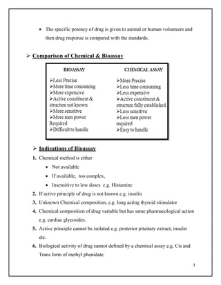
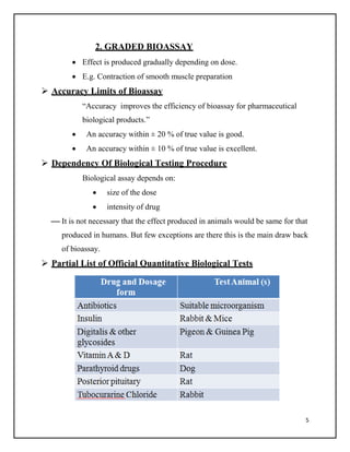
2. 2
Assay:
An assay is an investigative (analytic) procedure in laboratory
medicine, pharmacology, environmental biology, and molecular biology for
qualitatively assessing or quantitatively measuring the presence or amount or the
functional activity of a target entity (the analyte) which can be
a drug or biochemical substance or organic sample.
Types Of Assay
1. Chemical Assay
2. Immunoassay
3. Bioassay
Chemical Assay:
It is the study of the separation, identification and quantification of
the chemical components of natural and artificial materials.
Immunoassay
A technique that makes use of the binding between an antigen and its
homologous antibody in order to identify and quantify the specific antigen or
antibody in a sample
Biological Assay
Biological testing procedure for estimating the concentration of a
pharmaceutical drug substance in a formulated drug product or bulk
material
3. 3
The specific potency of drug is given to animal or human volunteers and
then drug response is compared with the standards.
Comparison of Chemical & Bioassay
Indications of Bioassay
1. Chemical method is either
Not available
If available, too complex,
Insensitive to low doses e.g. Histamine
2. If active principle of drug is not known e.g. insulin
3. Unknown Chemical composition, e.g. long acting thyroid stimulator
4. Chemical composition of drug variable but has same pharmacological action
e.g. cardiac glycosides.
5. Active principle cannot be isolated e.g. posterior pituitary extract, insulin
etc.
6. Biological activity of drug cannot defined by a chemical assay e.g. Cis and
Trans form of methyl phenidate.
4. 4
7. Not possible to separate interfering substance e.g. Vitamin D.
Principles of Bioassay
All bioassays should be comparative against a standard drug
Standard & new drug should be as far as possible identical to each other
Activity assayed should be the activity of interest
The degree of pharmacological response produced should be
reproducible under identical conditions. e.g. Adrenaline.
Method of comparison preferably (not essentially) test therapeutic
property of drug.
Individual variations must be minimised.
Classification of Bioassay
There are three types of bioassay:
1. Quantal
2. Graded
3. Effect produced in confined period
1. QUANTAL
All or none response in all individuals,
e.g. Digitalis induced cardiac arrest in guinea pigs
hypoglycemic convulsions in mice by insulin and
Calculation of LD50 in mice or rats
Not précised
Employed for:
Comparison of LD50 and ED50
Comparison of Threshold response
5. 5
2. GRADED BIOASSAY
Effect is produced gradually depending on dose.
E.g. Contraction of smooth muscle preparation
Accuracy Limits of Bioassay
“Accuracy improves the efficiency of bioassay for pharmaceutical
biological products.”
An accuracy within ± 20 % of true value is good.
An accuracy within ± 10 % of true value is excellent.
Dependency Of Biological Testing Procedure
Biological assay depends on:
size of the dose
intensity of drug
It is not necessary that the effect produced in animals would be same for that
produced in humans. But few exceptions are there this is the main draw back
of bioassay.
Partial List of Official Quantitative Biological Tests
6. 6
Purpose of Bioassay
1. Compare test sample with standard substance to determine quantity of test
sample required to produce an equivalent biological response to that of the
standard substance.
2. Measuring pharmacological activity of new or chemically undefined
substance.
3. Test method employed in measuring the response of living animals to
toxicity of chemical contaminant. Certain no. of individuals of sensitive
specie are exposed to specific conc. of contaminant for specific period to
examine toxic effects.
4. Investigating function of endogenous mediators.
5. Determine concentration as well as potency of unknown substance.
6. Improving and maintaining standards of basic environmental conditions
affecting well-being of people e.g. pollutants released by particular source
7. To determine specificity of compounds to be used e.g.
penicillin's are effective against G+ve but not on G-ve.
Preparation of Standard
A selective representative sample of a substance for which it is to serve
as a basis of the measurement is called standard preparation.
Uniform quality
Stable
Types of Standard Preparation
Two type of standard preparation:
1) International standard and reference standard
USP units(highly recognized-able and authorized
standard)
7. 7
2) British standard and reference standard
Potency
Measurement of drug activity expressed in terms of amount required to
produce an effect of given intensity.
EXAMPLES:
Highly potent drugs like morphine, alprazolam, chlorpromazine etc
produced high response at low conc. And low potent drugs like
ibuprofen and acetylsalicyclic acid produced low response at low
conc.
Advantages
Bioassays are procedures that can determine the concentration of purity or
biological activity of a substance such as vitamin, hormone, and plant
growth factor.
While measuring the effect on an organism, tissue cells, enzymes or the
receptor is preparing to be compared to a standard preparation.
Bioassays may be qualitative or quantitative. Qualitative bioassays are used
for assessing the physical effects of a substance that may not be quantified,
such as abnormal development or deformity.
8. 8
Quantitative bioassays involve estimation of the concentration or potency of
a substance by measurement of the biological response that it produces.
Quantitative bioassays are typically analyzed using the methods
of biostatistics.
They not only help to determine the concentration but also the potency of the
sample.
It is especially used to standardize drugs, vaccine, toxins or poisons,
disinfectants, antiseptics etc. as these are all used over biological system in
some or other form.
These also help determine the specificity of a compound to be used ex:
Penicillin's are effective against Gram+ve but not on Gram-ve. Testing of
infected patients sputum helps determine which anti-biotic be given for
quick recovery.
Certain complex compounds like Vitamin B-12 which can't be analyzed by
simple assay techniques can be effectively estimated by Bioassays.
Sometimes the chemical composition of samples are different but have same
biological activity.
For samples where no other methods of assays are available.
Biological products like toxin, anti-toxin, sera can be conveniently assayed.
Measure minute (Nano mole & Pico mole) quantities of active substances
can detect active substance without prior extraction or other treatment.
Disadvantages
Key problem is variability in response
Large number of animal to be used
Expertise in experimental design, execution of assay & analysis of data
required
9. 9
Leads to expensive & time consuming
Time related changes ins sensitivity of test organ.
Tachyphylactic responses of substance being assayed.
1. BIOASSAY OF ANTIBIOTICS
• The activity (potency) of antibiotics can be demonstrated by their inhibitory
effect on microorganisms under suitable conditions. A reduction in
antimicrobial activity may not be adequately demonstrated by chemical
methods. This chapter summarizes procedures for the antibiotics recognized
in the United States Pharmacopeia (USP) for which the microbiological
assay is the standard analytical method.
• Two general techniques are employed, the cylinder plate (or plate) assay and
the turbidimetric (or tube) assay.
10. 10
Techniques:
1.1. Cylinder-plate assay
The cylinder-plate assay depends on diffusion of the antibiotic from a
vertical cylinder through a solidified agar layer in a Petri dish or plate. The
growth of the specific microorganisms inoculated into the agar is prevented
in a circular area or zone around the cylinder containing the solution of the
antibiotic.
1.2. Turbidimetric assay
The turbidimetric assay depends on the inhibition of growth of a
microorganism in a uniform solution of the antibiotic in a fluid medium that
is favorable to the growth of the microorganism in the absence of the
antibiotic.
1.1. CYLINDER-PLATE METHOD
Temperature control:
Use appropriately qualified and calibrated equipment to obtain the
temperature ranges specified.
Apparatus
Plates:
Glass or disposable plastic Petri dishes (approximately 20 x 100
mm) with lids
Cylinders:
Stainless steel or porcelain cylinders; 8 mm + 0.1 mm o.d.; 6 mm +
0.1 mm i.d.; 10 mm+0.1 mm high
11. 11
Standard solutions
• To prepare a stock solution, dissolve a suitable quantity of the USP
Reference Standard of a given antibiotic, or the entire contents of a vial of
USP Reference Standard, where appropriate, in the solvent specified in and
dilute to the specified concentration. Store at 2–8, and use within the period
indicated. On the day of the assay, prepare from the stock solution five or
more test dilutions, in which the successive solutions increase stepwise in
concentration, usually in the ratio of 1:1.25.
Sample solutions
• Assign an assumed potency per unit weight or volume to the sample. On the
day of the assay prepare a stock solution in the same manner specified for
the USP Reference Standard. Dilute the sample stock solution in the
specified final diluent to obtain a nominal concentration equal to the median
concentration of the standard (S3).
Inocula:
• Suspend the test organism from a freshly grown slant or culture in 3 mL of
sterile saline TS. Glass beads can be used to facilitate the suspension. Spread
the saline suspension onto the surface of two or more agar plates (covering
the entire surface) or onto the surface of a Roux bottle containing 250 mL of
12. 12
the specified medium. Incubate for the specified time and at the temperature
as specified, or until growth is apparent.
• Prepare the inoculum by adding a portion of stock suspension to a sufficient
amount of agar medium that has been melted and cooled to 45–50 ºC. Swirl
the mixture without creating bubbles in order to obtain a homogeneous
suspension.
Analysis:
• Prepare the base layer for the required number of assay Petri plates, using
the medium and volume. Allow it to harden into a smooth base layer of
uniform depth. Prepare the appropriate amount of seed layer inoculum as
directed for the given antibiotic with any adjustments made based on the
preparatory trial analysis. Tilt the plate back and forth to spread the
inoculum evenly over the base layer surface, and allow it to harden.
• Incubate the plates for 16–18 h, and remove the cylinders. Measure and
record the diameter of each zone of growth inhibition to the nearest 0.1 mm.
1.2. TURBIDIMETRIC METHOD
Apparatus:
• Glass or plastic test tubes, e.g., 16 × 125 mm or 18 × 150 mm.
• Spectrophotometer: Measuring absorbance or transmittance within a fairly
narrow frequency band requires a suitable spectrophotometer in which the
wavelength can be varied or restricted by use of 580-nm or 530-nm filters.
Alternatively, a variable-wavelength spectrophotometer can be used and set
to a wavelength of 580 nm or 530 nm.
13. 13
Standard solutions:
• To prepare a stock solution, dissolve a quantity of the USP Reference
Standard of a given antibiotic or the entire contents of a vial of USP
Reference Standard, where appropriate, in the solvent specified, and dilute to
the required concentration. Store at 2–8, and use within the period indicated.
On the day of the assay, prepare from the stock solution five or more test
dilutions, the successive solutions increasing stepwise in concentration,
usually in the ratio of 1:1.25. Use the final diluent specified such that the
median level of the standard (S3)
Sample solutions:
• Assign an assumed potency per unit weight or volume to the unknown, and
on the day of the assay prepare a stock solution in the same manner specified
for the USP Reference Standard. Dilute the sample stock solution in the
specified final diluent at a nominal concentration equal to the median
concentration of the standard (S3)
Inocula:
• Suspend the test organism from a freshly grown slant or culture in 3 mL of
sterile saline TS. Glass beads can be used to facilitate the suspension.
Enterococcus hirae (ATCC 10541) and Staphylococcus aureus (ATCC
14. 14
9144) are grown in a liquid medium, not on agar. Spread the saline
suspension onto the surface of two or more agar plates (covering the entire
surface) or onto the surface of a Roux bottle containing 250 mL of the
specified medium. Incubate at the time and temperature specified, or until
growth is apparent. After incubation, harvest the organism from the plates or
Roux bottle with approximately 50 mL of sterile saline TS, using a sterile
bent glass rod or sterile glass beads. Pipet the suspension into a sterile glass
bottle. This is the harvest suspension.
Analysis:
• On the day of the assay, prepare the necessary concentration of antibiotic by
dilution of stock solutions of the standard and of each sample as specified
under Standard solutions and Sample solutions. Prepare five test levels, each
in triplicate, of the standard (S1–S5) and a single test level (U3), also in
triplicate, of up to 20 samples corresponding to S3 (median concentration) of
the standard.
• Place the tubes in test tube racks or other carriers. Include in each rack 1–2
control tubes containing 1 mL of the inoculum medium but no antibiotic.
Add the volumes of the standard and sample test dilutions as indicated.
Randomly distribute one complete set, including the controls, in a tube rack.
Add the volume of inoculum specified to each tube in the rack in turn, and
place the completed rack immediately in an incubator or a water bath
maintained at the temperature specified and for the time specified.
15. 15
2. BIOASSAY OF DIGITALIS
Principle:
• Potency of the test sample is compared with that of the standard preparation
by determining the action on the cardiac muscle. Any other equivalent
method, which gives results similar to those obtained by this method as also
valid.
Standard Preparation and Units:
• The standard preparation is a mixure of dried and powdered digitalis leaves
(1 unit = 76 mg.)
Preparation of Extracts
• Exact amount of the powder is extracted with dehydrated alcohol in a
continuous extraction apparatus for six hours. The final extract should
contain 10 ml. (5 ml. alcohol + 5 ml. water) per 10 g. of digitalis powder. It
should be stored in between 5 ºC and –5 ºC.
2.1. Guinea–pig Method (End point method)
• Standard and test sample extracts are diluted with normal saline in such a
way that 1 g of digitalis powder is diluted to 80 ml.
• A guinea pig is anaesthetized with a suitable anaesthetic. It is dissected on
the operation table. The jugular vein is traced out by removing adhering
tissues and cannulated by means of venous cannula.
• A pin is inserted in the heart, such that it gets inserted in the apex of the
heart. In this way, we can observe the heart beats by up and down
movements of the pin. The injection is continued through venous cannula
untill the heart is arrested in systole.
16. 16
• The amount of extract required to produce this effect is taken as the lethal
dose of the extract. Another set of 19 animals of the same species are used
for this experiment and the average lethal dose is determined. It is not
necessary to determine the lethal dose of the standard. during each time of
the experiment. But it should be occasionally checked.
• The lethal dose of the test sample is determined in a similar way using
minimum 6 guinea–pigs of the same strain.
The potency of the test sample is calculated in relation to that of the std.
preparation by dividing the average lethal dose of the sample to the test and
expressed as units per gram.
2.2. Pigeon Method
• Minimum 6 pigeons are used for testing each sample. They should be free
from gross evidence of disease or emaciation. The weight of the heaviest
pigeon should not exceed twice the weight of the lightest pigeon. Food is
withheld 16-28 hours before the experiment.
• Pigeons are divided on the basis of their sex, weight and breed, into two
groups. They are anaesthetized with anaesthetic ether.
17. 17
One side of the wing is dissected and the ulnar vein is cannulated by means
of a venous cannula. Dilutions are made with normal saline. Average lethal
dose of each sample is determined; results are tabulated and calculated as
per guinea pig method.
• The lethal dose per kg. of body weight is determined for each pigeon. The
potency of the test sample is
determined by dividing the mean lethal dose of standard by the mean lethal
dose of the test sample.
• In pigeons, stoppage of heart is associated with a characteristic vomiting
response called „emesis‟. The milk from the crop sac of pigeons is being
ejected out. This may be taken as the end point response of digitalis.
3. BIO-ASSAYS OF INSULINE
Mechanism of action
• Every pancreatic islet contains ~1,000 endocrine cells of which 75% are
insulin-producing beta-cells.
• Insulin is synthesised as pro-insulin in the endoplasmic reticulum and is
processed to the biologically active form inside the secretory granules.
18. 18
• The beta-cell is electrically excitable and uses changes in membrane
potential to couple variations in blood glucose to trigger insulin secretion.
• The beta-cell contains about 20 different ion channels proteins
• Two types of ion channels are particularly important for the initiation of
insulin secretion. The KATP-channels are active at low glucose
concentrations, because of the high intracellular ADP levels.
Bioassay of insulin
• Standard preparation and unit: It is pure, dry and crystalline insulin. One unit
contains 0.04082 mg. This unit is equivalent to international unit.
• Preparation of standard solution: Accurately weigh 20 units of insulin and
dissolve it in normal saline. Acidify it with HCl to pH 2.5. Add 0.5% phenol
as preservative. Add 1.4% to 1.8% glycerin. Final volume should contain 20
units/ml. Store the solution in a cool place and use it within six months.
• Preparation of test sample solution: The solution of the test sample is
prepared in the same way as the standard solution.
3.1. Rabbit method
Selection of rabbits: They should be healthy, weighing about 1800-3000
gms. They should then be maintained on uniform diet but are fasted for 18
hrs before assay. Water is withdrawn during the experiment.
Standard and Sample Dilutions: These are freshly prepared by diluting
with normal NaCl solution so as to contain 1 unit/ml. and 2 units/ml.
Doses: The dose which can produce suitable fall in blood sugar level is
calculated for the standard.
19. 19
Principle: The potency of a test sample is estimated by comparing the
hypoglycemic effect of the sample with that of the std. preparation of
insulin. Any other suitable method can also be used.
Experimental Procedure: Animals are divided into 4 groups of 3 rabbits
each. The rabbits are then put into an animal holder. They should be handled
with care to avoid excitement.
First part of the Test: A sample of blood is taken from the marginal ear
vein of each rabbit. Presence of reducing sugar is estimated per 100 ml. of
blood by a suitable chemical method. This concentration is called „Initial
Blood Sugar Level‟.
The four groups of rabbits are then given sc. injections of insulin as follows:
• From each rabbit, a sample of blood is withdrawn up to 5 hrs. at the interval
of 1 hr. each. Blood sugar is determined again. This is known as „Final
Blood Sugar Level‟.
• Second part of the test (Cross over test) : The same animals are used for the
second part. The experiment can be carried out after one week. Again they
are fasted and initial blood sugar is determined. The grouping is reversed,
that is to say, those animals which received the standard are given the test
and those which received the test are now given the standard. Those animals
20. 20
which received the less dose of the standard are given the higher dose of the
test sample and vice-versa. This test is known as „Twin Cross over Test‟.
3.2. Mouse Method
• Mice show characteristic convulsions after s.c. inj. of insulin at elevated
temperatures. The percentage convulsions produced by the test and standard
preparations are compared.
Experimental procedure: Minimum 100 mice weighing between 18-22
gms. of the same strain are used. They should be maintained on constant
diet. They should be fasted 18 hrs. prior to the experiment.
Standard and sample dilutions: Dilutions are prepared with sterile saline
solution, so as to contain 0.064 units/ml. (std dilution I) and 0.096 untis/ml.
(std. dilution II). Similarly, test sample solutions are also prepared.
• Mice are divided into 4 groups each containing 25 mice and insulin is
injected s.c. as follows
• Mice are put in an air incubator at 33ºC and observed for one and a half hr.
The mice which convulse or die are taken out of the incubator and observed.
These mice usually convulse severely but failure of the animal to upright
itself when placed on its back, should as well be considered as convulsion.
21. 21
3.3. Rat diaphragm method
• Sprague Dawley rats weighing 70–100 g are used. The animals are
sacrificed during anesthesia and the diaphragms still attached to the rib cages
are carefully removed, released from the rib cages and adhering connective
and fat tissues, washed in PBS, spread out and divided into two equal pieces
as described by Müller and coworkers (1994). For assaying the effects of
insulin/compounds/drugs, the hemidiaphragms are incubated in KRH buffer
gassed with carbogen (95% O2/5% CO2) in the presence of 5 mM glucose
Epididymal fat pad of rats:
• Insulin-like activity can be measured by the uptake of glucose into fat cells.
Adipose tissue from the epididymal fat pad of rats has been found to very
suitable.
• The difference of glucose concentration in the medium after incubation of
pieces of epididymal rat adipose tissue or measured oxygen consumption in
Warburg vessels, Radiolabelled 14C glucose, the 14CO2 is trapped and
counted.
• The concentration is determined by immuno-assay.
4. BIOASSAY OF VITAMIN D
• The biological assay of vitamin D comprises the recording and interpretation
of observations on groups of rats maintained on specified dietary regimens
throughout specified periods of their lives whereby the biological response
to the preparation under assay is compared with the response to USP
Vitamin D Capsules RS.
22. 22
Preliminary Period
• Throughout the preliminary period in the life of a rat, which is not longer
than 30 days and extends from birth to the first day of the depletion period,
maintain litters of rats under the immediate supervision of, or according to
the directions of, the individual responsible for the assay.
• During the preliminary period, use a dietary regimen that provides for
normal development but is limited in its content of vitamin D, so that when
placed upon the Rachitogenic Diet in the depletion period the rats develop
rickets. At the end of the preliminary period, reject any rat that weighs less
than 44 g or more than 60 g, or that shows evidence of injury, disease, or
anatomical abnormality.
Depletion Period
• Through the depletion period, which extends from the end of the preliminary
period to the first day of the assay period, provide each rat ad libitum with
the Rachitogenic Diet and water, and allow access to no other food or
dietary supplement.
Rachitogenic Diet
Assigning Rats to Groups
• Consider a litter suitable for the assay period when individual rats in the
litter show evidence of rickets such as enlarged joints and a distinctive
wobbly, rachitic gait, provided that the depletion period is not less than 19 or
Ingredient Parts by weight
Whole yellow corn, ground 76
Wheat gluten, ground 20
Calcium carbonate 3
Sodium chloride 1
23. 23
more than 25 days. The presence of rickets may be established also from the
width of the rachitic metaphysis upon X-ray examination or by applying the
Line Test to a leg bone of one member of each litter.
• Record the weight of each rat, and assign it to a group, in which each rat will
be fed a specified dose of the Reference Standard or of an assay sample that
is under examination for its vitamin D potency.
• The two standard groups may be used for the concurrent assay of more than
one assay sample.
• At Analysis for complete balance, whereby each litter is represented equally
in every group, use 7 or more litters containing at least as many depleted rats
as there are groups.
Assay Doses
• Select two dosage levels of the USP Cholecalciferol RS, spaced so that the
ratio of the larger to the smaller dose is not less than 1.5 or more than 2.5.
The dosage levels of the sample are equivalent to those of the standard or to
a mid-level equal to the square root of the product of the two dosage levels
of the standard.
• Select dosage levels such that, when fed to rachitic rats, they are expected to
produce degrees of calcification within the range specified under the test of
data acceptability.
• Before feeding, the Reference Standard and/or sample may be diluted with
cottonseed oil, provided that not more than 0.2 mL is fed on any one day.
Store the oil solutions in well-closed bottles, protected from light, at a
temperature not exceeding 10 , and use within 5 weeks.
• Assign one group of rats to each dosage level of the standard and of the one
or more samples
24. 24
Assay Period
• During the assay period, which extends from the end of the depletion period
for a fixed interval of 7 to 10 days, cage each rat individually and provide it
ad libitum with the Rachitogenic Diet and water. Supply a Rachitogenic Diet
prepared from the same lots of ingredients to all rats. On the first and on the
third (or fourth) day of the assay period, feed each rat one-half of its total
assigned dose.
• Throughout the assay period, maintain as uniform environmental conditions
as possible for all rats, and exclude exposure to antirachitic radiations. At the
end of a fixed period of 7 to 10 days, weigh and kill each rat. From those rats
that do not weigh less at the end than at the start of the assay period and that
have consumed each assigned dose within 24 hours of the time it was fed,
dissect out one or more leg bones for examination by the Line Test.
Line Test
Remove the proximal end of a tibia or the distal end of a radius, and clean
adhering tissue from it, in any one assay using the same bone from all
animals. With a clean, sharp blade cut a median, longitudinal section
through the juncture of the epiphysis and diaphysis at the same place on each
bone. Rinse both sections in purified water, immerse immediately in silver
nitrate solution (1 in 50) for 1 minute, and rinse again in purified water.
Expose the cut surface of bone, in water, to daylight or another source of
actinic light until the calcified areas develop a clearly defined stain without
marked discoloration of the uncalcified areas. The staining procedure may
be modified to differentiate more clearly between calcified and uncalcified
areas.
25. 25
Acceptability
• If the average score of the standard group on the high dosage level is not
greater than the average score of the standard group on the low dosage level,
discard the results, and repeat the assay.
• Assay groups that are not acceptable for measuring vitamin D potency, in
each of which the average score is less than the average score of the standard
group on the low dosage level or more than the average score of the standard
group on the high dosage level, its assayed content of vitamin D is
respectively less than that represented by the low dose or more than that
represented by the high dose of the Reference Standard.