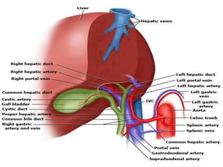
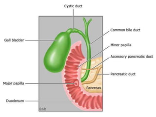
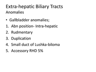
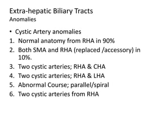
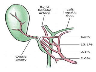
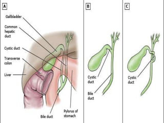
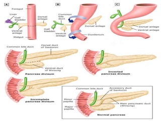
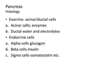
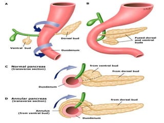
Hepatobiliary Anatomy
Dr Malith
The Liver
• Largest organ in the body
• Weighs about 1.5kg
• Located under diaphragm and protected by
the rib cage.
• Covered by Glisson’s capsule
Surgical Anatomy
Morphological-Surfaces
• Posterior
• Anterio-superior
• Inferior
Picture of surfaces of the liver
Anatomy
Morphological-ligaments
• Ligaments/Peritoneal Attachments
• Falciform ligament
• Anterior and posterior coronary ligaments
• Right triangular ligament-coronary layers
• Left triangular Ligament-falciform layers
• Round ligament-umbilical vein
• Fibrous ligamentum venosum-
• Hepatoduodenal Ligament-portal triad
Picture of portal triad
Surgical Anatomy
Morphological-segments
• Anatomic Lobes: Right and left lobe based on
Cantle's Line.
• Segmental Anatomy based on vascular supply
and ductal distribution (I-VIII) by Couniad
• Sectoral Anatomy based on hepatic vein
anatomy( Left lateral, left medial, right
anterior and right posterior) by Bismuth.
Picture for Segments
Surgical Anatomy
Morphological-sectors
• Caudate (I)
Three parts ;
1. Spiegal lobe
2. Paracval Portion
3. Caudate Process
• Segment II & III form lateral sector of left lobe
Surgical Anatomy
Morphological-sectors
• Segment IV: has IVa cephalad and IVb caudally
• Segment V-VIII
V and VIII form right anterior sector
VI &VII form right posterior sector
Picture of Surgical resections
Vascular Supply
Arterial and Venous
• Dual supply 75 % portal venin and 25 %
hepatic artery.
• Venous and biliary drainage
Picture of vascular supply
Vascular Supply
Arterial and Venous
• Hepatic proper artery is branch from celiac trunk
(splenic and left gastric).
• Its run to the left side in porta hepatis and divides
into right and left in 67% of people.
• Variations seen in 24% of population
1. Replaced RHA 11-21% from SMA
2. Replaced LHA 4-10% from left gastric artery
3. Replaced R & LHA 1-2%
4. Replaced CHA
Vascular Variation picture
Vascular Supply
Arterial and Venous
• Portal Vein, formed behind neck of pancreas
by SMV and SV.
• 5-8cm long and +/- 1 cm in diameter.
• Normal Pressure 3-5mmHg.
• Runs in Portal triad deep to the duct,
• Bifurcate into right(V-VIII) and left(II-IV) portal
veins.
• Variations: trifurcation/Aberrant from left PV.
Vascular Supply
Hepatic Veins and IVC
• Three in numbers
1. Right hepatic Vein (RHV) drains; V-VIII.
2. Middle Hepatic Vein (MHV) drains IV, V and
VIII. LHV and MHV form trunk in 95%.
3. LHV drains II and III.
4. Caudate drains directly to IVC
5. Small veins to IVC
• Inferior Accessory right hepatic vein 15-20%
Ductal anatomy
Classfications
• Intrahepatic Bile ducts
1. Canaliculi
2. Segmental bile ducts
3. Sectoral bile ducts
a. Right posterior sectoral duct RPSD (VI&VII)
b. Right anterior sectoral duct RASD (V&VIII)
c. Left lateral sectoral duct LLSD (II & III)
d. Left medial sectoral duct LMSD (Iva & Ivb)
3. LHD and RHD
Ductal anatomy
Classfications
• Extrahepatic bile ducts
1. CHD formed by left and right HDs
2. Cystic duct
3. CBD: CHD and cystic duct
Ductal anatomy
Variations-30-40%
• Bifurcation 57%
• Trifurcation 12%; Right anterior, right
posterior and LHDs
• RPSD drains into CHD in 16% or RASD into
CHD in 4%.
• RPSD into LHD in 5% or RASD into LHD in 1%.
• Absence of CHD confluence in 3%
• Absence of RHD in 2%.
Innervation of the Liver
• Parasympathetic –vagus
• Sympathetic from;
1. T7-10
2. Greater thoracic nerve
3. Caeliac ganglia
4. Splanchnic nerves
Form anterior and posterior hepatic plexuses of
nerves.
Lymphatic Drianage
• Lymph is formed at perisinusoidal space of
Dise and periportal cleft of Mall.
• Caudal drainage via Calot’s triangle node, CBD
nodes, Hepatic artery nodes, retropancreatic
nodes and last at celiac nodes
• Cephalad; cardiophrenic nodes
Surgical Anatomy
Microscopic Anatomy
• Functional unit is Lobule; central terminal
hepatic venule surrounded by 4-6 portal triads
• Blood flow from triad to terminal hepatic
venule.
• Bile is formed by hepatocytes and drain into
canaliculi and then to bile ducts
• Between terminal portal triad and central
hepatic venule are 3 zones,
Surgical Anatomy
Microscopic Anatomy
• Zone I (Periportal): increase O2 and blood
supply.
• Zone II (intermediate): equivocal supply
• Zone III( Perivenular): decrease o2 supply
Physiology of the liver
• Metabolism.
• Synthetic Function- Alb/Coagulations
factors/complements,
• Bilirubin metabolism
• Bile formation- primary/secondary bile acids
• Drug Metabolism
• Storage
Assessment of Liver function
• Laboratory: LFT/Clotting factors; abn include
1. Liver injury-raised AST/ALT
2. Abnormal Synthetic function-low Alb, raised
INR and decreased Vit-k dependant factors.
3. Cholestasis-bilirubin and fractionation, ALP
and GGT.
• Scoring Systems; Child-Pugh score, OKUDA
and MELD score.
Assessment of Liver function
• Child-Pugh prognostic indicator
• Class A; 5-6 10% MR
• Class B; 7-9 30% MR
• Class C; 10-15 75-80% MR
Variables Points
1 2 3
Bilirubin <2 mg 2-3 >3
Albumin >3.5g/dl 2.8-3.5 <2.8
INR <1.7 1.7-2.2 >2.2
Ascites None Mild Severe
Encephalopathy None Mild Severe
Assessment of Liver function
• Model of End-stage Liver Disease Scoring
(MELD) prognostic for organ donor
• Uses Bilirubin/Creatinine/INR/sodium
1. Score <10; elective surgery
2. 10-15 cautious surgery
3. > 15 no elective surgery
Assessment of Liver function
• OKUDA Staging Systems; staging and prognosis in
HCC.
• Stage I; no positives
• Stage II; < 2 positives
• Stage III; .3 positives
Criteria Positive Negative
Tumour Size >50% <50%
Ascites Present Absent
Albumin <3g/dl > 3g
Bilirubin >3mg <3mg/dl
Assessment of Liver function
• Functional Assessment; Clearance
test/Tolerance Test.
1. Indocyanine Green Clearance (ICG) 15% or
more of dye is retained mean impairment.
2. T-99 galactosyl-Human Serum albumin (GSA)
3. Monoethylglycine-xylidide lidocaine (MEGx)
for liver cirrhosis.
Assessment of Liver function
• Volumetric CT or U/S.
Gallbladder and Biliary Tree
Surgical Anatomy
• Pear-shaped organ located gallbaldder fossa
• 7-10cm long
• Parts; Fundus/Body/Infundibulum/Neck.
• Histology: Columnar cells/mucus cells at the
neck/infundibulum
• Layers: Mucosa, Lamina propria, no
Submucosa or muscularis mucosa, smooth
muscle, circular, longitudinal and oblique
Gallbladder and Biliary Tree
Surgical Anatomy
• Blood supply : Cystic artery frpm RHA in 90%
and LHA in 10%. Cystic drain into PV
• Lymphatics drainage via Cystic nodes.
• Innervation: sympathetic T7-9 and vagus
nerves.
Extra-hepatic Biliary Ducts
Surgical Anatomy
• CHD; 1-4cm long and 4mm in diameter.
• Cystic duct; variable length, union parallel or
spiral to CHD.
• CBD: 7-11cm long, 5-10 mm in diameter and
has three parts;
1. Supraduodenal- hepatoduodenal ligament
2. Retroduodenal; 1st part of D1.
3. Pancreatic Portionbehind HOP.
Extra-hepatic Biliary Tracts
Surgical Anatomy
• CBD and PD join in three configurations;
1. 70% outside duodenal wall ? APBJ anomalies
2. 20% in the wall of duodenum.
3. 10% separate opening
• CBD histology-columnar epithelium
• Blood supply; proximal RHA & Cystic artery
distally GDA and SMA forming networks from
3-9 O’clock
Extra-hepatic Biliary Tracts
Anomalies
• Gallbladder anomalies;
1. Abn position- Intra-hepatic
2. Rudmentary
3. Duplication
4. Small duct of Lushka-biloma
5. Accessory RHD 5%
Extra-hepatic Biliary Tracts
Anomalies
• Hepatic artery anomalies;
1. Two RHA; CHA and SMA in 5%
2. Replaced RHA from SMA
3. Abnormal Course; parallel to cystic duct
Extra-hepatic Biliary Tracts
Anomalies
• Cystic Artery anomalies
1. Normal anatomy from RHA in 90%
2. Both SMA and RHA (replaced /accessory) in
10%.
3. Two cystic arteries; RHA & CHA
4. Two cystic arteries; RHA & LHA
5. Abnormal Course; parallel/spiral
6. Two cystic arteries from RHA
Extra-hepatic Biliary Tracts
Anomalies
• Cystic Duct Anomalies
1. Abnormal union with CHD; high or low.
2. Adherent to CHD
3. Drainage to RHD
4. Abnormal Course; spiral anterior or posterior
5. Absence cystic duct.
Pancreas
Surgical Anatomy
• Pan=all, creas=flesh
• Topographic; retroperitoneal in C-loop of
duodenum.
• Weighs;75-100grams
• Length; 15-20cm
Pancreas
Parts
• Head; anterior TV colon and IVC posteriorly.
• Neck; PV and SMA/V.
• Body; SA/V.
• Tail; helium of the spleen.
Picture of the pancreas and parts
Pancreas
Vascular anatomy
• GDA; Anterior and Posterior superior PDA.
• SMA; Inferior PDA
• Splenic artery branches;
a. Dorsal pancreatic artery
b. Greater pancreatic artery
c. Caudal pancreatic artery
• Venous drainage via SMV/SV into PV.
Pancreas
Lymphatic drainage
• Head: via Subpyloric, portal, mesenteric,
mesocolic and aortocaval nodes.
• Body and Tail: Caeliac, aorto-caval, mesenteric
and mesocolic group of nodes.
Pancreas
Ductal Anatomy
• Embyrological; ventral and dorsal buds fuse.
• Ventral analge gives to Uncinate process and
inferior part of HOP.
• Dorsal analge gives to HOP, body and tail.
• Duct from ventral analge remains as main PD
and opens on to major ampulla of Vater
• Duct from dorsal analge remain as santorini
and opens on to minor papilla.
Pancreas
Neuroanatomy
• Autonomic Nervous system.
a. Sympathetic (inhibitory); greater, lesser and
least splanchnic nerves.
b. Parasympathetic(excitatory); caeliac branch
• Somatic fibres (afferent).
Pancreas
Composition
• Exocrine 85%
• Extracellular matrix; 10%
• Blood Vessels and ducts; 3-4%
• Endocrine 1-2%
• 20% of gland is required for normal function.
Pancreas
Histology
• Exocrine- acinar/ductal cells
a. Acinar cells; enzymes
b. Ductal water and electrolytes
• Endocrine cells
a. Alpha cells-glucagon
b. Beta cells insulin
c. Sigma cells-somatostatin etc.
Pancreas
Embyrological Anomalies
• Pancreas divisum; failure of ductal fusion.
Treated by minor papillotomy.
• Ectopic/Accessory pancreas; resection/bypass.
• Annular Pancreas; bypass or duodeno-
jejenostomy.
• Developmental Pancreatic Cysts;
congenital/duplication/dermoid cysts.
Pancreas divisum
Spleen
Surgical anatomy
• Embyrogenesis; outgrowth from dorsal
mesogastrium at 5th week gestation.
• Topographic; postero-lateral in LUQ
• Weighs; 150grams
• Length; 7-11cm
• Blood supply from Splenic artery an short
gastric arteries. Venous drainage via SV.
Spleen
Anomalies
• Asplenia-autosomal recessive
• Splenic hypolasia- autosomal dorminant
• Polysplenia
• Wandering spleen-ligament anomalies.
• Splenic cysts
• Accessory spleen x 7 sites: hilium/pedicle/tail
of pancreas/splenocolic ligament/greater
omentum/mesentery/left ovary.
Spleen
Histology and physiology
• Filtration of senescent RBCs.
• Extra-medullary hemopoiesis
• Host defense-opsinization.

Hepatobiliary anatomy

  • 1.
  • 2.
    The Liver • Largestorgan in the body • Weighs about 1.5kg • Located under diaphragm and protected by the rib cage. • Covered by Glisson’s capsule
  • 3.
  • 4.
    Picture of surfacesof the liver
  • 5.
    Anatomy Morphological-ligaments • Ligaments/Peritoneal Attachments •Falciform ligament • Anterior and posterior coronary ligaments • Right triangular ligament-coronary layers • Left triangular Ligament-falciform layers • Round ligament-umbilical vein • Fibrous ligamentum venosum- • Hepatoduodenal Ligament-portal triad
  • 6.
  • 7.
    Surgical Anatomy Morphological-segments • AnatomicLobes: Right and left lobe based on Cantle's Line. • Segmental Anatomy based on vascular supply and ductal distribution (I-VIII) by Couniad • Sectoral Anatomy based on hepatic vein anatomy( Left lateral, left medial, right anterior and right posterior) by Bismuth.
  • 8.
  • 9.
    Surgical Anatomy Morphological-sectors • Caudate(I) Three parts ; 1. Spiegal lobe 2. Paracval Portion 3. Caudate Process • Segment II & III form lateral sector of left lobe
  • 10.
    Surgical Anatomy Morphological-sectors • SegmentIV: has IVa cephalad and IVb caudally • Segment V-VIII V and VIII form right anterior sector VI &VII form right posterior sector
  • 11.
  • 12.
    Vascular Supply Arterial andVenous • Dual supply 75 % portal venin and 25 % hepatic artery. • Venous and biliary drainage
  • 13.
  • 14.
    Vascular Supply Arterial andVenous • Hepatic proper artery is branch from celiac trunk (splenic and left gastric). • Its run to the left side in porta hepatis and divides into right and left in 67% of people. • Variations seen in 24% of population 1. Replaced RHA 11-21% from SMA 2. Replaced LHA 4-10% from left gastric artery 3. Replaced R & LHA 1-2% 4. Replaced CHA
  • 15.
  • 17.
    Vascular Supply Arterial andVenous • Portal Vein, formed behind neck of pancreas by SMV and SV. • 5-8cm long and +/- 1 cm in diameter. • Normal Pressure 3-5mmHg. • Runs in Portal triad deep to the duct, • Bifurcate into right(V-VIII) and left(II-IV) portal veins. • Variations: trifurcation/Aberrant from left PV.
  • 19.
    Vascular Supply Hepatic Veinsand IVC • Three in numbers 1. Right hepatic Vein (RHV) drains; V-VIII. 2. Middle Hepatic Vein (MHV) drains IV, V and VIII. LHV and MHV form trunk in 95%. 3. LHV drains II and III. 4. Caudate drains directly to IVC 5. Small veins to IVC • Inferior Accessory right hepatic vein 15-20%
  • 21.
    Ductal anatomy Classfications • IntrahepaticBile ducts 1. Canaliculi 2. Segmental bile ducts 3. Sectoral bile ducts a. Right posterior sectoral duct RPSD (VI&VII) b. Right anterior sectoral duct RASD (V&VIII) c. Left lateral sectoral duct LLSD (II & III) d. Left medial sectoral duct LMSD (Iva & Ivb) 3. LHD and RHD
  • 23.
    Ductal anatomy Classfications • Extrahepaticbile ducts 1. CHD formed by left and right HDs 2. Cystic duct 3. CBD: CHD and cystic duct
  • 24.
    Ductal anatomy Variations-30-40% • Bifurcation57% • Trifurcation 12%; Right anterior, right posterior and LHDs • RPSD drains into CHD in 16% or RASD into CHD in 4%. • RPSD into LHD in 5% or RASD into LHD in 1%. • Absence of CHD confluence in 3% • Absence of RHD in 2%.
  • 25.
    Innervation of theLiver • Parasympathetic –vagus • Sympathetic from; 1. T7-10 2. Greater thoracic nerve 3. Caeliac ganglia 4. Splanchnic nerves Form anterior and posterior hepatic plexuses of nerves.
  • 27.
    Lymphatic Drianage • Lymphis formed at perisinusoidal space of Dise and periportal cleft of Mall. • Caudal drainage via Calot’s triangle node, CBD nodes, Hepatic artery nodes, retropancreatic nodes and last at celiac nodes • Cephalad; cardiophrenic nodes
  • 29.
    Surgical Anatomy Microscopic Anatomy •Functional unit is Lobule; central terminal hepatic venule surrounded by 4-6 portal triads • Blood flow from triad to terminal hepatic venule. • Bile is formed by hepatocytes and drain into canaliculi and then to bile ducts • Between terminal portal triad and central hepatic venule are 3 zones,
  • 31.
    Surgical Anatomy Microscopic Anatomy •Zone I (Periportal): increase O2 and blood supply. • Zone II (intermediate): equivocal supply • Zone III( Perivenular): decrease o2 supply
  • 32.
    Physiology of theliver • Metabolism. • Synthetic Function- Alb/Coagulations factors/complements, • Bilirubin metabolism • Bile formation- primary/secondary bile acids • Drug Metabolism • Storage
  • 33.
    Assessment of Liverfunction • Laboratory: LFT/Clotting factors; abn include 1. Liver injury-raised AST/ALT 2. Abnormal Synthetic function-low Alb, raised INR and decreased Vit-k dependant factors. 3. Cholestasis-bilirubin and fractionation, ALP and GGT. • Scoring Systems; Child-Pugh score, OKUDA and MELD score.
  • 34.
    Assessment of Liverfunction • Child-Pugh prognostic indicator • Class A; 5-6 10% MR • Class B; 7-9 30% MR • Class C; 10-15 75-80% MR Variables Points 1 2 3 Bilirubin <2 mg 2-3 >3 Albumin >3.5g/dl 2.8-3.5 <2.8 INR <1.7 1.7-2.2 >2.2 Ascites None Mild Severe Encephalopathy None Mild Severe
  • 35.
    Assessment of Liverfunction • Model of End-stage Liver Disease Scoring (MELD) prognostic for organ donor • Uses Bilirubin/Creatinine/INR/sodium 1. Score <10; elective surgery 2. 10-15 cautious surgery 3. > 15 no elective surgery
  • 36.
    Assessment of Liverfunction • OKUDA Staging Systems; staging and prognosis in HCC. • Stage I; no positives • Stage II; < 2 positives • Stage III; .3 positives Criteria Positive Negative Tumour Size >50% <50% Ascites Present Absent Albumin <3g/dl > 3g Bilirubin >3mg <3mg/dl
  • 37.
    Assessment of Liverfunction • Functional Assessment; Clearance test/Tolerance Test. 1. Indocyanine Green Clearance (ICG) 15% or more of dye is retained mean impairment. 2. T-99 galactosyl-Human Serum albumin (GSA) 3. Monoethylglycine-xylidide lidocaine (MEGx) for liver cirrhosis.
  • 38.
    Assessment of Liverfunction • Volumetric CT or U/S.
  • 39.
    Gallbladder and BiliaryTree Surgical Anatomy • Pear-shaped organ located gallbaldder fossa • 7-10cm long • Parts; Fundus/Body/Infundibulum/Neck. • Histology: Columnar cells/mucus cells at the neck/infundibulum • Layers: Mucosa, Lamina propria, no Submucosa or muscularis mucosa, smooth muscle, circular, longitudinal and oblique
  • 41.
    Gallbladder and BiliaryTree Surgical Anatomy • Blood supply : Cystic artery frpm RHA in 90% and LHA in 10%. Cystic drain into PV • Lymphatics drainage via Cystic nodes. • Innervation: sympathetic T7-9 and vagus nerves.
  • 42.
    Extra-hepatic Biliary Ducts SurgicalAnatomy • CHD; 1-4cm long and 4mm in diameter. • Cystic duct; variable length, union parallel or spiral to CHD. • CBD: 7-11cm long, 5-10 mm in diameter and has three parts; 1. Supraduodenal- hepatoduodenal ligament 2. Retroduodenal; 1st part of D1. 3. Pancreatic Portionbehind HOP.
  • 43.
    Extra-hepatic Biliary Tracts SurgicalAnatomy • CBD and PD join in three configurations; 1. 70% outside duodenal wall ? APBJ anomalies 2. 20% in the wall of duodenum. 3. 10% separate opening • CBD histology-columnar epithelium • Blood supply; proximal RHA & Cystic artery distally GDA and SMA forming networks from 3-9 O’clock
  • 44.
    Extra-hepatic Biliary Tracts Anomalies •Gallbladder anomalies; 1. Abn position- Intra-hepatic 2. Rudmentary 3. Duplication 4. Small duct of Lushka-biloma 5. Accessory RHD 5%
  • 45.
    Extra-hepatic Biliary Tracts Anomalies •Hepatic artery anomalies; 1. Two RHA; CHA and SMA in 5% 2. Replaced RHA from SMA 3. Abnormal Course; parallel to cystic duct
  • 46.
    Extra-hepatic Biliary Tracts Anomalies •Cystic Artery anomalies 1. Normal anatomy from RHA in 90% 2. Both SMA and RHA (replaced /accessory) in 10%. 3. Two cystic arteries; RHA & CHA 4. Two cystic arteries; RHA & LHA 5. Abnormal Course; parallel/spiral 6. Two cystic arteries from RHA
  • 48.
    Extra-hepatic Biliary Tracts Anomalies •Cystic Duct Anomalies 1. Abnormal union with CHD; high or low. 2. Adherent to CHD 3. Drainage to RHD 4. Abnormal Course; spiral anterior or posterior 5. Absence cystic duct.
  • 50.
    Pancreas Surgical Anatomy • Pan=all,creas=flesh • Topographic; retroperitoneal in C-loop of duodenum. • Weighs;75-100grams • Length; 15-20cm
  • 51.
    Pancreas Parts • Head; anteriorTV colon and IVC posteriorly. • Neck; PV and SMA/V. • Body; SA/V. • Tail; helium of the spleen.
  • 52.
    Picture of thepancreas and parts
  • 53.
    Pancreas Vascular anatomy • GDA;Anterior and Posterior superior PDA. • SMA; Inferior PDA • Splenic artery branches; a. Dorsal pancreatic artery b. Greater pancreatic artery c. Caudal pancreatic artery • Venous drainage via SMV/SV into PV.
  • 56.
    Pancreas Lymphatic drainage • Head:via Subpyloric, portal, mesenteric, mesocolic and aortocaval nodes. • Body and Tail: Caeliac, aorto-caval, mesenteric and mesocolic group of nodes.
  • 57.
    Pancreas Ductal Anatomy • Embyrological;ventral and dorsal buds fuse. • Ventral analge gives to Uncinate process and inferior part of HOP. • Dorsal analge gives to HOP, body and tail. • Duct from ventral analge remains as main PD and opens on to major ampulla of Vater • Duct from dorsal analge remain as santorini and opens on to minor papilla.
  • 59.
    Pancreas Neuroanatomy • Autonomic Nervoussystem. a. Sympathetic (inhibitory); greater, lesser and least splanchnic nerves. b. Parasympathetic(excitatory); caeliac branch • Somatic fibres (afferent).
  • 61.
    Pancreas Composition • Exocrine 85% •Extracellular matrix; 10% • Blood Vessels and ducts; 3-4% • Endocrine 1-2% • 20% of gland is required for normal function.
  • 62.
    Pancreas Histology • Exocrine- acinar/ductalcells a. Acinar cells; enzymes b. Ductal water and electrolytes • Endocrine cells a. Alpha cells-glucagon b. Beta cells insulin c. Sigma cells-somatostatin etc.
  • 63.
    Pancreas Embyrological Anomalies • Pancreasdivisum; failure of ductal fusion. Treated by minor papillotomy. • Ectopic/Accessory pancreas; resection/bypass. • Annular Pancreas; bypass or duodeno- jejenostomy. • Developmental Pancreatic Cysts; congenital/duplication/dermoid cysts.
  • 64.
  • 65.
    Spleen Surgical anatomy • Embyrogenesis;outgrowth from dorsal mesogastrium at 5th week gestation. • Topographic; postero-lateral in LUQ • Weighs; 150grams • Length; 7-11cm • Blood supply from Splenic artery an short gastric arteries. Venous drainage via SV.
  • 67.
    Spleen Anomalies • Asplenia-autosomal recessive •Splenic hypolasia- autosomal dorminant • Polysplenia • Wandering spleen-ligament anomalies. • Splenic cysts • Accessory spleen x 7 sites: hilium/pedicle/tail of pancreas/splenocolic ligament/greater omentum/mesentery/left ovary.
  • 68.
    Spleen Histology and physiology •Filtration of senescent RBCs. • Extra-medullary hemopoiesis • Host defense-opsinization.