Histology of liver ppt
•Download as PPTX, PDF•
2 likes•1,858 views

pls share very easy to study histology of liver prepared by dr. sandeep k sharma contact no .9335136978

Recommended
More Related Content
What's hot
What's hot (20)
Similar to Histology of liver ppt
Similar to Histology of liver ppt (20)
Recently uploaded
Recently uploaded (20)
Histology of liver ppt
- 1. HISTOLOGY OF THE LIVER
- 2. THE LIVER • Largest gland of the body. • 1500 grams and 2.5% of total body weight. • Location: - Right hypochondrium - Epigastric region - Left hypochondrium
- 3. VASCULAR SUPPLY OF THE LIVER • Receives dual vascular supply: -Hepatic Portal Vein (75%) - Hepatic Artery(25%) • Both vessels enter the liver via Porta hepatis.
- 4. HISTOLOGICAL STRUCTURE OF THE LIVER
- 5. THE LIVER STROMA Consists of: - 1- Connective Tissue Capsule 2-Trabeculae 3-Reticular fibers 1.Connective Tissue Capsule: Serous layer: Visceral Peritoneum Fibrous layer: Glisson’s capsule
- 6. 2-Trabeculae: The Glisson’s capsule extends into the interior of liver as numerous branching trabeculae and septa.
- 7. 3- Reticular fibers: - Supporting connective tissue of the liver. - Line the sinusoids, support the endothelial cells, and form a denser network of reticular fibers in the wall of the central vein. - Also merge with the collagen fibers in the interlobular septum, where they surround the portal vein and the bile duct.
- 8. THE LIVER PARENCHYMA • Organized as thousands of small hepatic lobules. • Hepatic Lobules: Structural units of Liver. Roughly hexagonal arrangement of irregular plates or cords of hepatocytes radiating outward from a central vein.
- 10. CONCEPTS OF LIVER LOBULES • Classical Hepatic Lobule • Portal Lobule • Hepatic Acinus of Rappaport
- 11. Classical Hepatic Lobule • Each lobule consists of a hexagonal mass of liver cells. • Central axis occupied by: Central vein: • Is an independent venous unit. • From the central vein, hepatocytes radiate irregularly as plates known as Hepatic lamina. • Spaces between the hepatic lamina are called Hepatic lacunae, occupied by Hepatic sinusoids.
- 14. CLASSICAL HEPATIC LOBULE Contd.. • Hepatic Sinusoids: - Wide diameter capillaries. - Their walls are fenestrated and made up of flattened endothelial cells. - Kupffer cells and pit cells are attached to the endothelial surface.
- 15. CLASSICAL HEPATIC LOBULE Contd.. • Hepatic sinusoids receive a mixture of blood from the portal vein and the hepatic artery of adjacent portal area. • They are interlaminar and centripetal in direction. • The blood flows towards Central vein →Sublobular vein →Hepatic vein →Inferior Venacava.
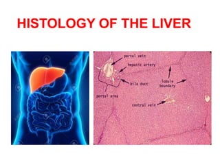
- 16. THE PORTAL SPACE Peripherally, each lobule has 3 to 6 portal areas with more fibrous connective tissue, each of which contains interlobular structures that comprise the portal triad. They include: 1-A venule branch of the portal vein, with blood rich in nutrients but low in O2. 2-An arteriole branch of the hepatic artery that supplies O2. 3-One or two small bile ductules of cuboidal epithelium, branches of the bile conducting system.
- 18. BILE CANALICULI • Formed by spaces present between plasma membranes of adjacent liver cells. • Form hexagonal networks around the liver cells. • Borders around the canaliculi are sealed by tight junctions. This forms the blood-bile barrier.
- 19. BILE CANALICULI • The canaliculi pass to periphery of the hepatic lobules where they form intralobular canal of Herring, that finally drains into the interlobular duct of the portal area. • Bile canaliculi are intralaminar and centrifugal in direction
- 20. SPACE OF DISSE / PERISINUSOIDAL SPACE • Potential space between the wall of sinusoids and laminae of the liver cells. • Filled with blood plasma and chylomicrons that percolate through the wall of sinusoids. • Presence of Ito cells.
- 22. Ito cells • Irregular outline with numerous lipid vesicles. • Function of Ito cells: - Secrete collagenous matrix - Provide growth factor for regeneration of damaged liver cells. - Store Vitamin A in their lipid vesicles.
- 23. SPACE OF MALL • Potential space, between the glisson’s capsule of portal area and the hepatic plates of the cells. • Lymphatics of liver begin here.
- 24. THE PORTAL LOBULE • Territory of liver tissue centered around a portal triad. • Drawn by joining the central veins of three adjacent lobules. • Nutritional lobule of the liver.
- 26. Hepatic acinus of Rappaport • Diamond shaped area of liver parenchyma. • Forms structural and metabolic functions of the liver. •Numerous branches arise at right angles from the blood vessels of portal area, these terminal vessels form backbone of the liver acinus.
- 28. Hepatic acinus of Rappaport contd.. The acinus can be divided into 3 zones based on the gradient of blood supply: Zone 1: Around the vascular backbone, is well oxygenated. Zone 2: Intermediate zone, moderately xygenated. Zone 3: Close to the central vein and the least oxygenated; most susceptible to anoxic injury.
- 29. THE HEPATOCYTES • Large cuboidal or polyhedral epithelial cells, with large, round central nuclei and eosinophilic cytoplasm rich in mitochondria.
- 30. THANK YOU!!