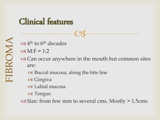
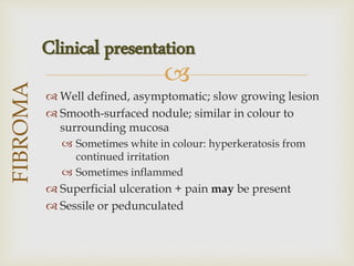
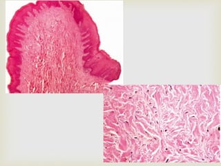
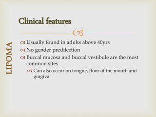
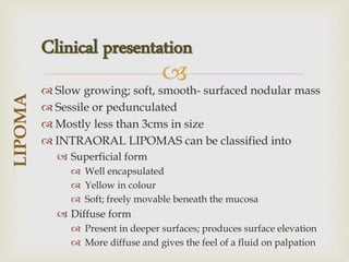
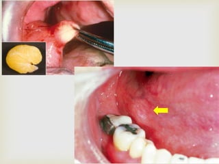
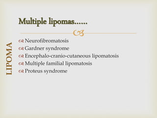
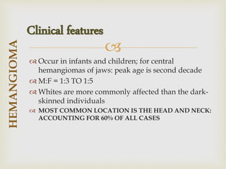
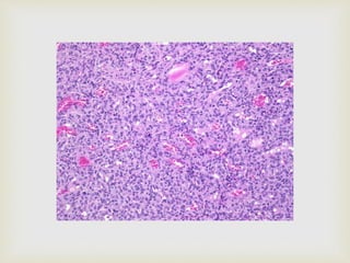
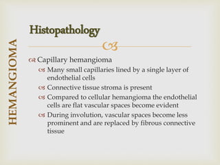
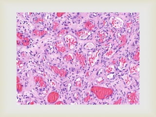
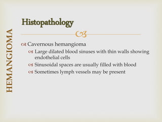
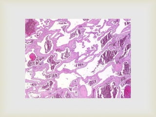
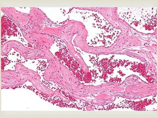
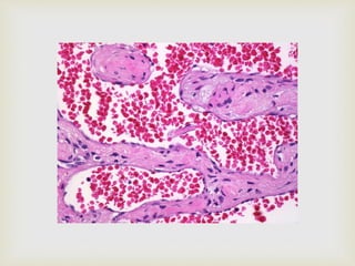
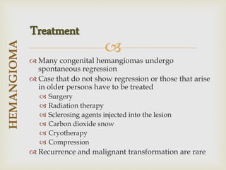
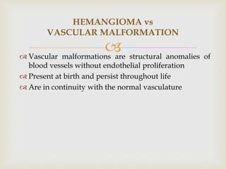
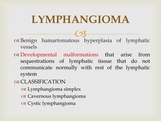
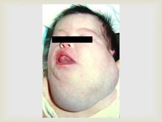
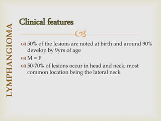
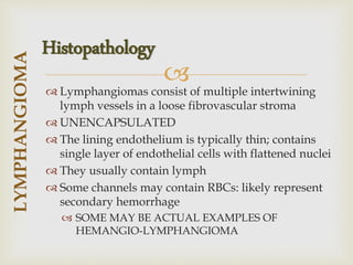
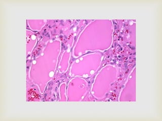
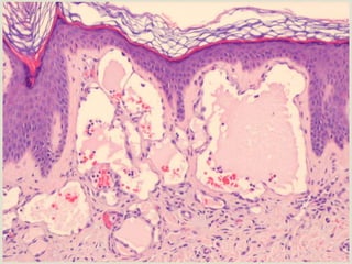
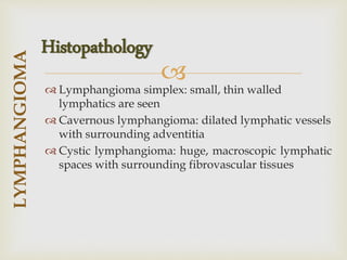
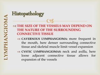
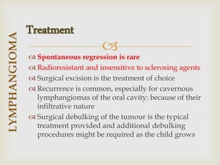
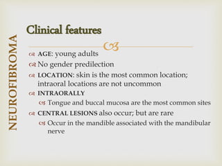
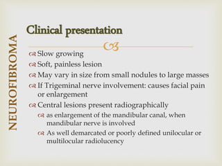
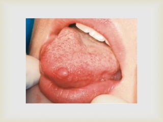
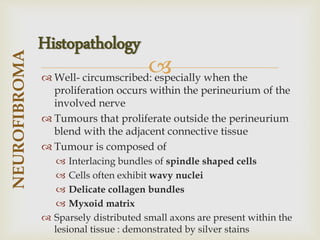
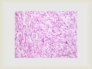
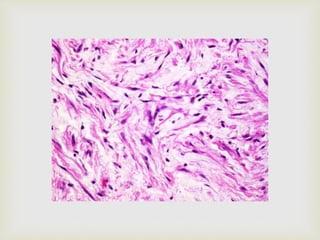
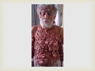
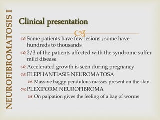
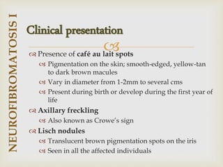
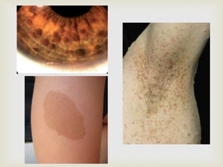
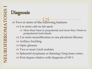
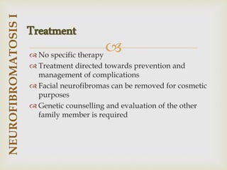
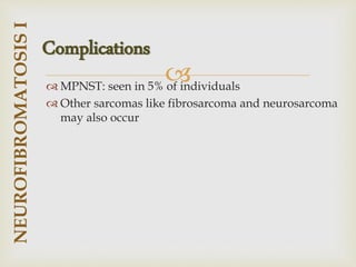
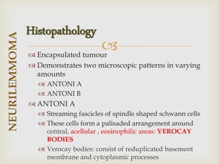
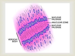
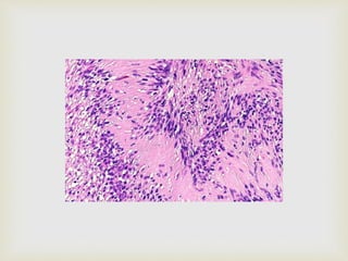
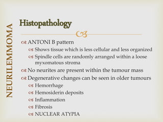
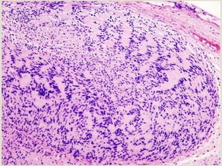
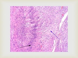
DR. AMRUTHA

 BENIGN TUMOURS
 MALIGNANT TUMOURS
 TUMOUR-LIKE LESIONS
CONTENTS

 FIBROMA
 PERIPHERAL OSSIFYING FIBROMA
 LIPOMA
 HEMANGIOMA
 LYMPHANGIOMA
 NEURILEMMOMA
 NEUROFIBROMA
BENIGN TUMOURS

 FIBROSARCOMA
 OSTEOSARCOMA
 HODGKIN’S LYMPHOMA
 BURKITT’S LYMPHOMA
 KAPOSI’S SARCOMA
 PLASMACYTOMA
 MULTIPLE MYELOMA
MALIGNANT TUMOURS

 PERIPHERAL GIANT CELL GRANULOMA
 CENTRAL GIANT CELL GRANULOMA
 ANEURYSMAL BONE CYST
TUMOUR-LIKE LESIONS

 FIBROMA
 PERIPHERAL OSSIFYING FIBROMA
 LIPOMA
 HEMANGIOMA
 LYMPHANGIOMA
 NEURILEMMOMA
 NEUROFIBROMA
BENIGN TUMOURS

 FIBROMA
 PERIPHERAL OSSIFYING FIBROMA
 LIPOMA
 HEMANGIOMA
 LYMPHANGIOMA
 NEURILEMMOMA
 NEUROFIBROMA
BENIGN TUMOURS

 Irritational fibroma, Traumatic fibroma, Focal fibrous
hyperplasia, Fibrous nodule
 Most common benign soft tissue neoplasm of oral
cavity
 Mostly reactive focal fibrous hyperplasia secondary
to trauma
FIBROMA

 4th to 6th decades
 M:F = 1:2
 Can occur anywhere in the mouth but common sites
are:
 Buccal mucosa, along the bite line
 Gingiva
 Labial mucosa
 Tongue
 Size: from few mm to several cms. Mostly > 1.5cms
FIBROMA
Clinical features

 Well defined, asymptomatic; slow growing lesion
 Smooth-surfaced nodule; similar in colour to
surrounding mucosa
 Sometimes white in colour: hyperkeratosis from
continued irritation
 Sometimes inflammed
 Superficial ulceration + pain may be present
 Sessile or pedunculated
FIBROMA
Clinical presentation

 Nodular mass of fibrous connective tissue covered with
stratified squamous epithelium
 Non-encapsulated lesion; fibrous tissue blends into the
surrounding connective tissue
 CONNECTIVE TISSUE
 Dense and collagenized; scattered inflammation
 Collagen fibres are arranged in radiating, circular or
haphazard pattern
 EPITHELIUM
 Atrophy;flat rete ridges or thin and elongated rete
ridges
 Sometimes hyperkeratosis (clinically white)
FIBROMA
Histopathology

 Conservative surgical excision
 Recurrence is extremely rare
FIBROMA
Treatment

 FIBROMA
 PERIPHERAL OSSIFYING FIBROMA
 LIPOMA
 HEMANGIOMA
 LYMPHANGIOMA
 NEURILEMMOMA
 NEUROFIBROMA
BENIGN TUMOURS

 Calcifying or ossifying fibroid epulis; peripheral
fibroma with calcifications
 Relatively common gingival growth
 Reactive rather than neoplastic
 Mineralised product: origin from cells of periosteum
or periodontal ligament
PERIPHERAL OSSIFYING FIBROMA

 Common in children and young adults; 10-19 yrs
 M:F = 1:2 to 2:3
 Occurs exclusively in the gingiva
 Slight predilection for the maxillary arch
 More than 50% occur in incisor cuspid region
 Adjacent teeth: usually unaffected; sometimes can
cause migration and loosening
PERIPHERAL
OSSIFYING
FIBROMA
Clinical features

 Nodular mass
 Pedunculated or sessile
 Usually arises from the interdental papilla
 Red to pink in colour; usually ulcerated
 Mostly less than 2cms
 DIFFERENTIAL DIAGNOSIS
 Red lesions: pyogenic granuloma
 Pink non-ulcerated lesions: irritational fibroma
 RADIOGRAPHICALLY: superficial erosion
PERIPHERAL
OSSIFYING
FIBROMA
Clinical presentation

 Fibrous proliferation associated with the formation
of mineralised product
 EPITHELIUM
 Intact or ulcerated layer of stratified squamous
epithelium
 When ulcerated: surface is covered by fibrinopurulent
membrane + subjacent zone of granulation tissue
 CONNECTIVE TISSUE
 Cellular mass of connective tissue showing large
numbers of proliferating fibroblasts
 Has delicate fibrillar stroma
PERIPHERAL
OSSIFYING
FIBROMA
Histopathology

 Several forms of CALCIFICATION occur
 Bone: in the form of single or multiple interconnecting
trabeculae of bone or osteoid; older lesions
demonstrate mature lamellar bone
 Cementum-like material: ovoid droplets of basophilic
cementum-like material which closely resembles
acellular cementum
 Dystrophic calcifications: present as multiple granules,
tiny globules or irregular masses of basophilic
mineralised material; these calcifications are more in
early ulcerated lesions
 Occasionally GIANT CELLS may be present
PERIPHERAL
OSSIFYING
FIBROMA
Histopathology

 Local surgical excision down to the periosteum
 Should be submitted for histopathologic examination
 Adjacent teeth should be thoroughly scaled to
prevent further irritation
 Recurrence rate of 8-16% is reported
PERIPHERAL
OSSIFYING
FIBROMA
Treatment

 FIBROMA
 PERIPHERAL OSSIFYING FIBROMA
 LIPOMA
 HEMANGIOMA
 LYMPHANGIOMA
 NEURILEMMOMA
 NEUROFIBROMA
BENIGN TUMOURS

 Benign tumour of fat tissue
 First described by ROUX (in 1848) as yellow epulis
 Relatively rare intraoral tumour; more frequent in
subcutaneous tissues of the neck
 The cells of lipoma differ metabolically from the
normal fat cells even though they are histologically
similar
LIPOMA

 Usually found in adults above 40yrs
 No gender predilection
 Buccal mucosa and buccal vestibule are the most
common sites
 Can also occur on tongue, floor of the mouth and
gingiva
LIPOMA
Clinical features

 Slow growing; soft, smooth- surfaced nodular mass
 Sessile or pedunculated
 Mostly less than 3cms in size
 INTRAORAL LIPOMAS can be classified into
 Superficial form
 Well encapsulated
 Yellow in colour
 Soft; freely movable beneath the mucosa
 Diffuse form
 Present in deeper surfaces; produces surface elevation
 More diffuse and gives the feel of a fluid on palpation
LIPOMA
Clinical presentation

 Neurofibromatosis
 Gardner syndrome
 Encephalo-cranio-cutaneous lipomatosis
 Multiple familial lipomatosis
 Proteus syndrome
LIPOMA
Multiple lipomas……

 Lipoma is composed predominantly of mature
adipocytes or fat cells
 Well demarcated from the surrounding connective
tissue by a fibrous capsule
 Lobular pattern is seen: collagenous streaks can be
seen seperating the fat cells into lobules
 Sometimes lesional fat cells infiltrate surrounding
tissues in the form of long thin extensions
 Extensive involvement of a wide area:
LIPOMATOSIS
LIPOMA
Histopathology

 Conservative local excision
 Recurrence is rare
LIPOMA
Treatment

 FIBROMA
 PERIPHERAL OSSIFYING FIBROMA
 LIPOMA
 HEMANGIOMA
 LYMPHANGIOMA
 NEURILEMMOMA
 NEUROFIBROMA
BENIGN TUMOURS

 A hemangioma is a benign and usually self-
involuting tumor (swelling or growth) of the
endothelial cells.
HEMANGIOMA

 Occur in infants and children; for central
hemangiomas of jaws: peak age is second decade
 M:F = 1:3 TO 1:5
 Whites are more commonly affected than the dark-
skinned individuals
 MOST COMMON LOCATION IS THE HEAD AND NECK:
ACCOUNTING FOR 60% OF ALL CASES
HEMANGIOMA
Clinical features

 Superficial tumours: appear raised and bosselated
with a bright red colour; firm and rubbery on
palpation
 Deep tumours: slightly raised with a bluish hue
HEMANGIOMA
Clinical presentation

 Fully developed hemangiomas are rare at birth
 It presents as a pale macule with thread like
telangiectasias on the skin
 Proliferative phase
 This phase lasts for a few weeks where rapid pace of
growth is observed
 6-10 months after the proliferative phase the tumour
begins to involute
 Colour changes to deep- purple hue; by the age of 5
yrs most of the red colour is gone
 Lesion feels less firm in palpation
HEMANGIOMA
Course of hemangioma…

 50% of the tumours show complete resolution by 5
yrs; 90% resolve by 9 yrs
 AFTER TUMOUR REGRESSION
 Normal skin might be restored
 Or permanent changes might be skin which include
 Atrophy
 Scarring
 Wrinkling
 Telangiectasias
 Complications might also occur which include
ulceration and hemorrhage
HEMANGIOMA
Course of hemangioma…

 Rendu- Osler- Weber syndrome
 Sturge- Weber syndrome
 Maffuci syndrome
 von Hippel Lindau syndrome
HEMANGIOMA
Syndromes associated…

 Flat/ raised lesion of the mucosa
 Deep red/ bluish red in colour
 Readily compressible and fills slowly when released
 Common sites: lips, tongue, buccal mucosa and
palate
 Usually traumatised, undergoes ulceration and
secondary infection
 INTRAMUSCULAR AND CENTRAL
HEMANGIOMAS are also reported in the oral cavity
HEMANGIOMA
Oral manifestations…

 Central hemangioma:
 Occur in maxilla and mandible; 2/3rds in mandible
 First 2 decades of life
 bone destructive lesion: honey comb appearance in
radiograph
 Root resorption is seen in some cases; vitality is not
affected
HEMANGIOMA
Oral manifestations…

 Angiography
 Ultrasonography
 Contrast enhanced MRI: can differentiate between
hemangioma and lymphangioma
 MRI
HEMANGIOMA
Radiographic imaging

 Three common types
 Cellular hemangioma
 Capillary hemangioma
 Cavernous hemangioma
 Cellular hemangioma
 Extensive endothelial proliferation
 Numerous plump endothelial cells
 Indistinct vascular lumina
 It may develop into a simple hemangioma or involute
HEMANGIOMA
Histopathology

 Capillary hemangioma
 Many small capillaries lined by a single layer of
endothelial cells
 Connective tissue stroma is present
 Compared to cellular hemangioma the endothelial
cells are flat vascular spaces become evident
 During involution, vascular spaces become less
prominent and are replaced by fibrous connective
tissue
HEMANGIOMA
Histopathology

 Cavernous hemangioma
 Large dilated blood sinuses with thin walls showing
endothelial cells
 Sinusoidal spaces are usually filled with blood
 Sometimes lymph vessels may be present
HEMANGIOMA
Histopathology

 Many congenital hemangiomas undergo
spontaneous regression
 Case that do not show regression or those that arise
in older persons have to be treated
 Surgery
 Radiation therapy
 Sclerosing agents injected into the lesion
 Carbon dioxide snow
 Cryotherapy
 Compression
 Recurrence and malignant transformation are rare
HEMANGIOMA
Treatment

 Vascular malformations are structural anomalies of
blood vessels without endothelial proliferation
 Present at birth and persist throughout life
 Are in continuity with the normal vasculature
HEMANGIOMA vs
VASCULAR MALFORMATION

 FIBROMA
 PERIPHERAL OSSIFYING FIBROMA
 LIPOMA
 HEMANGIOMA
 LYMPHANGIOMA
 NEURILEMMOMA
 NEUROFIBROMA
BENIGN TUMOURS

 Benign hamartomatous hyperplasia of lymphatic
vessels
 Developmental malformations that arise from
sequestrations of lymphatic tissue that do not
communicate normally with rest of the lymphatic
system
 CLASSIFICATION
 Lymphangioma simplex
 Cavernous lymphangioma
 Cystic lymphangioma
LYMPHANGIOMA

 50% of the lesions are noted at birth and around 90%
develop by 9yrs of age
 M = F
 50-70% of lesions occur in head and neck; most
common location being the lateral neck
LYMPHANGIOMA
Clinical features

 Most common location is the anterior two-thirds of
the tongue
 Other locations: palate, buccal mucosa, gingiva and
lips
 Usually superficial in location
 Demonstrates pebbly surface which resembles a
cluster of translucent vesicles
 The clinical appearance is simulated to tapioca
pudding or frog eggs
 Secondary haemorrhage may cause the vesicles to
appear purple
 Deep tumours present as soft ill-defined masses
LYMPHANGIOMA
Oral manifestations

 Small lymphangiomas less than 1cm occur on the
alveolar ridge: common in black neonates
 These lesions occur bilaterally on the mandibular
ridge
 M:F = 2:1
 They resolve spontaneously
 Central lymphangiomas are also reported : not
common in the oral cavity
LYMPHANGIOMA
Oral manifestations

 Lymphangiomas consist of multiple intertwining
lymph vessels in a loose fibrovascular stroma
 UNENCAPSULATED
 The lining endothelium is typically thin; contains
single layer of endothelial cells with flattened nuclei
 They usually contain lymph
 Some channels may contain RBCs: likely represent
secondary hemorrhage
 SOME MAY BE ACTUAL EXAMPLES OF
HEMANGIO-LYMPHANGIOMA
LYMPHANGIOMA
Histopathology

 In INTRAORAL TUMOURS
 Lymphatic vessels are characteristically located
beneath the epithelial surface
 They replace the connective tissue papillae: little or no
connective tissue is present between the lymph vessels
and the epithelium
 This superficial location results in the appearance of
translucent vesicle- like appearance
 Extension into deeper tissues might also be seen
LYMPHANGIOMA
Histopathology

 Lymphangioma simplex: small, thin walled
lymphatics are seen
 Cavernous lymphangioma: dilated lymphatic vessels
with surrounding adventitia
 Cystic lymphangioma: huge, macroscopic lymphatic
spaces with surrounding fibrovascular tissues
LYMPHANGIOMA
Histopathology

 THE SIZE OF THE VESSELS MAY DEPEND ON
THE NATURE OF THE SURROUNDING
CONNECTIVE TISSUE
 CAVERNOUS LYMPHANGIOMA: more frequent in
the mouth, here denser surrounding connective
tissue and skeletal muscle limit vessel expansion
 CYSTIC LYMPHANGIOMAS: neck and axilla, here
loose adjacent connective tissue allows for
expansion of the vessels
LYMPHANGIOMA
Histopathology

 Spontaneous regression is rare
 Radioresistant and insensitive to sclerosing agents
 Surgical excision is the treatment of choice
 Recurrence is common, especially for cavernous
lymphangiomas of the oral cavity: because of their
infiltrative nature
 Surgical debulking of the tumour is the typical
treatment provided and additional debulking
procedures might be required as the child grows
LYMPHANGIOMA
Treatment

 FIBROMA
 PERIPHERAL OSSIFYING FIBROMA
 LIPOMA
 HEMANGIOMA
 LYMPHANGIOMA
 NEUROFIBROMA
 NEURILEMMOMA
BENIGN TUMOURS

 Neurofibroma is a benign tumour of nerve tissue
origin
 Most common type of peripheral nerve neoplasm
 ORIGIN
 cells that constitute the nerve sheath, including
SCHWANN CELLS and PERINEURAL FIBROBLASTS
 It can occur either as a solitary lesion or as a part of
the syndrome: NEUROFIBROMATOSIS I
NEUROFIBROMA

 AGE: young adults
 No gender predilection
 LOCATION: skin is the most common location;
intraoral locations are not uncommon
 INTRAORALLY
 Tongue and buccal mucosa are the most common sites
 CENTRAL LESIONS also occur; but are rare
 Occur in the mandible associated with the mandibular
nerve
NEUROFIBROMA
Clinical features

 Slow growing
 Soft, painless lesion
 May vary in size from small nodules to large masses
 If Trigeminal nerve involvement: causes facial pain
or enlargement
 Central lesions present radiographically
 as enlargement of the mandibular canal, when
mandibular nerve is involved
 As well demarcated or poorly defined unilocular or
multilocular radiolucency
NEUROFIBROMA
Clinical presentation

 Well- circumscribed: especially when the
proliferation occurs within the perineurium of the
involved nerve
 Tumours that proliferate outside the perineurium
blend with the adjacent connective tissue
 Tumour is composed of
 Interlacing bundles of spindle shaped cells
 Cells often exhibit wavy nuclei
 Delicate collagen bundles
 Myxoid matrix
 Sparsely distributed small axons are present within the
lesional tissue : demonstrated by silver stains
NEUROFIBROMA
Histopathology

 Local surgical excision
 Recurrence is rare
 Malignant transformation is less but not rare
NEUROFIBROMA
Treatment

 Also known as von Recklinghausen’s disease
 Incidence: 1 in 3000 individuals
 Etiology
 Mutation in neurofibromin gene
 Inherited as an autosomal dominant trait
NEUROFIBROMATOSIS I

 Patients have multiple neurofibromas that can occur
anywhere on the body
 Most common on the skin
 Tumours may be present at birth; often appear
during puberty
 They continue to develop slowly through adulthood
 Size varies from small papules to large nodules
NEUROFIBROMATOSIS
I
Clinical features

 Some patients have few lesions ; some have
hundreds to thousands
 2/3 of the patients affected with the syndrome suffer
mild disease
 Accelerated growth is seen during pregnancy
 ELEPHANTIASIS NEUROMATOSA
 Massive baggy pendulous masses present on the skin
 PLEXIFORM NEUROFIBROMA
 On palpation gives the feeling of a bag of worms
NEUROFIBROMATOSIS
I
Clinical presentation

 Presence of café au lait spots
 Pigmentation on the skin; smooth-edged, yellow-tan
to dark brown macules
 Vary in diameter from 1-2mm to several cms
 Present during birth or develop during the first year of
life
 Axillary freckling
 Also known as Crowe’s sign
 Lisch nodules
 Translucent brown pigmentation spots on the iris
 Seen in all the affected individuals
NEUROFIBROMATOSIS
I
Clinical presentation

 7-20% show oral manifestations
 Discrete non-ulcerated nodules which are of same
colour of the mucosa
 Location: buccal mucosa, palate, alveolar ridge,
vestibule and tongue
 Sometimes may present as diffuse masses involving
tongue (presents as macroglossia) and alveolar ridge
 Enlargement of fungiform papillae is seen in 50% of
patients
 Central lesions can also occur
NEUROFIBROMATOSIS
I
Oral manifestations

 Two or more of the following features
 6 or more café au lait spots
 More than 5mm in prepubertal and more than 15mm in
postpubertal individuals
 2 or more neurofibromas or one plexiform fibroma
 Axillary freckling
 Optic gliomas
 Two or more Lisch nodules
 Sphenoid dysplasias or thinning f long bone cortex
 First degree relative with diagnosis of NF-1
NEUROFIBROMATOSIS
I
Diagnosis

 No specific therapy
 Treatment directed towards prevention and
management of complications
 Facial neurofibromas can be removed for cosmetic
purposes
 Genetic counselling and evaluation of the other
family member is required
NEUROFIBROMATOSIS
I
Treatment

 MPNST: seen in 5% of individuals
 Other sarcomas like fibrosarcoma and neurosarcoma
may also occur
NEUROFIBROMATOSIS
I
Complications

 FIBROMA
 PERIPHERAL OSSIFYING FIBROMA
 LIPOMA
 HEMANGIOMA
 LYMPHANGIOMA
 NEUROFIBROMA
 NEURILEMMOMA
BENIGN TUMOURS

 Neurolemmoma, Schwannoma, Perineural
Fibroblastoma, Neurinoma, Lemmoma
 Benign neural neoplasm of Schwann cell origin
NEURILEMMOMA

 Most common in young and middle aged adults; but
can arise in any age, even during the first year of life
 M = F
 Few mm to several cm
NEURILEMMOMA
Clinical features

 Encapsulated
 Slow- growing tumour
 Typically arises in association with a nerve trunk
 As it grows it pushes the nerve aside
 Tenderness and pain is not a common condition and
usually occurs due to pressure on adjacent nerves
NEURILEMMOMA
Clinical presentation

 Tongue is the most common location
 May also arise centrally and cause bony destruction
with expansion of cortical plates
 Pain and paraesthesia accompany central lesions
NEURILEMMOMA
Oral manifestations

 Encapsulated tumour
 Demonstrates two microscopic patterns in varying
amounts
 ANTONI A
 ANTONI B
 ANTONI A
 Streaming fascicles of spindle shaped schwann cells
 These cells form a palisaded arrangement around
central, acellular , eosinophilic areas: VEROCAY
BODIES
 Verocay bodies: consist of reduplicated basement
membrane and cytoplasmic processes
NEURILEMMOMA
Histopathology

 ANTONI B pattern
 Shows tissue which is less cellular and less organized
 Spindle cells are randomly arranged within a loose
myxomatous stroma
 No neurites are present within the tumour mass
 Degenerative changes can be seen in older tumours
 Hemorrhage
 Hemosiderin deposits
 Inflammation
 Fibrosis
 NUCLEAR ATYPIA
NEURILEMMOMA
Histopathology

 Surgical excision
 Recurrence and malignant transformation are rare
NEURILEMMOMA
Treatment

 Autosomal dominant condition
 Caused by mutation of gene producing the protein
merlin
 CHARACTERISTIC FEATURES
 Bilateral neurilemmomas of auditory vestibular
nerve
 Neurilemmomas of peripheral nerves
 Meningiomas of CNS
 SYMPTOMS
 Sensorineural deafness
 Dizziness
 Tinnitus
NEURILEMMOMA
Neurofibromatosis II

Benign Connective Tissue Tumors.pptx

  • 1.
  • 2.
      BENIGN TUMOURS MALIGNANT TUMOURS  TUMOUR-LIKE LESIONS CONTENTS
  • 3.
      FIBROMA  PERIPHERALOSSIFYING FIBROMA  LIPOMA  HEMANGIOMA  LYMPHANGIOMA  NEURILEMMOMA  NEUROFIBROMA BENIGN TUMOURS
  • 4.
      FIBROSARCOMA  OSTEOSARCOMA HODGKIN’S LYMPHOMA  BURKITT’S LYMPHOMA  KAPOSI’S SARCOMA  PLASMACYTOMA  MULTIPLE MYELOMA MALIGNANT TUMOURS
  • 5.
      PERIPHERAL GIANTCELL GRANULOMA  CENTRAL GIANT CELL GRANULOMA  ANEURYSMAL BONE CYST TUMOUR-LIKE LESIONS
  • 6.
      FIBROMA  PERIPHERALOSSIFYING FIBROMA  LIPOMA  HEMANGIOMA  LYMPHANGIOMA  NEURILEMMOMA  NEUROFIBROMA BENIGN TUMOURS
  • 7.
      FIBROMA  PERIPHERALOSSIFYING FIBROMA  LIPOMA  HEMANGIOMA  LYMPHANGIOMA  NEURILEMMOMA  NEUROFIBROMA BENIGN TUMOURS
  • 8.
      Irritational fibroma,Traumatic fibroma, Focal fibrous hyperplasia, Fibrous nodule  Most common benign soft tissue neoplasm of oral cavity  Mostly reactive focal fibrous hyperplasia secondary to trauma FIBROMA
  • 9.
      4th to6th decades  M:F = 1:2  Can occur anywhere in the mouth but common sites are:  Buccal mucosa, along the bite line  Gingiva  Labial mucosa  Tongue  Size: from few mm to several cms. Mostly > 1.5cms FIBROMA Clinical features
  • 10.
      Well defined,asymptomatic; slow growing lesion  Smooth-surfaced nodule; similar in colour to surrounding mucosa  Sometimes white in colour: hyperkeratosis from continued irritation  Sometimes inflammed  Superficial ulceration + pain may be present  Sessile or pedunculated FIBROMA Clinical presentation
  • 13.
      Nodular massof fibrous connective tissue covered with stratified squamous epithelium  Non-encapsulated lesion; fibrous tissue blends into the surrounding connective tissue  CONNECTIVE TISSUE  Dense and collagenized; scattered inflammation  Collagen fibres are arranged in radiating, circular or haphazard pattern  EPITHELIUM  Atrophy;flat rete ridges or thin and elongated rete ridges  Sometimes hyperkeratosis (clinically white) FIBROMA Histopathology
  • 15.
      Conservative surgicalexcision  Recurrence is extremely rare FIBROMA Treatment
  • 16.
      FIBROMA  PERIPHERALOSSIFYING FIBROMA  LIPOMA  HEMANGIOMA  LYMPHANGIOMA  NEURILEMMOMA  NEUROFIBROMA BENIGN TUMOURS
  • 17.
      Calcifying orossifying fibroid epulis; peripheral fibroma with calcifications  Relatively common gingival growth  Reactive rather than neoplastic  Mineralised product: origin from cells of periosteum or periodontal ligament PERIPHERAL OSSIFYING FIBROMA
  • 18.
      Common inchildren and young adults; 10-19 yrs  M:F = 1:2 to 2:3  Occurs exclusively in the gingiva  Slight predilection for the maxillary arch  More than 50% occur in incisor cuspid region  Adjacent teeth: usually unaffected; sometimes can cause migration and loosening PERIPHERAL OSSIFYING FIBROMA Clinical features
  • 19.
      Nodular mass Pedunculated or sessile  Usually arises from the interdental papilla  Red to pink in colour; usually ulcerated  Mostly less than 2cms  DIFFERENTIAL DIAGNOSIS  Red lesions: pyogenic granuloma  Pink non-ulcerated lesions: irritational fibroma  RADIOGRAPHICALLY: superficial erosion PERIPHERAL OSSIFYING FIBROMA Clinical presentation
  • 21.
      Fibrous proliferationassociated with the formation of mineralised product  EPITHELIUM  Intact or ulcerated layer of stratified squamous epithelium  When ulcerated: surface is covered by fibrinopurulent membrane + subjacent zone of granulation tissue  CONNECTIVE TISSUE  Cellular mass of connective tissue showing large numbers of proliferating fibroblasts  Has delicate fibrillar stroma PERIPHERAL OSSIFYING FIBROMA Histopathology
  • 22.
      Several formsof CALCIFICATION occur  Bone: in the form of single or multiple interconnecting trabeculae of bone or osteoid; older lesions demonstrate mature lamellar bone  Cementum-like material: ovoid droplets of basophilic cementum-like material which closely resembles acellular cementum  Dystrophic calcifications: present as multiple granules, tiny globules or irregular masses of basophilic mineralised material; these calcifications are more in early ulcerated lesions  Occasionally GIANT CELLS may be present PERIPHERAL OSSIFYING FIBROMA Histopathology
  • 24.
      Local surgicalexcision down to the periosteum  Should be submitted for histopathologic examination  Adjacent teeth should be thoroughly scaled to prevent further irritation  Recurrence rate of 8-16% is reported PERIPHERAL OSSIFYING FIBROMA Treatment
  • 25.
      FIBROMA  PERIPHERALOSSIFYING FIBROMA  LIPOMA  HEMANGIOMA  LYMPHANGIOMA  NEURILEMMOMA  NEUROFIBROMA BENIGN TUMOURS
  • 26.
      Benign tumourof fat tissue  First described by ROUX (in 1848) as yellow epulis  Relatively rare intraoral tumour; more frequent in subcutaneous tissues of the neck  The cells of lipoma differ metabolically from the normal fat cells even though they are histologically similar LIPOMA
  • 27.
      Usually foundin adults above 40yrs  No gender predilection  Buccal mucosa and buccal vestibule are the most common sites  Can also occur on tongue, floor of the mouth and gingiva LIPOMA Clinical features
  • 28.
      Slow growing;soft, smooth- surfaced nodular mass  Sessile or pedunculated  Mostly less than 3cms in size  INTRAORAL LIPOMAS can be classified into  Superficial form  Well encapsulated  Yellow in colour  Soft; freely movable beneath the mucosa  Diffuse form  Present in deeper surfaces; produces surface elevation  More diffuse and gives the feel of a fluid on palpation LIPOMA Clinical presentation
  • 30.
      Neurofibromatosis  Gardnersyndrome  Encephalo-cranio-cutaneous lipomatosis  Multiple familial lipomatosis  Proteus syndrome LIPOMA Multiple lipomas……
  • 31.
      Lipoma iscomposed predominantly of mature adipocytes or fat cells  Well demarcated from the surrounding connective tissue by a fibrous capsule  Lobular pattern is seen: collagenous streaks can be seen seperating the fat cells into lobules  Sometimes lesional fat cells infiltrate surrounding tissues in the form of long thin extensions  Extensive involvement of a wide area: LIPOMATOSIS LIPOMA Histopathology
  • 33.
      Conservative localexcision  Recurrence is rare LIPOMA Treatment
  • 34.
      FIBROMA  PERIPHERALOSSIFYING FIBROMA  LIPOMA  HEMANGIOMA  LYMPHANGIOMA  NEURILEMMOMA  NEUROFIBROMA BENIGN TUMOURS
  • 35.
      A hemangiomais a benign and usually self- involuting tumor (swelling or growth) of the endothelial cells. HEMANGIOMA
  • 36.
      Occur ininfants and children; for central hemangiomas of jaws: peak age is second decade  M:F = 1:3 TO 1:5  Whites are more commonly affected than the dark- skinned individuals  MOST COMMON LOCATION IS THE HEAD AND NECK: ACCOUNTING FOR 60% OF ALL CASES HEMANGIOMA Clinical features
  • 37.
      Superficial tumours:appear raised and bosselated with a bright red colour; firm and rubbery on palpation  Deep tumours: slightly raised with a bluish hue HEMANGIOMA Clinical presentation
  • 38.
      Fully developedhemangiomas are rare at birth  It presents as a pale macule with thread like telangiectasias on the skin  Proliferative phase  This phase lasts for a few weeks where rapid pace of growth is observed  6-10 months after the proliferative phase the tumour begins to involute  Colour changes to deep- purple hue; by the age of 5 yrs most of the red colour is gone  Lesion feels less firm in palpation HEMANGIOMA Course of hemangioma…
  • 39.
      50% ofthe tumours show complete resolution by 5 yrs; 90% resolve by 9 yrs  AFTER TUMOUR REGRESSION  Normal skin might be restored  Or permanent changes might be skin which include  Atrophy  Scarring  Wrinkling  Telangiectasias  Complications might also occur which include ulceration and hemorrhage HEMANGIOMA Course of hemangioma…
  • 41.
      Rendu- Osler-Weber syndrome  Sturge- Weber syndrome  Maffuci syndrome  von Hippel Lindau syndrome HEMANGIOMA Syndromes associated…
  • 42.
      Flat/ raisedlesion of the mucosa  Deep red/ bluish red in colour  Readily compressible and fills slowly when released  Common sites: lips, tongue, buccal mucosa and palate  Usually traumatised, undergoes ulceration and secondary infection  INTRAMUSCULAR AND CENTRAL HEMANGIOMAS are also reported in the oral cavity HEMANGIOMA Oral manifestations…
  • 44.
      Central hemangioma: Occur in maxilla and mandible; 2/3rds in mandible  First 2 decades of life  bone destructive lesion: honey comb appearance in radiograph  Root resorption is seen in some cases; vitality is not affected HEMANGIOMA Oral manifestations…
  • 45.
      Angiography  Ultrasonography Contrast enhanced MRI: can differentiate between hemangioma and lymphangioma  MRI HEMANGIOMA Radiographic imaging
  • 46.
      Three commontypes  Cellular hemangioma  Capillary hemangioma  Cavernous hemangioma  Cellular hemangioma  Extensive endothelial proliferation  Numerous plump endothelial cells  Indistinct vascular lumina  It may develop into a simple hemangioma or involute HEMANGIOMA Histopathology
  • 48.
      Capillary hemangioma Many small capillaries lined by a single layer of endothelial cells  Connective tissue stroma is present  Compared to cellular hemangioma the endothelial cells are flat vascular spaces become evident  During involution, vascular spaces become less prominent and are replaced by fibrous connective tissue HEMANGIOMA Histopathology
  • 52.
      Cavernous hemangioma Large dilated blood sinuses with thin walls showing endothelial cells  Sinusoidal spaces are usually filled with blood  Sometimes lymph vessels may be present HEMANGIOMA Histopathology
  • 56.
      Many congenitalhemangiomas undergo spontaneous regression  Case that do not show regression or those that arise in older persons have to be treated  Surgery  Radiation therapy  Sclerosing agents injected into the lesion  Carbon dioxide snow  Cryotherapy  Compression  Recurrence and malignant transformation are rare HEMANGIOMA Treatment
  • 57.
      Vascular malformationsare structural anomalies of blood vessels without endothelial proliferation  Present at birth and persist throughout life  Are in continuity with the normal vasculature HEMANGIOMA vs VASCULAR MALFORMATION
  • 58.
      FIBROMA  PERIPHERALOSSIFYING FIBROMA  LIPOMA  HEMANGIOMA  LYMPHANGIOMA  NEURILEMMOMA  NEUROFIBROMA BENIGN TUMOURS
  • 59.
      Benign hamartomatoushyperplasia of lymphatic vessels  Developmental malformations that arise from sequestrations of lymphatic tissue that do not communicate normally with rest of the lymphatic system  CLASSIFICATION  Lymphangioma simplex  Cavernous lymphangioma  Cystic lymphangioma LYMPHANGIOMA
  • 61.
      50% ofthe lesions are noted at birth and around 90% develop by 9yrs of age  M = F  50-70% of lesions occur in head and neck; most common location being the lateral neck LYMPHANGIOMA Clinical features
  • 62.
      Most commonlocation is the anterior two-thirds of the tongue  Other locations: palate, buccal mucosa, gingiva and lips  Usually superficial in location  Demonstrates pebbly surface which resembles a cluster of translucent vesicles  The clinical appearance is simulated to tapioca pudding or frog eggs  Secondary haemorrhage may cause the vesicles to appear purple  Deep tumours present as soft ill-defined masses LYMPHANGIOMA Oral manifestations
  • 64.
      Small lymphangiomasless than 1cm occur on the alveolar ridge: common in black neonates  These lesions occur bilaterally on the mandibular ridge  M:F = 2:1  They resolve spontaneously  Central lymphangiomas are also reported : not common in the oral cavity LYMPHANGIOMA Oral manifestations
  • 66.
      Lymphangiomas consistof multiple intertwining lymph vessels in a loose fibrovascular stroma  UNENCAPSULATED  The lining endothelium is typically thin; contains single layer of endothelial cells with flattened nuclei  They usually contain lymph  Some channels may contain RBCs: likely represent secondary hemorrhage  SOME MAY BE ACTUAL EXAMPLES OF HEMANGIO-LYMPHANGIOMA LYMPHANGIOMA Histopathology
  • 69.
      In INTRAORALTUMOURS  Lymphatic vessels are characteristically located beneath the epithelial surface  They replace the connective tissue papillae: little or no connective tissue is present between the lymph vessels and the epithelium  This superficial location results in the appearance of translucent vesicle- like appearance  Extension into deeper tissues might also be seen LYMPHANGIOMA Histopathology
  • 71.
      Lymphangioma simplex:small, thin walled lymphatics are seen  Cavernous lymphangioma: dilated lymphatic vessels with surrounding adventitia  Cystic lymphangioma: huge, macroscopic lymphatic spaces with surrounding fibrovascular tissues LYMPHANGIOMA Histopathology
  • 72.
      THE SIZEOF THE VESSELS MAY DEPEND ON THE NATURE OF THE SURROUNDING CONNECTIVE TISSUE  CAVERNOUS LYMPHANGIOMA: more frequent in the mouth, here denser surrounding connective tissue and skeletal muscle limit vessel expansion  CYSTIC LYMPHANGIOMAS: neck and axilla, here loose adjacent connective tissue allows for expansion of the vessels LYMPHANGIOMA Histopathology
  • 73.
      Spontaneous regressionis rare  Radioresistant and insensitive to sclerosing agents  Surgical excision is the treatment of choice  Recurrence is common, especially for cavernous lymphangiomas of the oral cavity: because of their infiltrative nature  Surgical debulking of the tumour is the typical treatment provided and additional debulking procedures might be required as the child grows LYMPHANGIOMA Treatment
  • 74.
      FIBROMA  PERIPHERALOSSIFYING FIBROMA  LIPOMA  HEMANGIOMA  LYMPHANGIOMA  NEUROFIBROMA  NEURILEMMOMA BENIGN TUMOURS
  • 75.
      Neurofibroma isa benign tumour of nerve tissue origin  Most common type of peripheral nerve neoplasm  ORIGIN  cells that constitute the nerve sheath, including SCHWANN CELLS and PERINEURAL FIBROBLASTS  It can occur either as a solitary lesion or as a part of the syndrome: NEUROFIBROMATOSIS I NEUROFIBROMA
  • 76.
      AGE: youngadults  No gender predilection  LOCATION: skin is the most common location; intraoral locations are not uncommon  INTRAORALLY  Tongue and buccal mucosa are the most common sites  CENTRAL LESIONS also occur; but are rare  Occur in the mandible associated with the mandibular nerve NEUROFIBROMA Clinical features
  • 77.
      Slow growing Soft, painless lesion  May vary in size from small nodules to large masses  If Trigeminal nerve involvement: causes facial pain or enlargement  Central lesions present radiographically  as enlargement of the mandibular canal, when mandibular nerve is involved  As well demarcated or poorly defined unilocular or multilocular radiolucency NEUROFIBROMA Clinical presentation
  • 79.
      Well- circumscribed:especially when the proliferation occurs within the perineurium of the involved nerve  Tumours that proliferate outside the perineurium blend with the adjacent connective tissue  Tumour is composed of  Interlacing bundles of spindle shaped cells  Cells often exhibit wavy nuclei  Delicate collagen bundles  Myxoid matrix  Sparsely distributed small axons are present within the lesional tissue : demonstrated by silver stains NEUROFIBROMA Histopathology
  • 82.
      Local surgicalexcision  Recurrence is rare  Malignant transformation is less but not rare NEUROFIBROMA Treatment
  • 83.
      Also knownas von Recklinghausen’s disease  Incidence: 1 in 3000 individuals  Etiology  Mutation in neurofibromin gene  Inherited as an autosomal dominant trait NEUROFIBROMATOSIS I
  • 84.
      Patients havemultiple neurofibromas that can occur anywhere on the body  Most common on the skin  Tumours may be present at birth; often appear during puberty  They continue to develop slowly through adulthood  Size varies from small papules to large nodules NEUROFIBROMATOSIS I Clinical features
  • 86.
      Some patientshave few lesions ; some have hundreds to thousands  2/3 of the patients affected with the syndrome suffer mild disease  Accelerated growth is seen during pregnancy  ELEPHANTIASIS NEUROMATOSA  Massive baggy pendulous masses present on the skin  PLEXIFORM NEUROFIBROMA  On palpation gives the feeling of a bag of worms NEUROFIBROMATOSIS I Clinical presentation
  • 87.
      Presence ofcafé au lait spots  Pigmentation on the skin; smooth-edged, yellow-tan to dark brown macules  Vary in diameter from 1-2mm to several cms  Present during birth or develop during the first year of life  Axillary freckling  Also known as Crowe’s sign  Lisch nodules  Translucent brown pigmentation spots on the iris  Seen in all the affected individuals NEUROFIBROMATOSIS I Clinical presentation
  • 89.
      7-20% showoral manifestations  Discrete non-ulcerated nodules which are of same colour of the mucosa  Location: buccal mucosa, palate, alveolar ridge, vestibule and tongue  Sometimes may present as diffuse masses involving tongue (presents as macroglossia) and alveolar ridge  Enlargement of fungiform papillae is seen in 50% of patients  Central lesions can also occur NEUROFIBROMATOSIS I Oral manifestations
  • 90.
      Two ormore of the following features  6 or more café au lait spots  More than 5mm in prepubertal and more than 15mm in postpubertal individuals  2 or more neurofibromas or one plexiform fibroma  Axillary freckling  Optic gliomas  Two or more Lisch nodules  Sphenoid dysplasias or thinning f long bone cortex  First degree relative with diagnosis of NF-1 NEUROFIBROMATOSIS I Diagnosis
  • 91.
      No specifictherapy  Treatment directed towards prevention and management of complications  Facial neurofibromas can be removed for cosmetic purposes  Genetic counselling and evaluation of the other family member is required NEUROFIBROMATOSIS I Treatment
  • 92.
      MPNST: seenin 5% of individuals  Other sarcomas like fibrosarcoma and neurosarcoma may also occur NEUROFIBROMATOSIS I Complications
  • 93.
      FIBROMA  PERIPHERALOSSIFYING FIBROMA  LIPOMA  HEMANGIOMA  LYMPHANGIOMA  NEUROFIBROMA  NEURILEMMOMA BENIGN TUMOURS
  • 94.
      Neurolemmoma, Schwannoma,Perineural Fibroblastoma, Neurinoma, Lemmoma  Benign neural neoplasm of Schwann cell origin NEURILEMMOMA
  • 95.
      Most commonin young and middle aged adults; but can arise in any age, even during the first year of life  M = F  Few mm to several cm NEURILEMMOMA Clinical features
  • 96.
      Encapsulated  Slow-growing tumour  Typically arises in association with a nerve trunk  As it grows it pushes the nerve aside  Tenderness and pain is not a common condition and usually occurs due to pressure on adjacent nerves NEURILEMMOMA Clinical presentation
  • 97.
      Tongue isthe most common location  May also arise centrally and cause bony destruction with expansion of cortical plates  Pain and paraesthesia accompany central lesions NEURILEMMOMA Oral manifestations
  • 98.
      Encapsulated tumour Demonstrates two microscopic patterns in varying amounts  ANTONI A  ANTONI B  ANTONI A  Streaming fascicles of spindle shaped schwann cells  These cells form a palisaded arrangement around central, acellular , eosinophilic areas: VEROCAY BODIES  Verocay bodies: consist of reduplicated basement membrane and cytoplasmic processes NEURILEMMOMA Histopathology
  • 101.
      ANTONI Bpattern  Shows tissue which is less cellular and less organized  Spindle cells are randomly arranged within a loose myxomatous stroma  No neurites are present within the tumour mass  Degenerative changes can be seen in older tumours  Hemorrhage  Hemosiderin deposits  Inflammation  Fibrosis  NUCLEAR ATYPIA NEURILEMMOMA Histopathology
  • 104.
      Surgical excision Recurrence and malignant transformation are rare NEURILEMMOMA Treatment
  • 105.
      Autosomal dominantcondition  Caused by mutation of gene producing the protein merlin  CHARACTERISTIC FEATURES  Bilateral neurilemmomas of auditory vestibular nerve  Neurilemmomas of peripheral nerves  Meningiomas of CNS  SYMPTOMS  Sensorineural deafness  Dizziness  Tinnitus NEURILEMMOMA Neurofibromatosis II

Editor's Notes

  • #10 Buccal mucosa along the biteline: presumably this is the consequence of trauma from cheek biting Gingiva: most gingival fibromas represent fibrous maturation of a pre-existing pyogenic granuloma