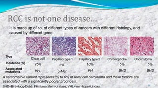
Renal cellcarcinoma
Account for approx. 3% ofadult solidmalignancy
51,000 cases diagnosed andmore than 12,900
deaths annually in theUS
2
RenalCellCarcinoma
3
First described by Konig in1826.
All these tumors arise from Renal tubular epithelium.
Accounts for 80–85% of kidneycancer
2 % to 4% increase in incidence per year
Epidemiology
4
Male predominance (1.6:1.0M:F(
Highest incidence between age 50-70
-Median age of diagnosis is 66 years
-Median age of death is70 years
Majority of RCC occurssporadically
Highest incidence in Scandinavia and North America,
lowest inAfrica
RiskFactors
5
Tobacco smoking contributes to24-30% of RCCcases
- Tobacco results in a 2-fold increased risk
Environmental:
Cadmium, thorium-dioxide, petroleum and phenacetin analgesics.
Occupational:
leather tanners, shoe workers, asbestos workers.
Hormonal:
diethylistillbestrol,
Obesity, HTN
35% - 47% pt on long term dialysis develop Acquired polycystic kidney
disease, out of which 5.8% develops Renal cancer.
NaturalHistory
6
27% diagnosed incidentally
Of them 45 % present with localized disease, 25%
with locally advanced disease, 30 % with metastatic
disease
Lymph node metastases- 9 % to 27% (renal hilar, para-
aortic and paracaval(
Renal vein – 21%& IVC4%
Distant metastases- lung (75%), bone (20%), liver (18%),
skin (8%) and CNS 8%))
ClinicalPresentation
7
Classic triad occur only in 5- 10% of patients
flank pain
hematuria
palpable abdominal mass
Systemic symptoms
Anaemia, Fatigue, Cachexia, Wt. Loss, Hypercalcemia, Hepatic
Dysfunction
Paraneoplastic Syndrome
Parathyroid like hormones, erythropoietin, renin, gonadotropins,
placental lactogen, prolactin,enteroglucagon, insulin like hormones,
adrenocorticotropic hormone and prostaglandins identified in RCC pt.
Hereditary Renal Cancer Syndromes
 Inherited forms of renal cancer are estimated to account
for 2%–6% of all kidney cancer .
 Currently, 10 inherited cancer susceptibility syndromes
are definitively associated with an increased risk of renal
cancer
 . Patients with these inherited syndromes develop
kidney cancer at an earlier age; furthermore, the lesions
can be multifocal, bilateral, and heterogeneous.
 Several syndromes have renal cancer as a primary feature,
including :
 Birt-Hogg-Dubé syndrome (BHD)
 Familial clear cell renal cancer due to chromosome 3 translocation
 Hereditary papillary renal cancer
 Hereditary leiomyomatosis and renal cell cancer (HLRCC)
 Von Hippel-Lindau disease (vHL)
DiagnosticWork-Up
10
General-History, Physical examination
Laboratory studies
CBC, LFT's, alkaline phosphotase, BUN, creatinine, urinalysis
Radiographic studies- Increased use of imaging has increased the detection of
renal lesions
Ultrasonography- Excellent in distinguishing cystic from solid masses
Computed tomography- Provides an excellent assessment of the parenchyma
andnodal status.
Magnetic Resonance Imaging - excellent demonstration of solid renal masses and is
image test of choice to demonstrate extent of vena caval involvement with tumor.
Useful in patients with renal insufficiency
MetastaticWork-Up
11
Chest CT
CT/MRI scan of abdomen or pelvis
Bone scan with plan films)only for patients with elevated
alkaline phosphatase or bone pain(.
Figure:Computedtomographydemonstrates a
right renalcarcinoma(m) with alarge
contralateral adrenalmetastasis(a).
Figure: CTscan shows large left renal mass
with calcification (m) invading the left renal
vein (arrow).
12
TNMstaging
22
T - primary tumour
TX Primary tumour cannot be assessed
T0 No evidence of primary tumour
T1 Tumour confined to kidney, <7cm
T1a ≤4cm, confined to kidney
T1b >4cm but <7cm, confined to kidney
T2 Tumour >7cm, confined to kidney
T3 Tumour extends into major veins or adrenal or perinephric tissue but not beyond
Gerota’s fascia
T3a Direct invasion of adrenal gland, perirenal and/or sinus fat
T3b Gross extension into renal vein or IVC
T3c Extends into IVC above diaphragm or wall of IVC
T4 Invasion beyond Gerota’s fascia
N - regional lymph nodes
NX Nodes cannot be assessed
N0 Regional lymph nodes not involved
N1 Metastasis in a single regional lymph node
N2 Metastases in >1regional lymph node
M - distant metastases
MX Metastases cannot be assessed
M0 No distant metastases
M1 Distant metastases
Stagingof RCC
15
Tumor Stage Description
I Renal cell carcinoma is confined to the kidneys
II Renal cell carcinoma extends through the renal capsule but is confined to
Gerota’sfascia
III Renal cell carcinoma involves the renal vein or inferior vena cava (IIIA)
or the renal hilar lymph nodes (IIIB)
IV Renal cell carcinoma has spread to local adjacent organs
(other than adrenal gland) or to distant sites
Staging
1
6
I T1 N0 M0
II T2 N0 M0
III
T3 N0 M0
T1 N1 M0
T2 N1 M0
IV
T4 N0 M0
T4 N1 M0
Any T N2 M0
Any T Any N M1
- Stage I-III: Localized disease
- Stage IV:Advanced, metastatic
disease
Prognosticfactors forRCC
1
7
Pathologic stage 5yr survival
T1-2organconfined 70-90%
T3 50-70%
N+, M1
Tumour size
5-30%
< 4 cm > 90%
4 - 10cm 50%
> 10cm
Histological type
0%
Clear cell 70%
Papillary, Chromophobe 85%
Multilocular cystic 100%
Medullary, Collecting duct 0%
A sarcomatoid variant represents1% to 6% of renal cell carcinoma and these tumors are
associated with a significantly poorer prognosis.
BHD=Birt-Hogg-Dubé; FH=fumarate hydratase; VHL=von Hippel-Lindau.
RCC is not one disease…
Clear cell
Type
Incidence (%)
Associated
mutations
75%
VHL
Papillary type 1
5%
c-Met
Papillary type 2
10%
FH
Chromophobe
5%
BHD
Oncocytoma
5%
BHD
It is made up of no. of different types of cancers with different histology, and
caused by different gene.
18
Management
1
9
Localized disease
Metastatic disease
Management
of
Localizeddisease
2
0
The gold standard treatment for localized RCC
Indications
1.Bilateral RCC
2. RCC in asolitary functioning kidney
3.Unilateral RCC with contralateral kidney under threat of its future function
(Renal artery stenosis, Chronic pyelonephritis , Hydronephrosis, Ureteral
reflux, Calculus disease, Systemic disease such as diabetes )
4. Tumor less than 4cms with normal opposite kidney.
Five year survival rate 75% to 85%
Local tumor recurrenceof 10%is reported.
NephronSparingSurgery
2
1
Radicalnephrectomy
2
2
Principles of Surgery- Early ligation of renal artery
and vein , removal of kidney including Gerota’s fascia
,
/+- removal of ipsilateral adrenal gland, regional
lymphadenectomy from crus of diaphragm to aortic
bifurcation.
Roleof RT
2
3
Potential benefit of RT in selected cases where there is a highrisk of local failure
Pre-op RTin unresectable, locally-advanced tumours (“downstaging”),
including T3a/T3c
T3b (vena cava invasion) doesn’t necessarily increase risk of local
failure
Post op in cases with incomplete resection with positive margins
Or found to have lymph node involvement
RTin these cases may improve local control but probably not
overall survival
Clear role in palliation
Management of MetastaticDisease
2
4
Metastatic
Disease
Surgery Radio
Therapy
Chemo
Therapy
Targeted
Therapy
Immuno
Therapy
Surgery
2
5
Palliative Nephrectomy – Indicated in patients with
Severe hemorrhage
Severe pain
compression of adjacent viscera
Solitary metastasis can be resected and may show
some survival advantage
Therapeutic:
Not curative but may produce some long-term survival.
The possibility of disease-free survival increases after resection of
primary tumor and isolated metastasis excision.
to decrease tumor burden in preparation for subsequent therapy
Resection of met’s
in pt. not relieved from palliative RT
In solitary mets.
Surgery…
RadioTherapy
2
7
Palliative only
Used for local or symptomatic metastatic disease, such as
painful osseous lesions or brain metastasis.
Treatment field encompasses metastatic deposit (or local
recurrence) with 2-3cmmargins
Higher doses (up to 35-40Gy) may be required to
overcome radioresistance
Symptomatic relief in 64-84% of patients
Chemotherapy
2
8
RCCis a chemo resistant tumor.
Phenomenon due to presence of multi drug
resistant glycoprotein (MDR) in tumor cell -
causes extrusion of the drug
Conventional therapy has little to offer
5-FU alone has a response rate of 10%,
On-going clinical trials of combination
TargetedMolecularTherapy
2
9
New treatment approach that targets only the cancer
cells .
In renal cell carcinoma patients, this type of therapy
uses drugs that stop the new blood vessels from
growing, and targets certain factors that cause the cells
to grow.
Tyrosine kinase (TK) inhibitors block the intracellular domain of the
VGEF receptor
- Sunitinib (Sutent) - Sorafenib (Nexavar)
TargetedMolecularTherapy…
3
0
 Monoclonal antibodythat binds circulating VEGF
preventing the activation of the VEGF receptor
 -Bevacizumab (Avastin)
 Mammalian target of rapamycin (mTor) inhibitors
 Temsirolimus (TMSR(
Immunotherapy
3
1
Systemic type of treatment used to improve the body’s
natural defenses.
Boosts the immune system and slows down the cancer
growth
Clinical response to immunotherapy seen in patients with
1.Good performance status
2. Hada prior nephrectomy
3. Non bulky pulmonary or soft tissue metastasis
4. Asymptomatic patient
Summary
3
2
RCC is relatively uncommon but increasing incidence
Associated with tobacco and inherited disorders
Surgery is the only curative modality for Stage I, II, and
III
RCC is radio resistant, RT’s role in paliation
Stage IV disease holds poor prognosis despite
advancements in molecular understanding
IL-2, Sorafenib, Sunitinib, and Temsirolimus are FDA
approved treatments for advanced RCC
Refrences
 Smith & Tanagho's General Urology
 The Oxford Handbook of Urology
 up-to-date
Thankyou
3
4

Renal cell carcinoma

  • 2.
    Renal cellcarcinoma Account forapprox. 3% ofadult solidmalignancy 51,000 cases diagnosed andmore than 12,900 deaths annually in theUS 2
  • 3.
    RenalCellCarcinoma 3 First described byKonig in1826. All these tumors arise from Renal tubular epithelium. Accounts for 80–85% of kidneycancer 2 % to 4% increase in incidence per year
  • 4.
    Epidemiology 4 Male predominance (1.6:1.0M:F( Highestincidence between age 50-70 -Median age of diagnosis is 66 years -Median age of death is70 years Majority of RCC occurssporadically Highest incidence in Scandinavia and North America, lowest inAfrica
  • 5.
    RiskFactors 5 Tobacco smoking contributesto24-30% of RCCcases - Tobacco results in a 2-fold increased risk Environmental: Cadmium, thorium-dioxide, petroleum and phenacetin analgesics. Occupational: leather tanners, shoe workers, asbestos workers. Hormonal: diethylistillbestrol, Obesity, HTN 35% - 47% pt on long term dialysis develop Acquired polycystic kidney disease, out of which 5.8% develops Renal cancer.
  • 6.
    NaturalHistory 6 27% diagnosed incidentally Ofthem 45 % present with localized disease, 25% with locally advanced disease, 30 % with metastatic disease Lymph node metastases- 9 % to 27% (renal hilar, para- aortic and paracaval( Renal vein – 21%& IVC4% Distant metastases- lung (75%), bone (20%), liver (18%), skin (8%) and CNS 8%))
  • 7.
    ClinicalPresentation 7 Classic triad occuronly in 5- 10% of patients flank pain hematuria palpable abdominal mass Systemic symptoms Anaemia, Fatigue, Cachexia, Wt. Loss, Hypercalcemia, Hepatic Dysfunction Paraneoplastic Syndrome Parathyroid like hormones, erythropoietin, renin, gonadotropins, placental lactogen, prolactin,enteroglucagon, insulin like hormones, adrenocorticotropic hormone and prostaglandins identified in RCC pt.
  • 8.
    Hereditary Renal CancerSyndromes  Inherited forms of renal cancer are estimated to account for 2%–6% of all kidney cancer .  Currently, 10 inherited cancer susceptibility syndromes are definitively associated with an increased risk of renal cancer  . Patients with these inherited syndromes develop kidney cancer at an earlier age; furthermore, the lesions can be multifocal, bilateral, and heterogeneous.
  • 9.
     Several syndromeshave renal cancer as a primary feature, including :  Birt-Hogg-Dubé syndrome (BHD)  Familial clear cell renal cancer due to chromosome 3 translocation  Hereditary papillary renal cancer  Hereditary leiomyomatosis and renal cell cancer (HLRCC)  Von Hippel-Lindau disease (vHL)
  • 10.
    DiagnosticWork-Up 10 General-History, Physical examination Laboratorystudies CBC, LFT's, alkaline phosphotase, BUN, creatinine, urinalysis Radiographic studies- Increased use of imaging has increased the detection of renal lesions Ultrasonography- Excellent in distinguishing cystic from solid masses Computed tomography- Provides an excellent assessment of the parenchyma andnodal status. Magnetic Resonance Imaging - excellent demonstration of solid renal masses and is image test of choice to demonstrate extent of vena caval involvement with tumor. Useful in patients with renal insufficiency
  • 11.
    MetastaticWork-Up 11 Chest CT CT/MRI scanof abdomen or pelvis Bone scan with plan films)only for patients with elevated alkaline phosphatase or bone pain(.
  • 12.
    Figure:Computedtomographydemonstrates a right renalcarcinoma(m)with alarge contralateral adrenalmetastasis(a). Figure: CTscan shows large left renal mass with calcification (m) invading the left renal vein (arrow). 12
  • 13.
    TNMstaging 22 T - primarytumour TX Primary tumour cannot be assessed T0 No evidence of primary tumour T1 Tumour confined to kidney, <7cm T1a ≤4cm, confined to kidney T1b >4cm but <7cm, confined to kidney T2 Tumour >7cm, confined to kidney T3 Tumour extends into major veins or adrenal or perinephric tissue but not beyond Gerota’s fascia T3a Direct invasion of adrenal gland, perirenal and/or sinus fat T3b Gross extension into renal vein or IVC T3c Extends into IVC above diaphragm or wall of IVC T4 Invasion beyond Gerota’s fascia
  • 14.
    N - regionallymph nodes NX Nodes cannot be assessed N0 Regional lymph nodes not involved N1 Metastasis in a single regional lymph node N2 Metastases in >1regional lymph node M - distant metastases MX Metastases cannot be assessed M0 No distant metastases M1 Distant metastases
  • 15.
    Stagingof RCC 15 Tumor StageDescription I Renal cell carcinoma is confined to the kidneys II Renal cell carcinoma extends through the renal capsule but is confined to Gerota’sfascia III Renal cell carcinoma involves the renal vein or inferior vena cava (IIIA) or the renal hilar lymph nodes (IIIB) IV Renal cell carcinoma has spread to local adjacent organs (other than adrenal gland) or to distant sites
  • 16.
    Staging 1 6 I T1 N0M0 II T2 N0 M0 III T3 N0 M0 T1 N1 M0 T2 N1 M0 IV T4 N0 M0 T4 N1 M0 Any T N2 M0 Any T Any N M1 - Stage I-III: Localized disease - Stage IV:Advanced, metastatic disease
  • 17.
    Prognosticfactors forRCC 1 7 Pathologic stage5yr survival T1-2organconfined 70-90% T3 50-70% N+, M1 Tumour size 5-30% < 4 cm > 90% 4 - 10cm 50% > 10cm Histological type 0% Clear cell 70% Papillary, Chromophobe 85% Multilocular cystic 100% Medullary, Collecting duct 0%
  • 18.
    A sarcomatoid variantrepresents1% to 6% of renal cell carcinoma and these tumors are associated with a significantly poorer prognosis. BHD=Birt-Hogg-Dubé; FH=fumarate hydratase; VHL=von Hippel-Lindau. RCC is not one disease… Clear cell Type Incidence (%) Associated mutations 75% VHL Papillary type 1 5% c-Met Papillary type 2 10% FH Chromophobe 5% BHD Oncocytoma 5% BHD It is made up of no. of different types of cancers with different histology, and caused by different gene. 18
  • 19.
  • 20.
  • 21.
    The gold standardtreatment for localized RCC Indications 1.Bilateral RCC 2. RCC in asolitary functioning kidney 3.Unilateral RCC with contralateral kidney under threat of its future function (Renal artery stenosis, Chronic pyelonephritis , Hydronephrosis, Ureteral reflux, Calculus disease, Systemic disease such as diabetes ) 4. Tumor less than 4cms with normal opposite kidney. Five year survival rate 75% to 85% Local tumor recurrenceof 10%is reported. NephronSparingSurgery 2 1
  • 22.
    Radicalnephrectomy 2 2 Principles of Surgery-Early ligation of renal artery and vein , removal of kidney including Gerota’s fascia , /+- removal of ipsilateral adrenal gland, regional lymphadenectomy from crus of diaphragm to aortic bifurcation.
  • 23.
    Roleof RT 2 3 Potential benefitof RT in selected cases where there is a highrisk of local failure Pre-op RTin unresectable, locally-advanced tumours (“downstaging”), including T3a/T3c T3b (vena cava invasion) doesn’t necessarily increase risk of local failure Post op in cases with incomplete resection with positive margins Or found to have lymph node involvement RTin these cases may improve local control but probably not overall survival Clear role in palliation
  • 24.
    Management of MetastaticDisease 2 4 Metastatic Disease SurgeryRadio Therapy Chemo Therapy Targeted Therapy Immuno Therapy
  • 25.
    Surgery 2 5 Palliative Nephrectomy –Indicated in patients with Severe hemorrhage Severe pain compression of adjacent viscera Solitary metastasis can be resected and may show some survival advantage
  • 26.
    Therapeutic: Not curative butmay produce some long-term survival. The possibility of disease-free survival increases after resection of primary tumor and isolated metastasis excision. to decrease tumor burden in preparation for subsequent therapy Resection of met’s in pt. not relieved from palliative RT In solitary mets. Surgery…
  • 27.
    RadioTherapy 2 7 Palliative only Used forlocal or symptomatic metastatic disease, such as painful osseous lesions or brain metastasis. Treatment field encompasses metastatic deposit (or local recurrence) with 2-3cmmargins Higher doses (up to 35-40Gy) may be required to overcome radioresistance Symptomatic relief in 64-84% of patients
  • 28.
    Chemotherapy 2 8 RCCis a chemoresistant tumor. Phenomenon due to presence of multi drug resistant glycoprotein (MDR) in tumor cell - causes extrusion of the drug Conventional therapy has little to offer 5-FU alone has a response rate of 10%, On-going clinical trials of combination
  • 29.
    TargetedMolecularTherapy 2 9 New treatment approachthat targets only the cancer cells . In renal cell carcinoma patients, this type of therapy uses drugs that stop the new blood vessels from growing, and targets certain factors that cause the cells to grow. Tyrosine kinase (TK) inhibitors block the intracellular domain of the VGEF receptor - Sunitinib (Sutent) - Sorafenib (Nexavar)
  • 30.
    TargetedMolecularTherapy… 3 0  Monoclonal antibodythatbinds circulating VEGF preventing the activation of the VEGF receptor  -Bevacizumab (Avastin)  Mammalian target of rapamycin (mTor) inhibitors  Temsirolimus (TMSR(
  • 31.
    Immunotherapy 3 1 Systemic type oftreatment used to improve the body’s natural defenses. Boosts the immune system and slows down the cancer growth Clinical response to immunotherapy seen in patients with 1.Good performance status 2. Hada prior nephrectomy 3. Non bulky pulmonary or soft tissue metastasis 4. Asymptomatic patient
  • 32.
    Summary 3 2 RCC is relativelyuncommon but increasing incidence Associated with tobacco and inherited disorders Surgery is the only curative modality for Stage I, II, and III RCC is radio resistant, RT’s role in paliation Stage IV disease holds poor prognosis despite advancements in molecular understanding IL-2, Sorafenib, Sunitinib, and Temsirolimus are FDA approved treatments for advanced RCC
  • 33.
    Refrences  Smith &Tanagho's General Urology  The Oxford Handbook of Urology  up-to-date
  • 34.