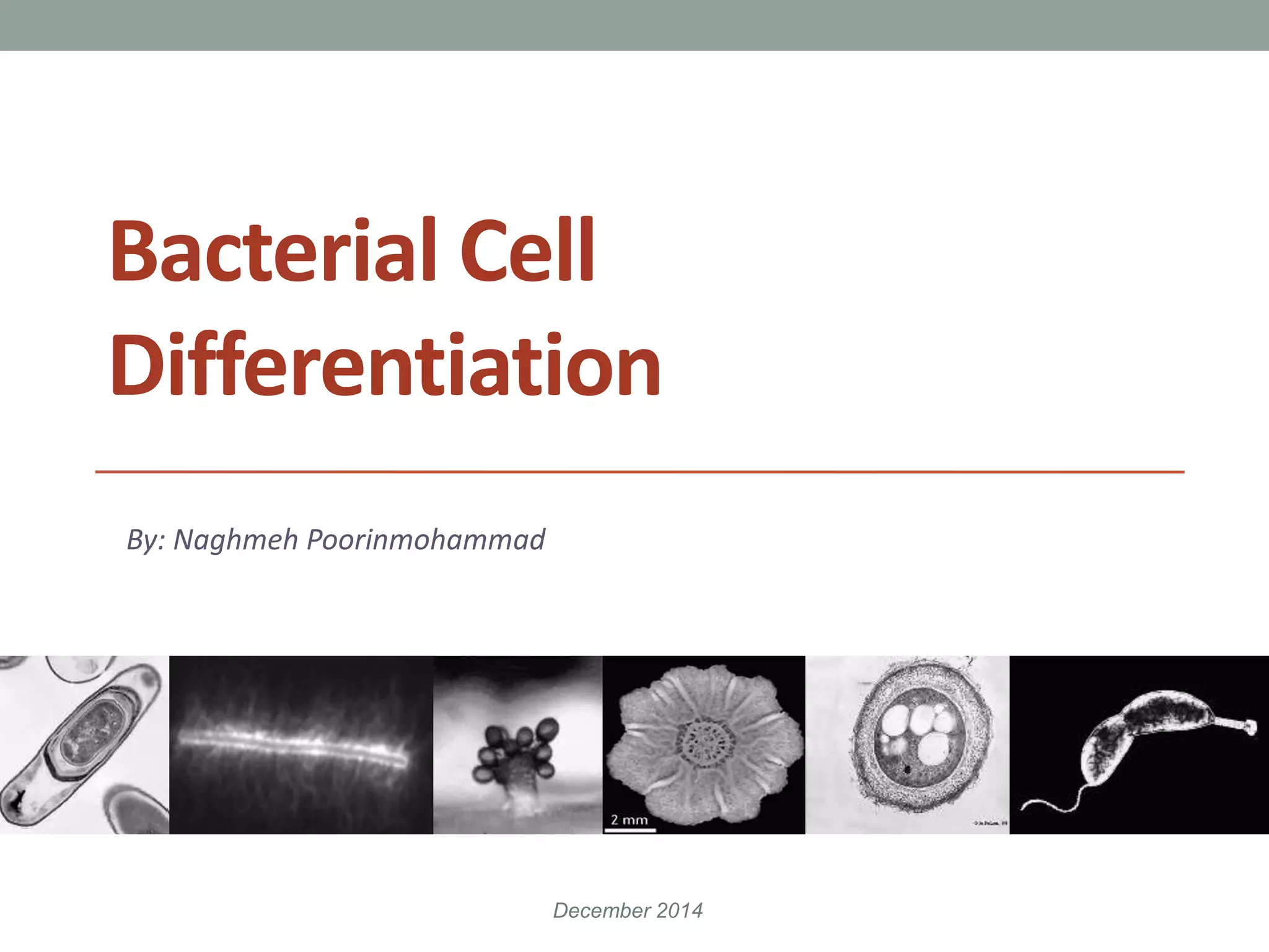
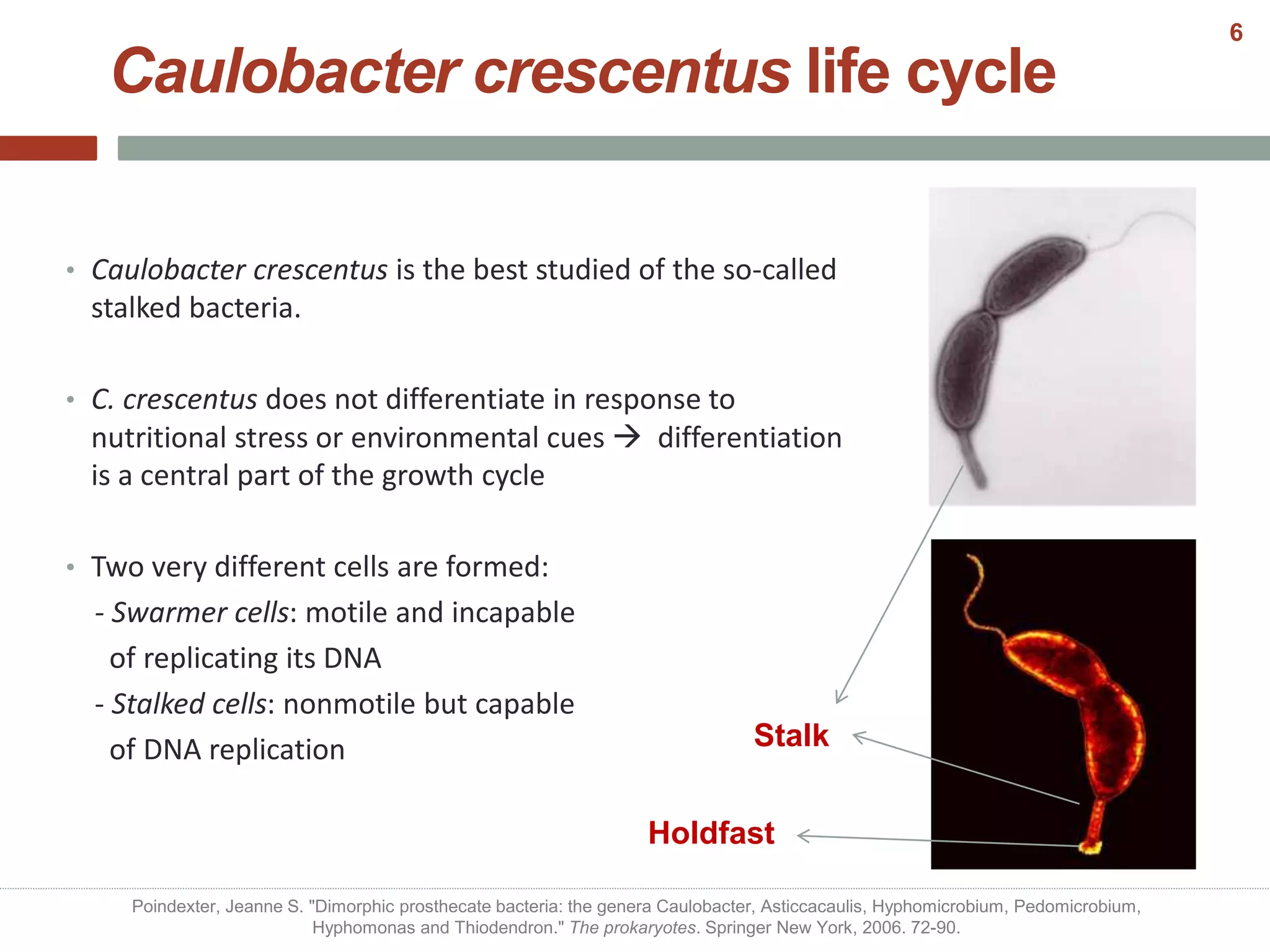
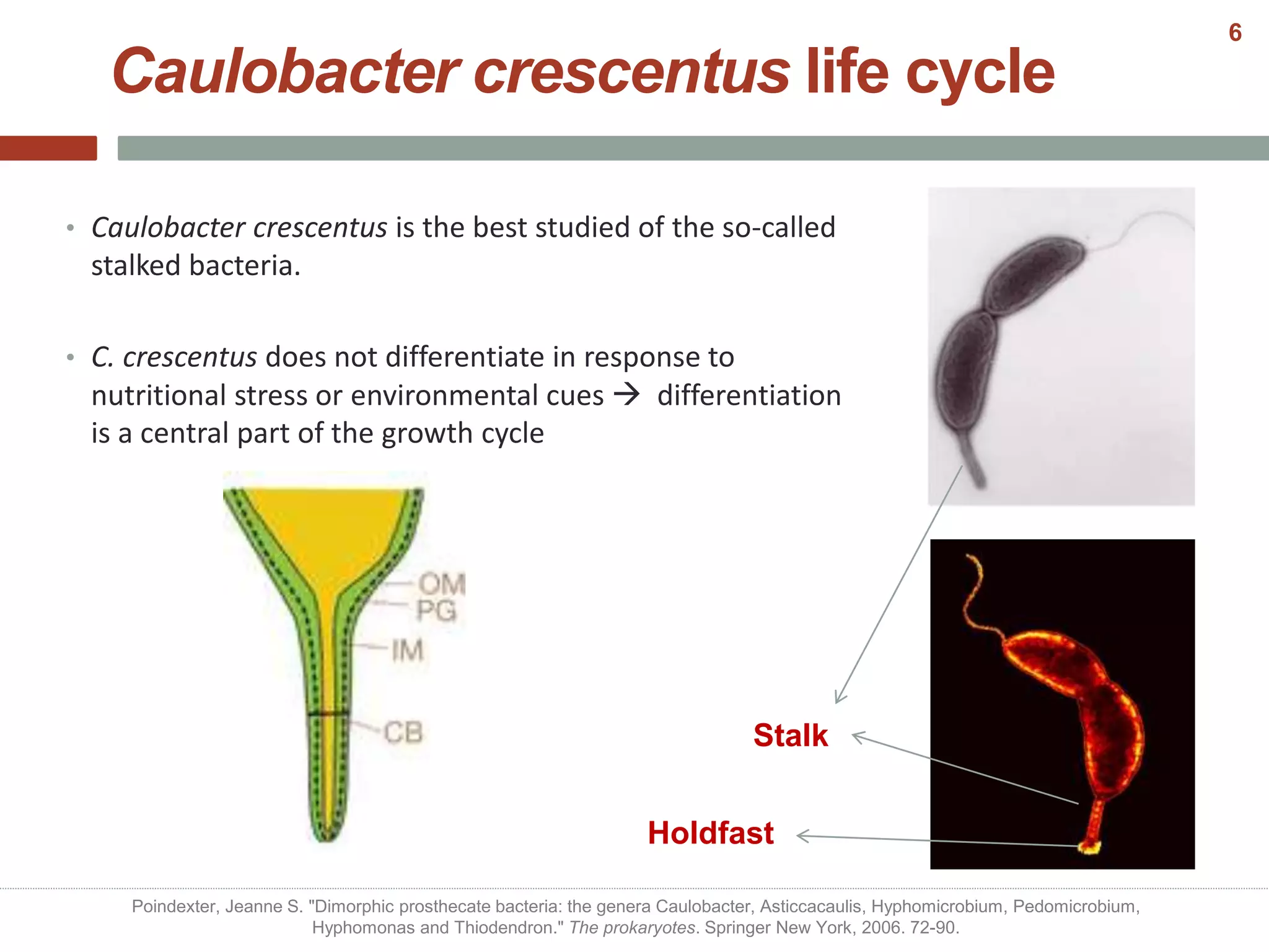
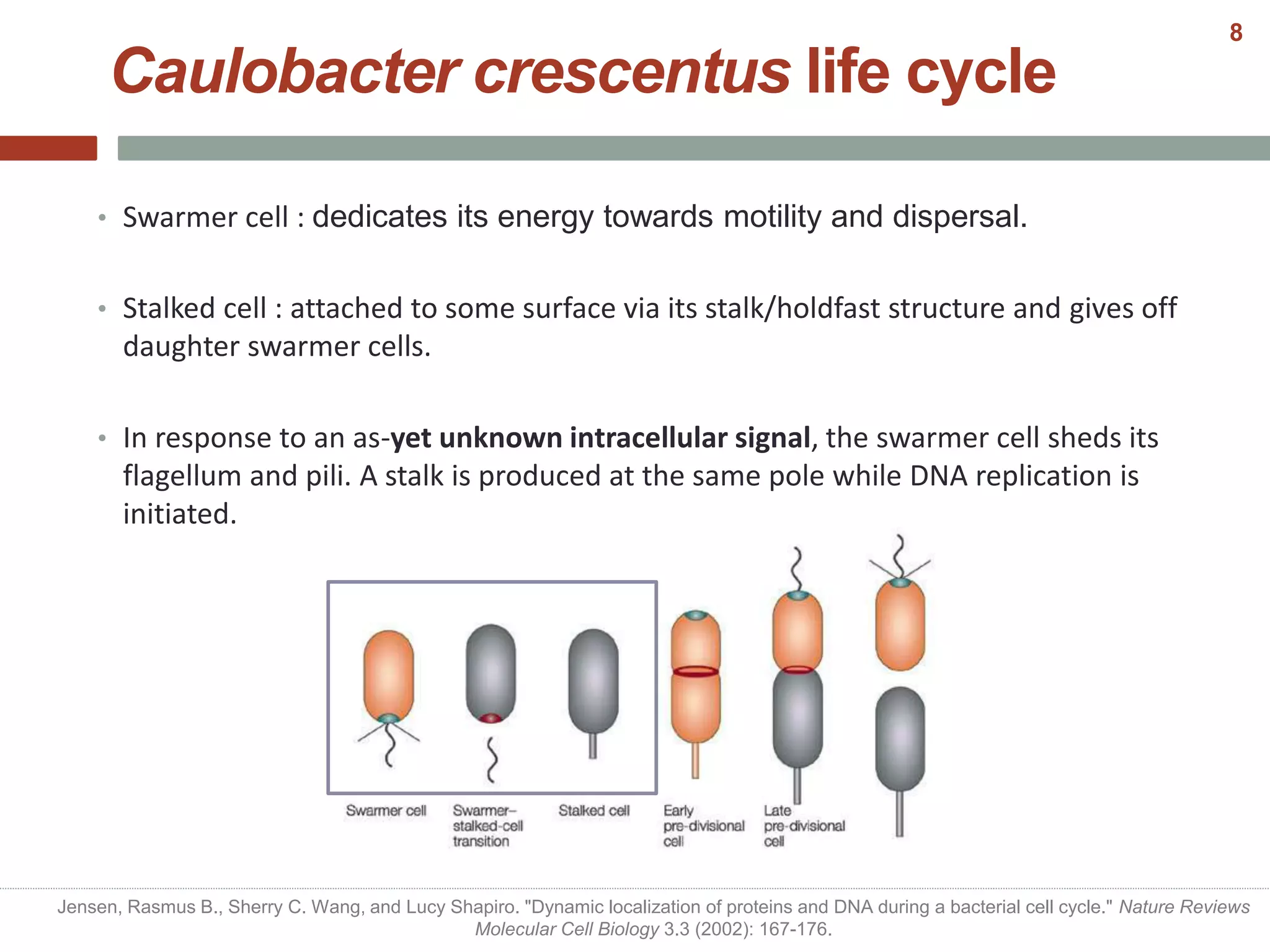
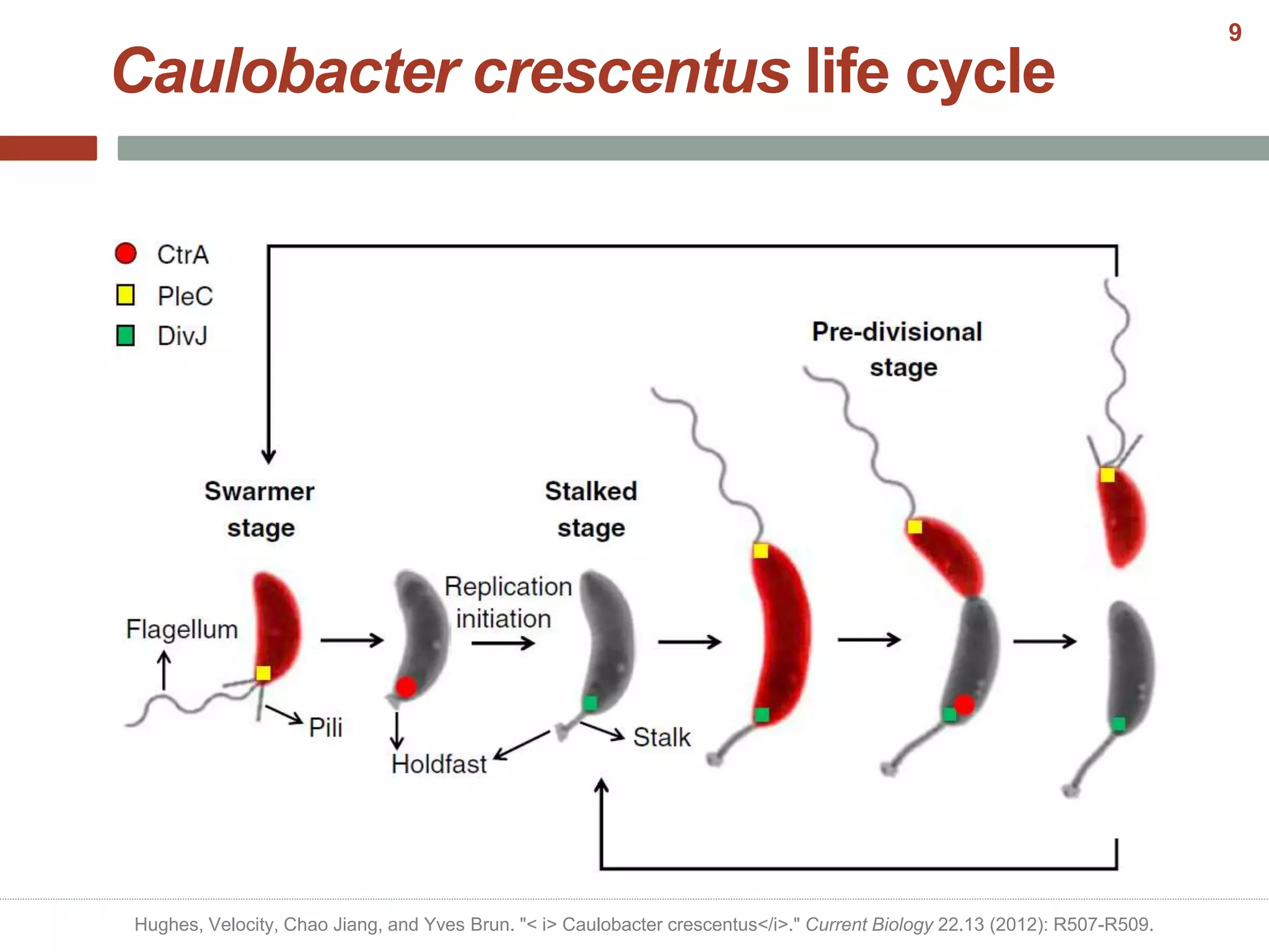
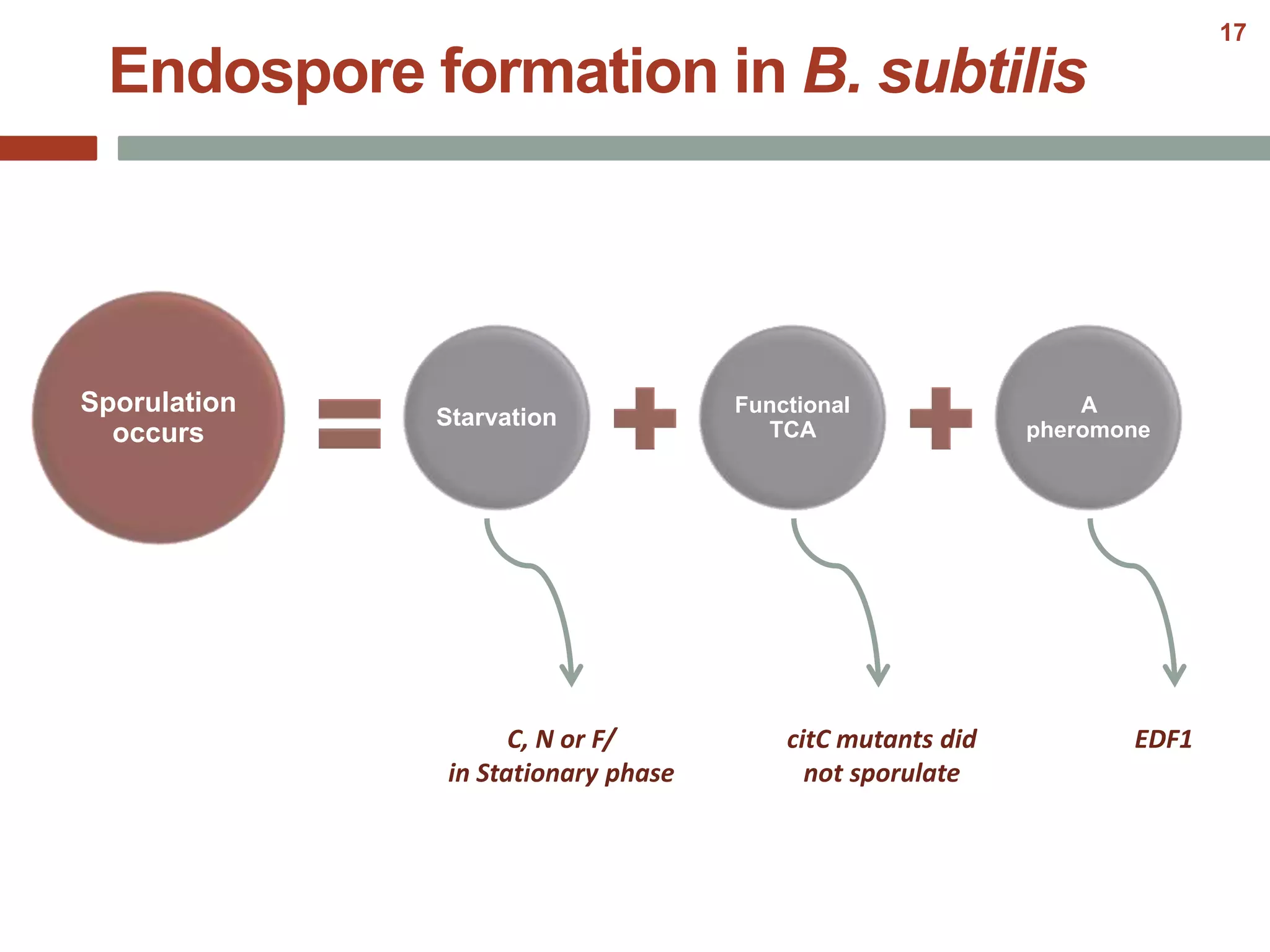
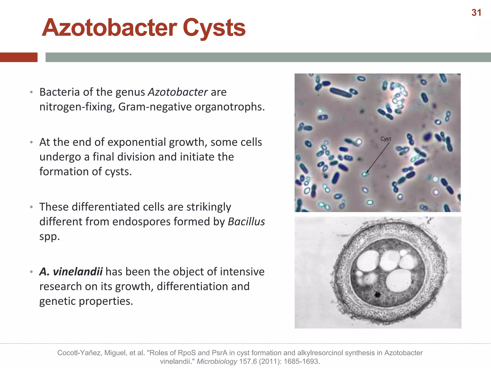
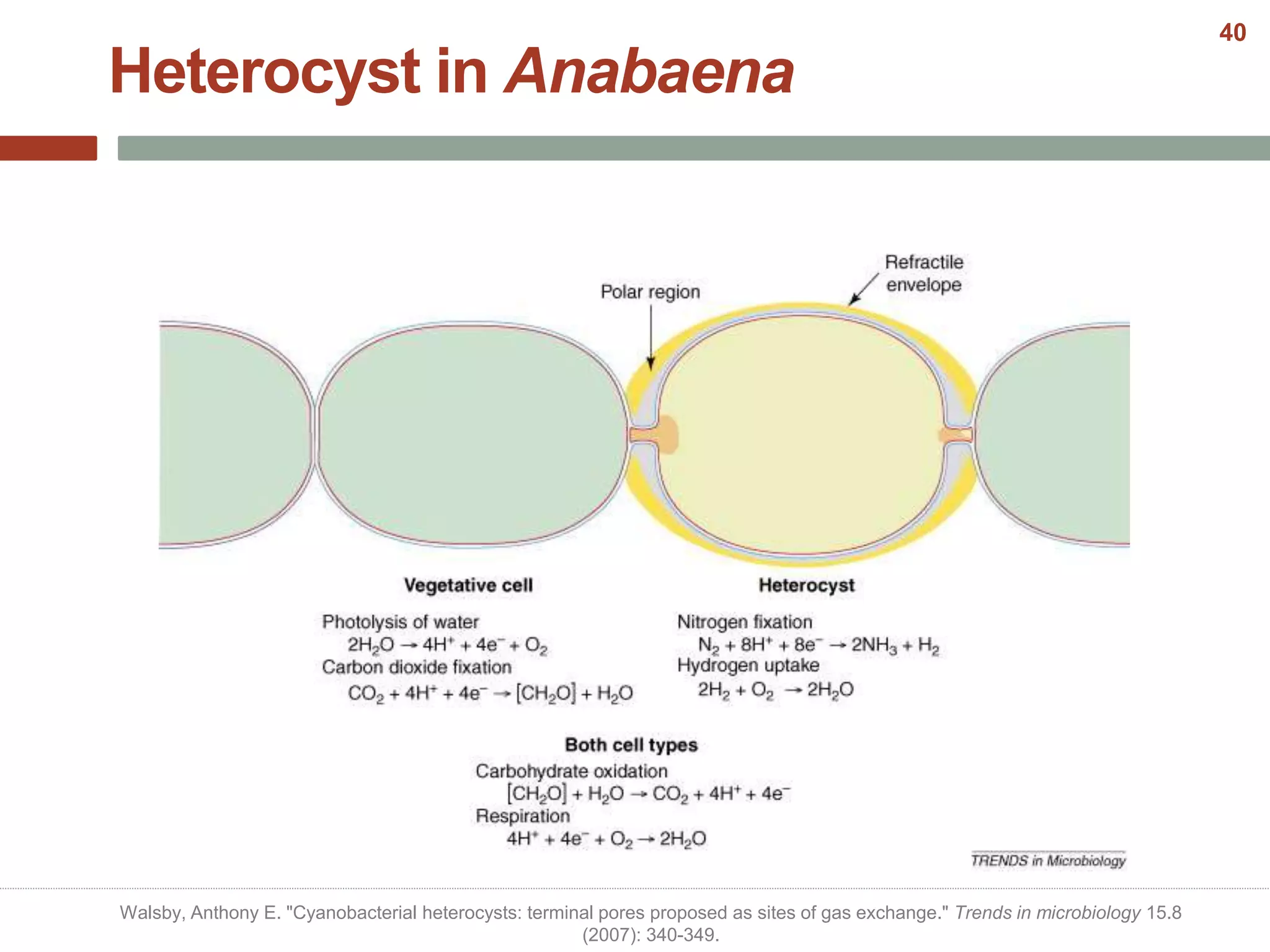
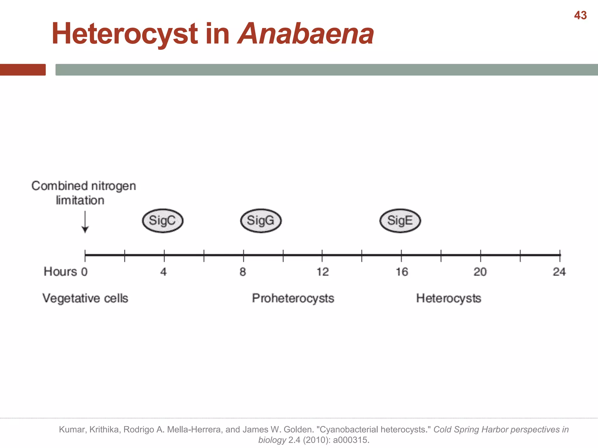
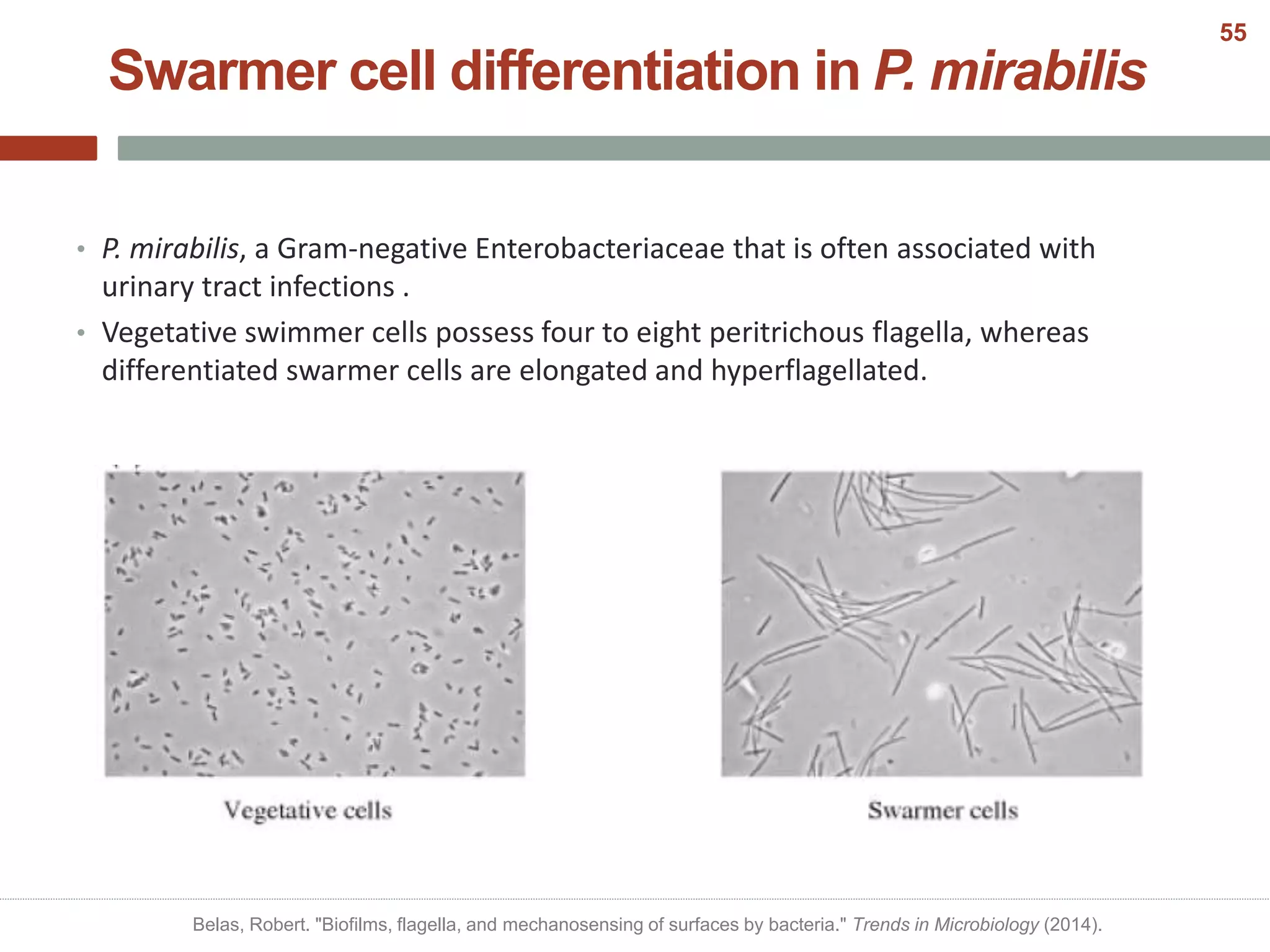
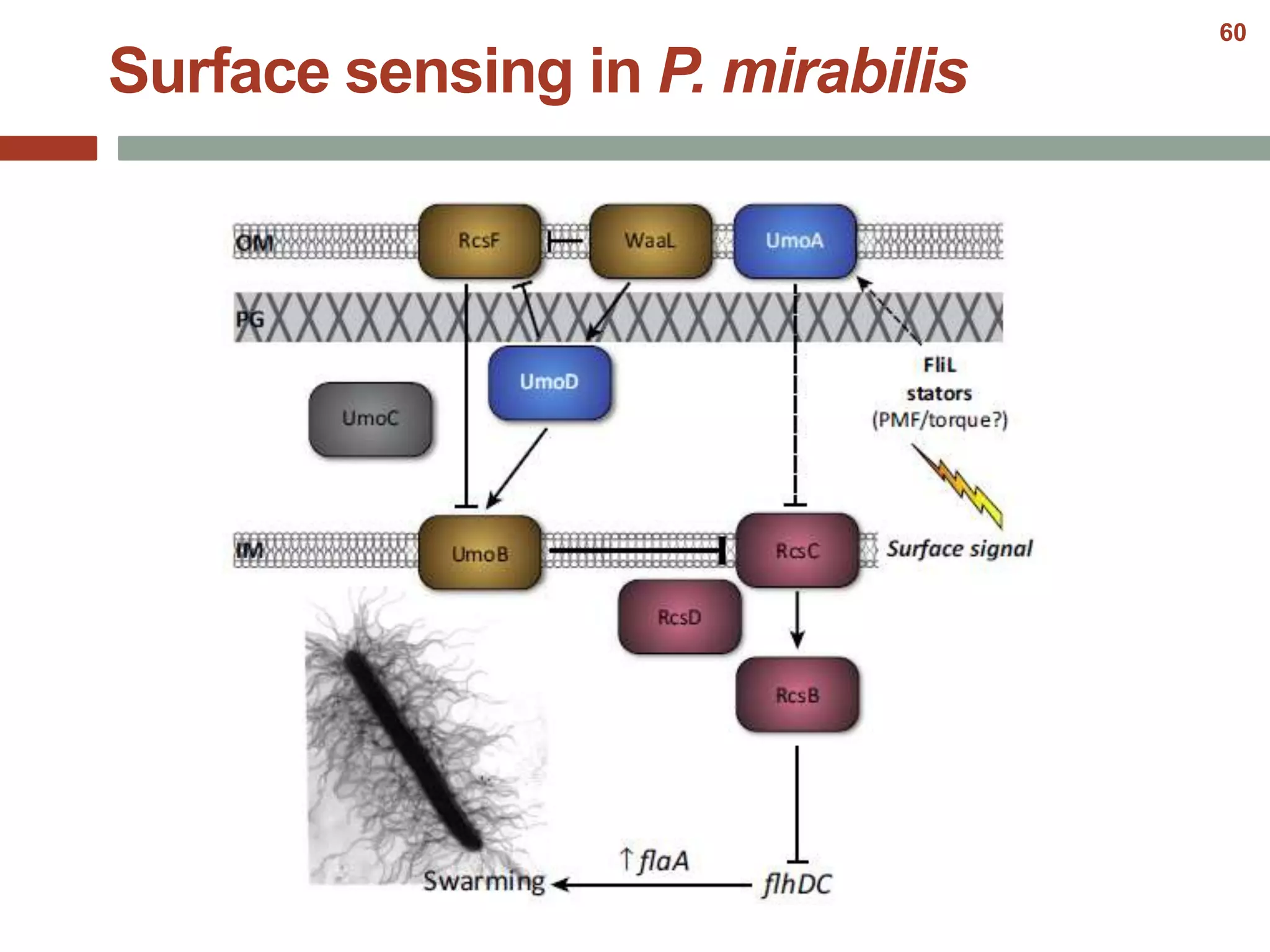
SlideShare a Scribd company logo
Bacterial Cell
Differentiation
By: Naghmeh Poorinmohammad
December 2014
Outline
• Introduction
• Differentiation in bacteria
I. Single-cell Differentiations
II. Multicellular Differentiations
• Summary
• References
Introduction
• Living cells can change morphologically.
• Important reasons for differentiation:
1. Adaption to environmental conditions
2. Expressing different functions at different times in the life cycle
• Thus differentiation can be triggered by:
1. Environmental signal
2. No external signal  inherent to the growth
• The specialized cell types is the outcome of complex regulatory pathways:
1. Altered gene expression
2. differential protein stability
3. differential protein localization
Chater, Keith F. "Bacterial Cell Differentiation." eLS (2005).
3
I. Single-Cell Differentiations
1. Dimorphic life cycle in Caulobacter crescentus
2. Endospore formation in Bacillus sibtilis
3. Actinomycetes spores
4. Cysts in Cyanobacteria
5. Heterocysts in Azotobacter
4
Dimorphic life cycle in
Caulobacter crescentus
Caulobacter crescentus life cycle
• Caulobacter crescentus is the best studied of the so-called
stalked bacteria.
• C. crescentus does not differentiate in response to
nutritional stress or environmental cues  differentiation
is a central part of the growth cycle
• Two very different cells are formed:
- Swarmer cells: motile and incapable
of replicating its DNA
- Stalked cells: nonmotile but capable
of DNA replication Stalk
Holdfast
Poindexter, Jeanne S. "Dimorphic prosthecate bacteria: the genera Caulobacter, Asticcacaulis, Hyphomicrobium, Pedomicrobium,
Hyphomonas and Thiodendron." The prokaryotes. Springer New York, 2006. 72-90.
6
Caulobacter crescentus life cycle
• Caulobacter crescentus is the best studied of the so-called
stalked bacteria.
• C. crescentus does not differentiate in response to
nutritional stress or environmental cues  differentiation
is a central part of the growth cycle
Stalk
Holdfast
Poindexter, Jeanne S. "Dimorphic prosthecate bacteria: the genera Caulobacter, Asticcacaulis, Hyphomicrobium, Pedomicrobium,
Hyphomonas and Thiodendron." The prokaryotes. Springer New York, 2006. 72-90.
6
Other Stalked Bacteria
Jiang, Chao, et al. "Sequential evolution of bacterial morphology by co-option of a developmental regulator." Nature (2014).
7
• Swarmer cell : dedicates its energy towards motility and dispersal.
• Stalked cell : attached to some surface via its stalk/holdfast structure and gives off
daughter swarmer cells.
• In response to an as-yet unknown intracellular signal, the swarmer cell sheds its
flagellum and pili. A stalk is produced at the same pole while DNA replication is
initiated.
Caulobacter crescentus life cycle
Jensen, Rasmus B., Sherry C. Wang, and Lucy Shapiro. "Dynamic localization of proteins and DNA during a bacterial cell cycle." Nature Reviews
Molecular Cell Biology 3.3 (2002): 167-176.
8
Caulobacter crescentus life cycle
Hughes, Velocity, Chao Jiang, and Yves Brun. "< i> Caulobacter crescentus</i>." Current Biology 22.13 (2012): R507-R509.
9
Jensen, Rasmus B., Sherry C. Wang, and Lucy Shapiro. "Dynamic localization of proteins and DNA during a bacterial cell cycle." Nature Reviews
Molecular Cell Biology 3.3 (2002): 167-176.
10
az http://2014.igem.org/Team:USTC-China/project/cimager
11
Endospore formation in
Bacillus subtilis
Endospore formation
• It is an extreme survival strategy employed by certain low G+C Gram-positive
bacteria.
• Spores are resistant to heat, cold, radiation, and other adverse environmental
conditions.
• The primary function of endospore formation appears to be the survival and
dissemination of the species.
• When the environment becomes more favorable, the endospore can reactivate
itself to the vegetative state.
• Time and effort has been expended in the study of this process in the genus
Bacillus, particularly Bacillus subtilis.
Higgins, Douglas, and Jonathan Dworkin. "Recent progress in Bacillus subtilis sporulation." FEMS microbiology reviews 36.1
(2012): 131-148.
13
Endospore
Formation
Desulfoto-
maculum
Thermo-
actinomycetes
Sporosarcina
Clostridium
Sporolacto-
bacillus
Bacillus
Endospore formation
14
• Actively growing cells of Bacillus subtilis are induced to differentiate into spores by
starvation for carbon, nitrogen or, in some circumstances a phosphorus
source.(piggot 2004)
• REMEBER: sporulation is a very energy-consuming process, it cannot proceed in the
complete absence of nutrients.
Endospore formation in B. subtilis
Higgins, Douglas, and Jonathan Dworkin. "Recent progress in Bacillus subtilis sporulation." FEMS microbiology reviews 36.1
(2012): 131-148.
15
• Two transcriptional regulators, σH
and Spo0A, play key roles in
initiation of sporulation.
• Several additional proteins
participate, mainly by controlling
the accumulated concentration of
Spo0A~P.
• Over 125 gene products govern the
complex morphological and
biochemical changes that take place
during sporulation.
• Spore formation takes about 7 h at
37 C.
Endospore formation in B. subtilis
Higgins, Douglas, and Jonathan Dworkin. "Recent progress in Bacillus subtilis sporulation." FEMS microbiology reviews 36.1
(2012): 131-148.
16
Endospore formation in B. subtilis
Starvation
Functional
TCA
A
pheromone
Sporulation
occurs
C, N or F/
in Stationary phase
citC mutants did
not sporulate
EDF1
17
Endospore formation in B. subtilis
Moat, Albert G., John W. Foster, and Michael P. Spector, eds. Microbial physiology. John Wiley & Sons, 2003.
18
Moat, Albert G., John W. Foster, and Michael P. Spector, eds. Microbial physiology. John Wiley & Sons, 2003.
19
Endospore formation in B. subtilis
Tojo, Shigeo, Kazutake Hirooka, and Yasutaro Fujita. "Expression of kinA and kinB of Bacillus subtilis, necessary for sporulation initiation, is under
positive stringent transcription control." Journal of bacteriology 195.8 (2013): 1656-1665.
20
Spores of Actinomycetes
(S. coelicolor)
Actinomycete Spores
• Under nutrient poor conditions filaments differentiate into spores.
• In contrast to endospores, these structures are part of the reproductive process.
• The developmental process to create an actinomycete spore is less complex than
that of the endospore. It involves the simple formation of cross walls that divide the
filament into sections, each containing a chromosome. These then differentiate into
mature spores.
• During this process a tougher cell wall is laid down and there is conversion of the
cytoplasm to a dormant state so that the spore becomes more resistant to heat and
chemicals, though not as hardy as an endospore.
• Actinomycete spores are capable of surviving for long periods of time (for years) and
can germinate into vegetative cells when appropriate growth conditions are
present.
Angert, Esther R. "Alternatives to binary fission in bacteria." Nature Reviews Microbiology 3.3 (2005): 214-224.
22
Actinomycete Spores
• During the life cycle of the filamentous bacteria Streptomyces, morphological
differentiation is closely integrated with fundamental growth and cell-cycle
processes.
Angert, Esther R. "Alternatives to binary fission in bacteria." Nature Reviews Microbiology 3.3 (2005): 214-224.
23
• In response to nutrient depletion and other signals, both production of secondary
metabolites and morphological differentiation are initiated.
Spore in S. coelicolor
Aeral hyphae
Switches from
extension to
septation
Controlled cell
division
Segregation of
chromosomes
Spore
maturation
24
Spore in S. coelicolor
Switch from
extension to
septation
Flärdh, Klas, and Mark J. Buttner. "Streptomyces morphogenetics: dissecting differentiation in a filamentous
bacterium." Nature Reviews Microbiology 7.1 (2009): 36-49.
25
Spore in S. coelicolor
Switch from
extension to
septation
Flärdh, Klas, and Mark J. Buttner. "Streptomyces morphogenetics: dissecting differentiation in a filamentous
bacterium." Nature Reviews Microbiology 7.1 (2009): 36-49.
25
Controlled
cell division
Switch from
extension to
septation
Spore in S. coelicolor
Flärdh, Klas, and Mark J. Buttner. "Streptomyces morphogenetics: dissecting differentiation in a filamentous
bacterium." Nature Reviews Microbiology 7.1 (2009): 36-49.
26
Controlled
cell division
Switch from
extension to
septation
Chromosom
segragation
Spore in S. coelicolor
Flärdh, Klas, and Mark J. Buttner. "Streptomyces morphogenetics: dissecting differentiation in a filamentous
bacterium." Nature Reviews Microbiology 7.1 (2009): 36-49.
27
• Spore maturation involves the production of a thick, lysozyme-
resistant spore wall.
• This wall is laid down after sporulation septation is complete.
• The correct assembly of the spore wall depends on mreB, which
may explain why Streptomyces and a few other sporulating
actinomycete genera contain genes for this ancestral actin,
whereas most other actinobacteria do not.
• The subcellular localization of MreB–EGFP is consistent with a
role for MreB in assembly of the spore wall.
• Correct is: first as bands that coincide with sporulation septa at
both poles after septal constriction, and then spreading out to
surround the spore completely, lining the inner surface of the
membrane before disappearing in the mature spores.
Controlled
cell division
Switch from
extension to
septation
Chromosom
segragation
Spore
maturation
Spore in S. coelicolor
Flärdh, Klas, and Mark J. Buttner. "Streptomyces morphogenetics: dissecting differentiation in a filamentous
bacterium." Nature Reviews Microbiology 7.1 (2009): 36-49.
28
Spore in S. coelicolor
"Encyclopedia of Life Science." Reference
Reviews 24, no. 5 (2010): 46-46.
29
Cysts in Azotobacter
Azotobacter Cysts
• Bacteria of the genus Azotobacter are
nitrogen-fixing, Gram-negative organotrophs.
• At the end of exponential growth, some cells
undergo a final division and initiate the
formation of cysts.
• These differentiated cells are strikingly
different from endospores formed by Bacillus
spp.
• A. vinelandii has been the object of intensive
research on its growth, differentiation and
genetic properties.
Cocotl-Yañez, Miguel, et al. "Roles of RpoS and PsrA in cyst formation and alkylresorcinol synthesis in Azotobacter
vinelandii." Microbiology 157.6 (2011): 1685-1693.
31
• When A. vinelandii experiences a lack of nutrients, it will develop into cyst . The cell
is then protected against desiccation by a multilayered coat, of which gel-forming
alginate is a necessary part.(j. bacteril 2009)
• Depending on the carbon source and the conditions under which growth occurs,
poly-b-hydroxybutyrate (PHB) accumulates in vegetative cells, and the extent of
encystment in such cultures is related to the intracellular concentration of that
polymer.
• The problem studying encystment genetics is the inability to generate useful
mutants of the organism.
• Azotobacter cells are refractory to mutagenesis except for those genes involved in
nitrogen fixation and resistance to certain antibiotics. The basis for this difficulty
may lie in the large amount of deoxyribonucleic acid (DNA) (6.610214 g per cell) that
these cells possess.
Azotobacter Cysts
Gimmestad, Martin, et al. "Characterization of three new Azotobacter vinelandii alginate lyases, one of which is involved in cyst
germination." Journal of bacteriology 191.15 (2009): 4845-4853.
32
Exine
Intine
Funa, Nobutaka, et al. "Phenolic lipid synthesis by type III polyketide synthases is essential for cyst formation in Azotobacter
vinelandii."Proceedings of the National Academy of Sciences 103.16 (2006): 6356-6361.
33
AlgU (sigma E)
algC cydR
RpoS
algD phbB
PsrA
Azotobacter Cysts
Alginate
synthesis
PHB
biosynthesis
flhDC cydAB
Loss of
flagella
Inhibits nitrogen
fixation
Alginate
synthesis
Cocotl-Yañez, Miguel, et al. "Roles of RpoS and PsrA in cyst formation and alkylresorcinol synthesis in Azotobacter vinelandii." Microbiology 157.6 (2011):
1685-1693.
34
Heterocyst in cyanobacteria
• Many cyanobacterial species are capable of nitrogen fixation. However, oxygenic
photosynthesis and nitrogen fixation are incompatible processes because
nitrogenase is inactivated by oxygen.
Mechanisms to
separate these
activities
Temporal
Spatialy
(differentiation)
Kumar, Krithika, Rodrigo A. Mella-Herrera, and James W. Golden. "Cyanobacterial heterocysts." Cold Spring Harbor perspectives in
biology 2.4 (2010): a000315.
36
• In the presence of a source of combined nitrogen such as nitrate or ammonium,
Anabaena grows as long filaments containing hundreds of photosynthetic vegetative
cells.
• In the absence of combined nitrogen, it produces heterocysts every ten to twenty
vegetative cells along filaments.
Heterocyst in cyanobacteria
Kumar, Krithika, Rodrigo A. Mella-Herrera, and James W. Golden. "Cyanobacterial heterocysts." Cold Spring Harbor perspectives in
biology 2.4 (2010): a000315.
37
• Oxygen-producing photosystem PSII is dismantled during differentiation and
heterocysts show an increased rate of respiration.
• Morphological changes include the deposition of two additional envelope layers
around the heterocyst: an inner “laminated” layer composed of two heterocyst
specific glycolipids (HGL)and an outer polysaccharide layer (HEP).
• Heterocysts and vegetative cells are mutually interdependent. Because they lack
photosystem II and carbon fixation, heterocysts are dependent on vegetative cells
for a source of reductant and carbon, which is probably partially supplied as sucrose.
• In Anabaena PCC 7120, vegetative cells must also supply glutamate to heterocysts,
which convert it to glutamine and other amino acids.
• In return, newly fixed nitrogen is rapidly exported from heterocysts and distributed
to nearby vegetative cells.
Heterocyst in Anabaena
Kumar, Krithika, Rodrigo A. Mella-Herrera, and James W. Golden. "Cyanobacterial heterocysts." Cold Spring Harbor perspectives in
biology 2.4 (2010): a000315.
38
• The timeline of heterocyst development begins with sensing combined-nitrogen
limitation and culminates with nitrogen fixation in the mature heterocyst.
• Heterocyst development is complete in about 20 hours at 30 ◦C reversibly when the
combined nitrogen source is available.
Heterocyst in Anabaena
 Kumar, Krithika, Rodrigo A. Mella-Herrera, and James W. Golden. "Cyanobacterial heterocysts." Cold Spring Harbor perspectives in
biology 2.4 (2010): a000315.
 Picture from: Cumino, Andrea C., et al. "Carbon cycling in Anabaena sp. PCC 7120. Sucrose synthesis in the heterocysts and possible
role in nitrogen fixation." Plant physiology 143.3 (2007): 1385-1397.
39
Heterocyst in Anabaena
Walsby, Anthony E. "Cyanobacterial heterocysts: terminal pores proposed as sites of gas exchange." Trends in microbiology 15.8
(2007): 340-349.
40
Heterocyst in Anabaena
Walsby, Anthony E. "Cyanobacterial heterocysts: terminal pores proposed as sites of gas exchange." Trends in microbiology 15.8
(2007): 340-349.
41
• In cyanobacteria, 2-oxoglutarate, an intermediate in the Krebs cycle, constitutes
the signal for nitrogen deprivation.
• The Krebs cycle in cyanobacteria is incomplete because of the lack of 2-oxoglutarate
dehydrogenase  2-oxoglutarate’s main function is to serve as a precursor in a
variety of biosynthetic reactions. It is the primary carbon skeleton for incorporation
of ammonium and is considered the metabolic junction between carbon and
nitrogen balance in cyanobacteria.
• Nitrogen limiting conditions result in an increase in the levels of 2-oxoglutarate.
• NtcA, a transcriptional regulator belonging to the CRP (cyclic AMP receptor protein)
family of proteins, senses 2-oxoglutarate levels.
• In Anabaena PCC 7120, NtcA is required for the expression of the genes in pathways
for ammonium and nitrate assimilation, as well as heterocyst development.
Heterocyst in Anabaena
Kumar, Krithika, Rodrigo A. Mella-Herrera, and James W. Golden. "Cyanobacterial heterocysts." Cold Spring Harbor perspectives in
biology 2.4 (2010): a000315.
42
Heterocyst in Anabaena
Kumar, Krithika, Rodrigo A. Mella-Herrera, and James W. Golden. "Cyanobacterial heterocysts." Cold Spring Harbor perspectives in
biology 2.4 (2010): a000315.
43
Heterocyst in Anabaena
Kumar, Krithika, Rodrigo A. Mella-Herrera, and James W. Golden. "Cyanobacterial heterocysts." Cold Spring Harbor perspectives in
biology 2.4 (2010): a000315.
44
II. Multicellular Differentiations
1. Fruiting body in Myxobacteria
2. Hyper flagellated swarmer cells
3. Biofilm formation
45
Fruiting body in
Myxobacteria
Fruiting body in Myxobacteria
• Myxobacteria are a group of Gram-negative bacteria that are predominantly found
in soil.
• During starvation, growth is arrested and a developmental program is initiated,
which culminates in the formation of spore-bearing fruiting bodies that have a well-
defined shape .
Claessen, Dennis, et al. "Bacterial solutions to multicellularity: a tale of biofilms, filaments and fruiting bodies." Nature Reviews
Microbiology 12.2 (2014): 115-124.
47
• The first signs of fruiting body formation are evident 4–6 hours after the exhaustion
of resources.
• This is accompanied by changes in cell motility and the formation of aggregation
centers.
• Within 24 hours, the aggregation process is complete, and the nascent fruiting
bodies each contain approximately 105 densely packed cells that differentiate into
spores.
Fruiting body in Myxobacteria
Claessen, Dennis, et al. "Bacterial solutions to multicellularity: a tale of biofilms, filaments and fruiting bodies." Nature Reviews
Microbiology 12.2 (2014): 115-124.
48
Fruiting
body
benefits
Spores can
exploit new
resources
Peripheral rods
can grow in
nutrient-limited
conditions
Lysed cells
provide
nutrients
Spores
promote
survival
Fruiting body in Myxobacteria
49
Myxococcus xanthus life cycle
• Nutrition is depleted
• Cell density is high
• Solid support is available
Kaiser, Dale, Mark Robinson, and Lee Kroos. "Myxobacteria, polarity, and multicellular morphogenesis." Cold Spring Harbor perspectives in
biology 2.8 (2010): a000380.
50
Myxococcus xanthus life cycle
Chater, Keith F. "Bacterial Cell Differentiation." eLS (2005).
51
Regulation of fruiting body formation
From: Kaiser et al. 2010
52
Myxococcus xanthus life cycle
Bryan et al. 2000
53
Hyper flagellated
Swarmer cells
Swarmer cell differentiation
• Swarming is the fastest known bacterial mode of surface translocation and enables
the rapid colonization of a nutrient-rich environment and host tissues.
• It requires functional flagella and is coupled to the production of a viscous slime
layer.
• It is widespread in many genera of Gram negative and Gram-positive flagellated
bacteria and is typically assayed on a solidified medium, containing 0.5–2% agar.
• Species such as Proteus mirabilis and Vibrio parahaemolyticus, which are capable of
vigorous swarming even on high-agar medium.
• Swarming has been studied extensively in P. mirabilis, in which elongated,
multinucleated and hyper-flagellated swarmer cells can spread as multicellular rafts
across surfaces.
Verstraeten, Natalie, et al. "Living on a surface: swarming and biofilm formation." Trends in microbiology 16.10 (2008): 496-506.
55
Swarmer cell differentiation
Verstraeten, Natalie, et al. "Living on a surface: swarming and biofilm formation." Trends in microbiology 16.10 (2008): 496-506.
56
Swarmer cell differentiation in P. mirabilis
• P. mirabilis, a Gram-negative Enterobacteriaceae that is often associated with
urinary tract infections .
• Vegetative swimmer cells possess four to eight peritrichous flagella, whereas
differentiated swarmer cells are elongated and hyperflagellated.
Vegetative P. mirabilis Differentiated to a swarmer P. mirabilis
Belas, Robert. "Biofilms, flagella, and mechanosensing of surfaces by bacteria." Trends in Microbiology (2014).
57
Swarmer cell differentiation in P. mirabilis
• P. mirabilis, a Gram-negative Enterobacteriaceae that is often associated with
urinary tract infections .
• Vegetative swimmer cells possess four to eight peritrichous flagella, whereas
differentiated swarmer cells are elongated and hyperflagellated.
Belas, Robert. "Biofilms, flagella, and mechanosensing of surfaces by bacteria." Trends in Microbiology (2014).
55
• Although many species swarm including Aeromonas, Azospirillum, B. subtilis, E.
coli, Rhodospirillum, Rhizobium, Salmonella, Serratia, and Yersinia, only a few (two
notable examples are V. parahaemolyticus and P. mirabilis) do so after a surface-
induced physiological differentiation that results in an elongated, highly flagellated
swarmer cell.
• P. mirabilis swarmer cell differentiation is triggered by physical conditions that
inhibit the rotation of the peritrichous flagella of the swimmer cell (belas 2014)
• P. mirabilis flagellum functions as a mechanosensor of the surface signal.
• Both lipopolysaccharide (LPS) and O-antigen play a part in P. mirabilis surface
sensing.
• Nutrient availability is crucial to sustain the energy demanding process of swarming
(verstraeten)
Swarmer cell differentiation in P. mirabilis
Morgenstein, Randy M., Bree Szostek, and Philip N. Rather. "Regulation of gene expression during swarmer cell differentiation in
Proteus mirabilis." FEMS microbiology reviews 34.5 (2010): 753-763.
58
Swarmer cell differentiation in P. mirabilis
Armbruster, Chelsie E., and Harry LT Mobley. "Merging mythology and morphology: the multifaceted lifestyle of Proteus mirabilis." Nature
Reviews Microbiology 10.11 (2012): 743-754.
59
Surface sensing in P. mirabilis
60
Summary
Why studying
bacterial cell
differentiations?
Basic science is important, isn’t it?
Fighting the bacs!
Making use of ‘em!
61
References to dig deeper!
• Chater, Keith F. "Bacterial Cell Differentiation." eLS (2005).
• Kaiser, Dale. "Cell-Cell Interactions." The Prokaryotes. Springer Berlin
Heidelberg, 2013. 511-528.
• Celiker, Hasan, and Jeff Gore. "Cellular cooperation: insights from
microbes."Trends in cell biology 23.1 (2013): 9-15.
62
Thank You

More Related Content

What's hot

Siderophores ppt
Siderophores pptSiderophores ppt
Siderophores ppt
Sundaresh Kalal
 
plant viruses- Geminivirus ppt
 plant viruses- Geminivirus ppt plant viruses- Geminivirus ppt
plant viruses- Geminivirus ppt
thirupathiSathya
 
Bergey's Manual of systematic bacteriology
Bergey's Manual of systematic bacteriologyBergey's Manual of systematic bacteriology
Bergey's Manual of systematic bacteriology
microbiology Notes
 
Siderophore: Secretion of Bacteria as a Chelating Agent
Siderophore: Secretion of Bacteria as a Chelating AgentSiderophore: Secretion of Bacteria as a Chelating Agent
Siderophore: Secretion of Bacteria as a Chelating Agent
Vasantrao Nail Marathwada Krishi Vidyapeeth, Parbhani
 
Halophiles (Introduction, Adaptations, Applications)
Halophiles (Introduction, Adaptations, Applications)Halophiles (Introduction, Adaptations, Applications)
Halophiles (Introduction, Adaptations, Applications)
Jamil Ahmad
 
Secondary screening of industrial important microbes
Secondary screening of industrial important microbes   Secondary screening of industrial important microbes
Secondary screening of industrial important microbes
DhruviSuvagiya
 
Photosynthesis in bacteria and its types
Photosynthesis in bacteria and its typesPhotosynthesis in bacteria and its types
Photosynthesis in bacteria and its types
Saajida Sultaana
 
Nodule formation and nod factors
Nodule formation and nod factorsNodule formation and nod factors
Nodule formation and nod factors
vibhakhanna1
 
Quorum Sensing
Quorum SensingQuorum Sensing
Quorum Sensing
Alizay Shahid
 
bacterial-bioluminescence
bacterial-bioluminescencebacterial-bioluminescence
bacterial-bioluminescence
Merlyn Denesia
 
bacteriophage.ppt
bacteriophage.pptbacteriophage.ppt
bacteriophage.ppt
NCRIMS, Meerut
 
Archaea Bacteria (Methanogens, Halophiles, Thermophiles)
Archaea Bacteria (Methanogens, Halophiles, Thermophiles)Archaea Bacteria (Methanogens, Halophiles, Thermophiles)
Archaea Bacteria (Methanogens, Halophiles, Thermophiles)
Dr. Mohammedazim Bagban
 
Bergey's classification presentation
Bergey's classification presentationBergey's classification presentation
Bergey's classification presentation
BIDISHA MANDAL
 
Animal viruses
Animal virusesAnimal viruses
Animal viruses
Monika K Raut
 
Industrial Microorganisms
Industrial MicroorganismsIndustrial Microorganisms
Industrial Microorganisms
M Rakibul Islam
 
Measurement of microbial growth
Measurement of microbial growthMeasurement of microbial growth
Measurement of microbial growth
NOOR ARSHIA
 
Microbial genetics mutation
Microbial genetics mutationMicrobial genetics mutation
Microbial genetics mutationLani Manahan
 
Host pathogen interactions
Host pathogen interactionsHost pathogen interactions
Host pathogen interactions
thuphan95
 
Microbial Diversity Part 1: Introduction to Diversity
Microbial Diversity Part 1: Introduction to DiversityMicrobial Diversity Part 1: Introduction to Diversity
Microbial Diversity Part 1: Introduction to Diversity
RachelMackelprang
 

What's hot (20)

Siderophores ppt
Siderophores pptSiderophores ppt
Siderophores ppt
 
plant viruses- Geminivirus ppt
 plant viruses- Geminivirus ppt plant viruses- Geminivirus ppt
plant viruses- Geminivirus ppt
 
Bergey's Manual of systematic bacteriology
Bergey's Manual of systematic bacteriologyBergey's Manual of systematic bacteriology
Bergey's Manual of systematic bacteriology
 
Siderophore: Secretion of Bacteria as a Chelating Agent
Siderophore: Secretion of Bacteria as a Chelating AgentSiderophore: Secretion of Bacteria as a Chelating Agent
Siderophore: Secretion of Bacteria as a Chelating Agent
 
Halophiles (Introduction, Adaptations, Applications)
Halophiles (Introduction, Adaptations, Applications)Halophiles (Introduction, Adaptations, Applications)
Halophiles (Introduction, Adaptations, Applications)
 
Secondary screening of industrial important microbes
Secondary screening of industrial important microbes   Secondary screening of industrial important microbes
Secondary screening of industrial important microbes
 
Photosynthesis in bacteria and its types
Photosynthesis in bacteria and its typesPhotosynthesis in bacteria and its types
Photosynthesis in bacteria and its types
 
Nodule formation and nod factors
Nodule formation and nod factorsNodule formation and nod factors
Nodule formation and nod factors
 
Quorum Sensing
Quorum SensingQuorum Sensing
Quorum Sensing
 
bacterial-bioluminescence
bacterial-bioluminescencebacterial-bioluminescence
bacterial-bioluminescence
 
bacteriophage.ppt
bacteriophage.pptbacteriophage.ppt
bacteriophage.ppt
 
Archaea Bacteria (Methanogens, Halophiles, Thermophiles)
Archaea Bacteria (Methanogens, Halophiles, Thermophiles)Archaea Bacteria (Methanogens, Halophiles, Thermophiles)
Archaea Bacteria (Methanogens, Halophiles, Thermophiles)
 
PGPR
PGPRPGPR
PGPR
 
Bergey's classification presentation
Bergey's classification presentationBergey's classification presentation
Bergey's classification presentation
 
Animal viruses
Animal virusesAnimal viruses
Animal viruses
 
Industrial Microorganisms
Industrial MicroorganismsIndustrial Microorganisms
Industrial Microorganisms
 
Measurement of microbial growth
Measurement of microbial growthMeasurement of microbial growth
Measurement of microbial growth
 
Microbial genetics mutation
Microbial genetics mutationMicrobial genetics mutation
Microbial genetics mutation
 
Host pathogen interactions
Host pathogen interactionsHost pathogen interactions
Host pathogen interactions
 
Microbial Diversity Part 1: Introduction to Diversity
Microbial Diversity Part 1: Introduction to DiversityMicrobial Diversity Part 1: Introduction to Diversity
Microbial Diversity Part 1: Introduction to Diversity
 

Viewers also liked

Cell differentiation
 Cell differentiation Cell differentiation
Cell differentiation
jmpettis10
 
Differentiation
DifferentiationDifferentiation
DifferentiationC Russo
 
Heterocyst differentiation
Heterocyst  differentiationHeterocyst  differentiation
Heterocyst differentiation
Sweksha Singh
 
Spore forming bacteria
Spore forming bacteriaSpore forming bacteria
Spore forming bacteriasundu1
 
Module 10 patterns of inheritance
Module 10 patterns of inheritanceModule 10 patterns of inheritance
Module 10 patterns of inheritanceCRCourseDev
 
Lesson 4.1 Introduction of Reproduction
Lesson 4.1 Introduction of ReproductionLesson 4.1 Introduction of Reproduction
Lesson 4.1 Introduction of ReproductionMailyn Morales
 
Chapter 9 notes
Chapter 9 notesChapter 9 notes
Chapter 9 notesmjnepa
 
Genome Editing Comes of Age; CRISPR, rAAV and the new landscape of molecular ...
Genome Editing Comes of Age; CRISPR, rAAV and the new landscape of molecular ...Genome Editing Comes of Age; CRISPR, rAAV and the new landscape of molecular ...
Genome Editing Comes of Age; CRISPR, rAAV and the new landscape of molecular ...
Candy Smellie
 
Cell differentiation notes
Cell differentiation notesCell differentiation notes
Cell differentiation notes
Personal
 
Bacterial anatomy, physiology, growth, nutrition, metabolism, toxin and bacte...
Bacterial anatomy, physiology, growth, nutrition, metabolism, toxin and bacte...Bacterial anatomy, physiology, growth, nutrition, metabolism, toxin and bacte...
Bacterial anatomy, physiology, growth, nutrition, metabolism, toxin and bacte...
Santosh Kumar Yadav
 
Review of CRISPR/Cas9
Review of CRISPR/Cas9Review of CRISPR/Cas9
Review of CRISPR/Cas9
Hub_lot
 
Receptor tyrosine kinase
Receptor tyrosine kinaseReceptor tyrosine kinase
Receptor tyrosine kinase
Sobia
 
Rewriting the Genome Using CRISPR and Synthetic Biology
Rewriting the Genome Using CRISPR and Synthetic Biology Rewriting the Genome Using CRISPR and Synthetic Biology
Rewriting the Genome Using CRISPR and Synthetic Biology
Integrated DNA Technologies
 
AIDS
AIDSAIDS
AGR 154 CHAPTER 4 - FUNGI
AGR 154 CHAPTER 4 - FUNGIAGR 154 CHAPTER 4 - FUNGI
AGR 154 CHAPTER 4 - FUNGI
AINA AFEEQA
 
Endospore
EndosporeEndospore
Endospore
Mumal Kanwar
 
the application of CRISPR/Cas9 system in genome editing
the application of CRISPR/Cas9 system in genome editingthe application of CRISPR/Cas9 system in genome editing
the application of CRISPR/Cas9 system in genome editing
Arash zolnori
 
Bacterial virulence factors
Bacterial virulence factorsBacterial virulence factors
Bacterial virulence factors
Ahmed Mohamed
 
An Introduction to Crispr Genome Editing
An Introduction to Crispr Genome EditingAn Introduction to Crispr Genome Editing
An Introduction to Crispr Genome Editing
Chris Thorne
 
Gene Therapy
Gene TherapyGene Therapy
Gene Therapy
Shrikesh Kumar Rai
 

Viewers also liked (20)

Cell differentiation
 Cell differentiation Cell differentiation
Cell differentiation
 
Differentiation
DifferentiationDifferentiation
Differentiation
 
Heterocyst differentiation
Heterocyst  differentiationHeterocyst  differentiation
Heterocyst differentiation
 
Spore forming bacteria
Spore forming bacteriaSpore forming bacteria
Spore forming bacteria
 
Module 10 patterns of inheritance
Module 10 patterns of inheritanceModule 10 patterns of inheritance
Module 10 patterns of inheritance
 
Lesson 4.1 Introduction of Reproduction
Lesson 4.1 Introduction of ReproductionLesson 4.1 Introduction of Reproduction
Lesson 4.1 Introduction of Reproduction
 
Chapter 9 notes
Chapter 9 notesChapter 9 notes
Chapter 9 notes
 
Genome Editing Comes of Age; CRISPR, rAAV and the new landscape of molecular ...
Genome Editing Comes of Age; CRISPR, rAAV and the new landscape of molecular ...Genome Editing Comes of Age; CRISPR, rAAV and the new landscape of molecular ...
Genome Editing Comes of Age; CRISPR, rAAV and the new landscape of molecular ...
 
Cell differentiation notes
Cell differentiation notesCell differentiation notes
Cell differentiation notes
 
Bacterial anatomy, physiology, growth, nutrition, metabolism, toxin and bacte...
Bacterial anatomy, physiology, growth, nutrition, metabolism, toxin and bacte...Bacterial anatomy, physiology, growth, nutrition, metabolism, toxin and bacte...
Bacterial anatomy, physiology, growth, nutrition, metabolism, toxin and bacte...
 
Review of CRISPR/Cas9
Review of CRISPR/Cas9Review of CRISPR/Cas9
Review of CRISPR/Cas9
 
Receptor tyrosine kinase
Receptor tyrosine kinaseReceptor tyrosine kinase
Receptor tyrosine kinase
 
Rewriting the Genome Using CRISPR and Synthetic Biology
Rewriting the Genome Using CRISPR and Synthetic Biology Rewriting the Genome Using CRISPR and Synthetic Biology
Rewriting the Genome Using CRISPR and Synthetic Biology
 
AIDS
AIDSAIDS
AIDS
 
AGR 154 CHAPTER 4 - FUNGI
AGR 154 CHAPTER 4 - FUNGIAGR 154 CHAPTER 4 - FUNGI
AGR 154 CHAPTER 4 - FUNGI
 
Endospore
EndosporeEndospore
Endospore
 
the application of CRISPR/Cas9 system in genome editing
the application of CRISPR/Cas9 system in genome editingthe application of CRISPR/Cas9 system in genome editing
the application of CRISPR/Cas9 system in genome editing
 
Bacterial virulence factors
Bacterial virulence factorsBacterial virulence factors
Bacterial virulence factors
 
An Introduction to Crispr Genome Editing
An Introduction to Crispr Genome EditingAn Introduction to Crispr Genome Editing
An Introduction to Crispr Genome Editing
 
Gene Therapy
Gene TherapyGene Therapy
Gene Therapy
 

Similar to Bacterial Cell Differentiation

MARPHOGENISIS of bacterial cell in bacillus .pptx
MARPHOGENISIS of bacterial cell in bacillus .pptxMARPHOGENISIS of bacterial cell in bacillus .pptx
MARPHOGENISIS of bacterial cell in bacillus .pptx
Vinuthana1
 
Bacteria ii
Bacteria  ii Bacteria  ii
Bacteria ii
Pharmacy Universe
 
Cellular differentiation
Cellular differentiationCellular differentiation
Cellular differentiation
sanamTulja1
 
bacteria.ppt
bacteria.pptbacteria.ppt
bacteria.ppt
sayed589913
 
Cell biology
Cell biologyCell biology
Cell biology
MahrukhShehzadi1
 
Microbial-Diversity.pptx 123333333333333333333
Microbial-Diversity.pptx 123333333333333333333Microbial-Diversity.pptx 123333333333333333333
Microbial-Diversity.pptx 123333333333333333333
petry2
 
Genome evolution discussion questions
Genome evolution discussion questionsGenome evolution discussion questions
Genome evolution discussion questions
abhiagphd
 
Spore: Potential of Invaluable Bacterial Wrap
Spore: Potential of Invaluable Bacterial Wrap  Spore: Potential of Invaluable Bacterial Wrap
Spore: Potential of Invaluable Bacterial Wrap
SSR Institute of International Journal of Life Sciences
 
Bacterial Endospore.ppt
Bacterial Endospore.pptBacterial Endospore.ppt
Bacterial Endospore.ppt
MdBabulAktar4
 
Sporulation & morphogenesis
Sporulation & morphogenesisSporulation & morphogenesis
Sporulation & morphogenesis
Pragya Tyagi
 
Genetics of Microorganisms. Forms of variation in microbes : Non-heredity and...
Genetics of Microorganisms. Forms of variation in microbes : Non-heredity and...Genetics of Microorganisms. Forms of variation in microbes : Non-heredity and...
Genetics of Microorganisms. Forms of variation in microbes : Non-heredity and...
Eneutron
 
Cytodifferentiation
CytodifferentiationCytodifferentiation
Micro final.pptx
Micro final.pptxMicro final.pptx
Micro final.pptx
PinuBhai
 
1st lec microbiology Dentistry-- .Dr.Olfet.pptx
1st lec microbiology Dentistry-- .Dr.Olfet.pptx1st lec microbiology Dentistry-- .Dr.Olfet.pptx
1st lec microbiology Dentistry-- .Dr.Olfet.pptx
raely0885
 
1 Curso Ingeniería Celular
1   Curso Ingeniería Celular1   Curso Ingeniería Celular
1 Curso Ingeniería Celular
Guillermo Garibay
 
Morphology of bacteria
Morphology of bacteriaMorphology of bacteria
Morphology of bacteria
Snehal Patel
 
bcterialultrastructureall-201118081937.pdf
bcterialultrastructureall-201118081937.pdfbcterialultrastructureall-201118081937.pdf
bcterialultrastructureall-201118081937.pdf
dawitg2
 
Bcterial ultrastructure all
Bcterial ultrastructure allBcterial ultrastructure all
Bcterial ultrastructure all
Vasundhara Kakade Pisal
 
Microbial Growth.pptx...............................
Microbial Growth.pptx...............................Microbial Growth.pptx...............................
Microbial Growth.pptx...............................
Shraddha Jaiswal
 
Cell structure I 2011 [Compatibility Mode].pdf
Cell structure  I  2011 [Compatibility Mode].pdfCell structure  I  2011 [Compatibility Mode].pdf
Cell structure I 2011 [Compatibility Mode].pdf
PaulTinarwo1
 

Similar to Bacterial Cell Differentiation (20)

MARPHOGENISIS of bacterial cell in bacillus .pptx
MARPHOGENISIS of bacterial cell in bacillus .pptxMARPHOGENISIS of bacterial cell in bacillus .pptx
MARPHOGENISIS of bacterial cell in bacillus .pptx
 
Bacteria ii
Bacteria  ii Bacteria  ii
Bacteria ii
 
Cellular differentiation
Cellular differentiationCellular differentiation
Cellular differentiation
 
bacteria.ppt
bacteria.pptbacteria.ppt
bacteria.ppt
 
Cell biology
Cell biologyCell biology
Cell biology
 
Microbial-Diversity.pptx 123333333333333333333
Microbial-Diversity.pptx 123333333333333333333Microbial-Diversity.pptx 123333333333333333333
Microbial-Diversity.pptx 123333333333333333333
 
Genome evolution discussion questions
Genome evolution discussion questionsGenome evolution discussion questions
Genome evolution discussion questions
 
Spore: Potential of Invaluable Bacterial Wrap
Spore: Potential of Invaluable Bacterial Wrap  Spore: Potential of Invaluable Bacterial Wrap
Spore: Potential of Invaluable Bacterial Wrap
 
Bacterial Endospore.ppt
Bacterial Endospore.pptBacterial Endospore.ppt
Bacterial Endospore.ppt
 
Sporulation & morphogenesis
Sporulation & morphogenesisSporulation & morphogenesis
Sporulation & morphogenesis
 
Genetics of Microorganisms. Forms of variation in microbes : Non-heredity and...
Genetics of Microorganisms. Forms of variation in microbes : Non-heredity and...Genetics of Microorganisms. Forms of variation in microbes : Non-heredity and...
Genetics of Microorganisms. Forms of variation in microbes : Non-heredity and...
 
Cytodifferentiation
CytodifferentiationCytodifferentiation
Cytodifferentiation
 
Micro final.pptx
Micro final.pptxMicro final.pptx
Micro final.pptx
 
1st lec microbiology Dentistry-- .Dr.Olfet.pptx
1st lec microbiology Dentistry-- .Dr.Olfet.pptx1st lec microbiology Dentistry-- .Dr.Olfet.pptx
1st lec microbiology Dentistry-- .Dr.Olfet.pptx
 
1 Curso Ingeniería Celular
1   Curso Ingeniería Celular1   Curso Ingeniería Celular
1 Curso Ingeniería Celular
 
Morphology of bacteria
Morphology of bacteriaMorphology of bacteria
Morphology of bacteria
 
bcterialultrastructureall-201118081937.pdf
bcterialultrastructureall-201118081937.pdfbcterialultrastructureall-201118081937.pdf
bcterialultrastructureall-201118081937.pdf
 
Bcterial ultrastructure all
Bcterial ultrastructure allBcterial ultrastructure all
Bcterial ultrastructure all
 
Microbial Growth.pptx...............................
Microbial Growth.pptx...............................Microbial Growth.pptx...............................
Microbial Growth.pptx...............................
 
Cell structure I 2011 [Compatibility Mode].pdf
Cell structure  I  2011 [Compatibility Mode].pdfCell structure  I  2011 [Compatibility Mode].pdf
Cell structure I 2011 [Compatibility Mode].pdf
 

More from N Poorin

Metabolic engineering approaches in medicinal plants
Metabolic engineering approaches in medicinal plantsMetabolic engineering approaches in medicinal plants
Metabolic engineering approaches in medicinal plants
N Poorin
 
Novel methods and materials in bioseparation 2015
Novel methods and materials in bioseparation 2015Novel methods and materials in bioseparation 2015
Novel methods and materials in bioseparation 2015
N Poorin
 
3D cell culture engineering
3D cell culture engineering3D cell culture engineering
3D cell culture engineering
N Poorin
 
Biomolecular interaction analysis (BIA) techniques
Biomolecular interaction analysis (BIA) techniquesBiomolecular interaction analysis (BIA) techniques
Biomolecular interaction analysis (BIA) techniques
N Poorin
 
Cancer Nanobiotechnology
Cancer NanobiotechnologyCancer Nanobiotechnology
Cancer Nanobiotechnology
N Poorin
 
Techniques in proteomics
Techniques in proteomicsTechniques in proteomics
Techniques in proteomics
N Poorin
 
Protein-protein interaction (PPI)
Protein-protein interaction (PPI)Protein-protein interaction (PPI)
Protein-protein interaction (PPI)
N Poorin
 
Carbon nanotubes
Carbon nanotubesCarbon nanotubes
Carbon nanotubes
N Poorin
 

More from N Poorin (8)

Metabolic engineering approaches in medicinal plants
Metabolic engineering approaches in medicinal plantsMetabolic engineering approaches in medicinal plants
Metabolic engineering approaches in medicinal plants
 
Novel methods and materials in bioseparation 2015
Novel methods and materials in bioseparation 2015Novel methods and materials in bioseparation 2015
Novel methods and materials in bioseparation 2015
 
3D cell culture engineering
3D cell culture engineering3D cell culture engineering
3D cell culture engineering
 
Biomolecular interaction analysis (BIA) techniques
Biomolecular interaction analysis (BIA) techniquesBiomolecular interaction analysis (BIA) techniques
Biomolecular interaction analysis (BIA) techniques
 
Cancer Nanobiotechnology
Cancer NanobiotechnologyCancer Nanobiotechnology
Cancer Nanobiotechnology
 
Techniques in proteomics
Techniques in proteomicsTechniques in proteomics
Techniques in proteomics
 
Protein-protein interaction (PPI)
Protein-protein interaction (PPI)Protein-protein interaction (PPI)
Protein-protein interaction (PPI)
 
Carbon nanotubes
Carbon nanotubesCarbon nanotubes
Carbon nanotubes
 

Recently uploaded

Leaf Initiation, Growth and Differentiation.pdf
Leaf Initiation, Growth and Differentiation.pdfLeaf Initiation, Growth and Differentiation.pdf
Leaf Initiation, Growth and Differentiation.pdf
RenuJangid3
 
Unveiling the Energy Potential of Marshmallow Deposits.pdf
Unveiling the Energy Potential of Marshmallow Deposits.pdfUnveiling the Energy Potential of Marshmallow Deposits.pdf
Unveiling the Energy Potential of Marshmallow Deposits.pdf
Erdal Coalmaker
 
ESR spectroscopy in liquid food and beverages.pptx
ESR spectroscopy in liquid food and beverages.pptxESR spectroscopy in liquid food and beverages.pptx
ESR spectroscopy in liquid food and beverages.pptx
PRIYANKA PATEL
 
Travis Hills' Endeavors in Minnesota: Fostering Environmental and Economic Pr...
Travis Hills' Endeavors in Minnesota: Fostering Environmental and Economic Pr...Travis Hills' Endeavors in Minnesota: Fostering Environmental and Economic Pr...
Travis Hills' Endeavors in Minnesota: Fostering Environmental and Economic Pr...
Travis Hills MN
 
Seminar of U.V. Spectroscopy by SAMIR PANDA
 Seminar of U.V. Spectroscopy by SAMIR PANDA Seminar of U.V. Spectroscopy by SAMIR PANDA
Seminar of U.V. Spectroscopy by SAMIR PANDA
SAMIR PANDA
 
Shallowest Oil Discovery of Turkiye.pptx
Shallowest Oil Discovery of Turkiye.pptxShallowest Oil Discovery of Turkiye.pptx
Shallowest Oil Discovery of Turkiye.pptx
Gokturk Mehmet Dilci
 
Eukaryotic Transcription Presentation.pptx
Eukaryotic Transcription Presentation.pptxEukaryotic Transcription Presentation.pptx
Eukaryotic Transcription Presentation.pptx
RitabrataSarkar3
 
Lateral Ventricles.pdf very easy good diagrams comprehensive
Lateral Ventricles.pdf very easy good diagrams comprehensiveLateral Ventricles.pdf very easy good diagrams comprehensive
Lateral Ventricles.pdf very easy good diagrams comprehensive
silvermistyshot
 
Mudde & Rovira Kaltwasser. - Populism - a very short introduction [2017].pdf
Mudde & Rovira Kaltwasser. - Populism - a very short introduction [2017].pdfMudde & Rovira Kaltwasser. - Populism - a very short introduction [2017].pdf
Mudde & Rovira Kaltwasser. - Populism - a very short introduction [2017].pdf
frank0071
 
Orion Air Quality Monitoring Systems - CWS
Orion Air Quality Monitoring Systems - CWSOrion Air Quality Monitoring Systems - CWS
Orion Air Quality Monitoring Systems - CWS
Columbia Weather Systems
 
Nutraceutical market, scope and growth: Herbal drug technology
Nutraceutical market, scope and growth: Herbal drug technologyNutraceutical market, scope and growth: Herbal drug technology
Nutraceutical market, scope and growth: Herbal drug technology
Lokesh Patil
 
Nucleic Acid-its structural and functional complexity.
Nucleic Acid-its structural and functional complexity.Nucleic Acid-its structural and functional complexity.
Nucleic Acid-its structural and functional complexity.
Nistarini College, Purulia (W.B) India
 
Toxic effects of heavy metals : Lead and Arsenic
Toxic effects of heavy metals : Lead and ArsenicToxic effects of heavy metals : Lead and Arsenic
Toxic effects of heavy metals : Lead and Arsenic
sanjana502982
 
SAR of Medicinal Chemistry 1st by dk.pdf
SAR of Medicinal Chemistry 1st by dk.pdfSAR of Medicinal Chemistry 1st by dk.pdf
SAR of Medicinal Chemistry 1st by dk.pdf
KrushnaDarade1
 
bordetella pertussis.................................ppt
bordetella pertussis.................................pptbordetella pertussis.................................ppt
bordetella pertussis.................................ppt
kejapriya1
 
Richard's aventures in two entangled wonderlands
Richard's aventures in two entangled wonderlandsRichard's aventures in two entangled wonderlands
Richard's aventures in two entangled wonderlands
Richard Gill
 
Chapter 12 - climate change and the energy crisis
Chapter 12 - climate change and the energy crisisChapter 12 - climate change and the energy crisis
Chapter 12 - climate change and the energy crisis
tonzsalvador2222
 
DERIVATION OF MODIFIED BERNOULLI EQUATION WITH VISCOUS EFFECTS AND TERMINAL V...
DERIVATION OF MODIFIED BERNOULLI EQUATION WITH VISCOUS EFFECTS AND TERMINAL V...DERIVATION OF MODIFIED BERNOULLI EQUATION WITH VISCOUS EFFECTS AND TERMINAL V...
DERIVATION OF MODIFIED BERNOULLI EQUATION WITH VISCOUS EFFECTS AND TERMINAL V...
Wasswaderrick3
 
platelets_clotting_biogenesis.clot retractionpptx
platelets_clotting_biogenesis.clot retractionpptxplatelets_clotting_biogenesis.clot retractionpptx
platelets_clotting_biogenesis.clot retractionpptx
muralinath2
 
Introduction to Mean Field Theory(MFT).pptx
Introduction to Mean Field Theory(MFT).pptxIntroduction to Mean Field Theory(MFT).pptx
Introduction to Mean Field Theory(MFT).pptx
zeex60
 

Recently uploaded (20)

Leaf Initiation, Growth and Differentiation.pdf
Leaf Initiation, Growth and Differentiation.pdfLeaf Initiation, Growth and Differentiation.pdf
Leaf Initiation, Growth and Differentiation.pdf
 
Unveiling the Energy Potential of Marshmallow Deposits.pdf
Unveiling the Energy Potential of Marshmallow Deposits.pdfUnveiling the Energy Potential of Marshmallow Deposits.pdf
Unveiling the Energy Potential of Marshmallow Deposits.pdf
 
ESR spectroscopy in liquid food and beverages.pptx
ESR spectroscopy in liquid food and beverages.pptxESR spectroscopy in liquid food and beverages.pptx
ESR spectroscopy in liquid food and beverages.pptx
 
Travis Hills' Endeavors in Minnesota: Fostering Environmental and Economic Pr...
Travis Hills' Endeavors in Minnesota: Fostering Environmental and Economic Pr...Travis Hills' Endeavors in Minnesota: Fostering Environmental and Economic Pr...
Travis Hills' Endeavors in Minnesota: Fostering Environmental and Economic Pr...
 
Seminar of U.V. Spectroscopy by SAMIR PANDA
 Seminar of U.V. Spectroscopy by SAMIR PANDA Seminar of U.V. Spectroscopy by SAMIR PANDA
Seminar of U.V. Spectroscopy by SAMIR PANDA
 
Shallowest Oil Discovery of Turkiye.pptx
Shallowest Oil Discovery of Turkiye.pptxShallowest Oil Discovery of Turkiye.pptx
Shallowest Oil Discovery of Turkiye.pptx
 
Eukaryotic Transcription Presentation.pptx
Eukaryotic Transcription Presentation.pptxEukaryotic Transcription Presentation.pptx
Eukaryotic Transcription Presentation.pptx
 
Lateral Ventricles.pdf very easy good diagrams comprehensive
Lateral Ventricles.pdf very easy good diagrams comprehensiveLateral Ventricles.pdf very easy good diagrams comprehensive
Lateral Ventricles.pdf very easy good diagrams comprehensive
 
Mudde & Rovira Kaltwasser. - Populism - a very short introduction [2017].pdf
Mudde & Rovira Kaltwasser. - Populism - a very short introduction [2017].pdfMudde & Rovira Kaltwasser. - Populism - a very short introduction [2017].pdf
Mudde & Rovira Kaltwasser. - Populism - a very short introduction [2017].pdf
 
Orion Air Quality Monitoring Systems - CWS
Orion Air Quality Monitoring Systems - CWSOrion Air Quality Monitoring Systems - CWS
Orion Air Quality Monitoring Systems - CWS
 
Nutraceutical market, scope and growth: Herbal drug technology
Nutraceutical market, scope and growth: Herbal drug technologyNutraceutical market, scope and growth: Herbal drug technology
Nutraceutical market, scope and growth: Herbal drug technology
 
Nucleic Acid-its structural and functional complexity.
Nucleic Acid-its structural and functional complexity.Nucleic Acid-its structural and functional complexity.
Nucleic Acid-its structural and functional complexity.
 
Toxic effects of heavy metals : Lead and Arsenic
Toxic effects of heavy metals : Lead and ArsenicToxic effects of heavy metals : Lead and Arsenic
Toxic effects of heavy metals : Lead and Arsenic
 
SAR of Medicinal Chemistry 1st by dk.pdf
SAR of Medicinal Chemistry 1st by dk.pdfSAR of Medicinal Chemistry 1st by dk.pdf
SAR of Medicinal Chemistry 1st by dk.pdf
 
bordetella pertussis.................................ppt
bordetella pertussis.................................pptbordetella pertussis.................................ppt
bordetella pertussis.................................ppt
 
Richard's aventures in two entangled wonderlands
Richard's aventures in two entangled wonderlandsRichard's aventures in two entangled wonderlands
Richard's aventures in two entangled wonderlands
 
Chapter 12 - climate change and the energy crisis
Chapter 12 - climate change and the energy crisisChapter 12 - climate change and the energy crisis
Chapter 12 - climate change and the energy crisis
 
DERIVATION OF MODIFIED BERNOULLI EQUATION WITH VISCOUS EFFECTS AND TERMINAL V...
DERIVATION OF MODIFIED BERNOULLI EQUATION WITH VISCOUS EFFECTS AND TERMINAL V...DERIVATION OF MODIFIED BERNOULLI EQUATION WITH VISCOUS EFFECTS AND TERMINAL V...
DERIVATION OF MODIFIED BERNOULLI EQUATION WITH VISCOUS EFFECTS AND TERMINAL V...
 
platelets_clotting_biogenesis.clot retractionpptx
platelets_clotting_biogenesis.clot retractionpptxplatelets_clotting_biogenesis.clot retractionpptx
platelets_clotting_biogenesis.clot retractionpptx
 
Introduction to Mean Field Theory(MFT).pptx
Introduction to Mean Field Theory(MFT).pptxIntroduction to Mean Field Theory(MFT).pptx
Introduction to Mean Field Theory(MFT).pptx
 

Bacterial Cell Differentiation

  • 1. Bacterial Cell Differentiation By: Naghmeh Poorinmohammad December 2014
  • 2. Outline • Introduction • Differentiation in bacteria I. Single-cell Differentiations II. Multicellular Differentiations • Summary • References
  • 3. Introduction • Living cells can change morphologically. • Important reasons for differentiation: 1. Adaption to environmental conditions 2. Expressing different functions at different times in the life cycle • Thus differentiation can be triggered by: 1. Environmental signal 2. No external signal  inherent to the growth • The specialized cell types is the outcome of complex regulatory pathways: 1. Altered gene expression 2. differential protein stability 3. differential protein localization Chater, Keith F. "Bacterial Cell Differentiation." eLS (2005). 3
  • 4. I. Single-Cell Differentiations 1. Dimorphic life cycle in Caulobacter crescentus 2. Endospore formation in Bacillus sibtilis 3. Actinomycetes spores 4. Cysts in Cyanobacteria 5. Heterocysts in Azotobacter 4
  • 5. Dimorphic life cycle in Caulobacter crescentus
  • 6. Caulobacter crescentus life cycle • Caulobacter crescentus is the best studied of the so-called stalked bacteria. • C. crescentus does not differentiate in response to nutritional stress or environmental cues  differentiation is a central part of the growth cycle • Two very different cells are formed: - Swarmer cells: motile and incapable of replicating its DNA - Stalked cells: nonmotile but capable of DNA replication Stalk Holdfast Poindexter, Jeanne S. "Dimorphic prosthecate bacteria: the genera Caulobacter, Asticcacaulis, Hyphomicrobium, Pedomicrobium, Hyphomonas and Thiodendron." The prokaryotes. Springer New York, 2006. 72-90. 6
  • 7. Caulobacter crescentus life cycle • Caulobacter crescentus is the best studied of the so-called stalked bacteria. • C. crescentus does not differentiate in response to nutritional stress or environmental cues  differentiation is a central part of the growth cycle Stalk Holdfast Poindexter, Jeanne S. "Dimorphic prosthecate bacteria: the genera Caulobacter, Asticcacaulis, Hyphomicrobium, Pedomicrobium, Hyphomonas and Thiodendron." The prokaryotes. Springer New York, 2006. 72-90. 6
  • 8. Other Stalked Bacteria Jiang, Chao, et al. "Sequential evolution of bacterial morphology by co-option of a developmental regulator." Nature (2014). 7
  • 9. • Swarmer cell : dedicates its energy towards motility and dispersal. • Stalked cell : attached to some surface via its stalk/holdfast structure and gives off daughter swarmer cells. • In response to an as-yet unknown intracellular signal, the swarmer cell sheds its flagellum and pili. A stalk is produced at the same pole while DNA replication is initiated. Caulobacter crescentus life cycle Jensen, Rasmus B., Sherry C. Wang, and Lucy Shapiro. "Dynamic localization of proteins and DNA during a bacterial cell cycle." Nature Reviews Molecular Cell Biology 3.3 (2002): 167-176. 8
  • 10. Caulobacter crescentus life cycle Hughes, Velocity, Chao Jiang, and Yves Brun. "< i> Caulobacter crescentus</i>." Current Biology 22.13 (2012): R507-R509. 9
  • 11. Jensen, Rasmus B., Sherry C. Wang, and Lucy Shapiro. "Dynamic localization of proteins and DNA during a bacterial cell cycle." Nature Reviews Molecular Cell Biology 3.3 (2002): 167-176. 10
  • 14. Endospore formation • It is an extreme survival strategy employed by certain low G+C Gram-positive bacteria. • Spores are resistant to heat, cold, radiation, and other adverse environmental conditions. • The primary function of endospore formation appears to be the survival and dissemination of the species. • When the environment becomes more favorable, the endospore can reactivate itself to the vegetative state. • Time and effort has been expended in the study of this process in the genus Bacillus, particularly Bacillus subtilis. Higgins, Douglas, and Jonathan Dworkin. "Recent progress in Bacillus subtilis sporulation." FEMS microbiology reviews 36.1 (2012): 131-148. 13
  • 16. • Actively growing cells of Bacillus subtilis are induced to differentiate into spores by starvation for carbon, nitrogen or, in some circumstances a phosphorus source.(piggot 2004) • REMEBER: sporulation is a very energy-consuming process, it cannot proceed in the complete absence of nutrients. Endospore formation in B. subtilis Higgins, Douglas, and Jonathan Dworkin. "Recent progress in Bacillus subtilis sporulation." FEMS microbiology reviews 36.1 (2012): 131-148. 15
  • 17. • Two transcriptional regulators, σH and Spo0A, play key roles in initiation of sporulation. • Several additional proteins participate, mainly by controlling the accumulated concentration of Spo0A~P. • Over 125 gene products govern the complex morphological and biochemical changes that take place during sporulation. • Spore formation takes about 7 h at 37 C. Endospore formation in B. subtilis Higgins, Douglas, and Jonathan Dworkin. "Recent progress in Bacillus subtilis sporulation." FEMS microbiology reviews 36.1 (2012): 131-148. 16
  • 18. Endospore formation in B. subtilis Starvation Functional TCA A pheromone Sporulation occurs C, N or F/ in Stationary phase citC mutants did not sporulate EDF1 17
  • 19. Endospore formation in B. subtilis Moat, Albert G., John W. Foster, and Michael P. Spector, eds. Microbial physiology. John Wiley & Sons, 2003. 18
  • 20. Moat, Albert G., John W. Foster, and Michael P. Spector, eds. Microbial physiology. John Wiley & Sons, 2003. 19
  • 21. Endospore formation in B. subtilis Tojo, Shigeo, Kazutake Hirooka, and Yasutaro Fujita. "Expression of kinA and kinB of Bacillus subtilis, necessary for sporulation initiation, is under positive stringent transcription control." Journal of bacteriology 195.8 (2013): 1656-1665. 20
  • 23. Actinomycete Spores • Under nutrient poor conditions filaments differentiate into spores. • In contrast to endospores, these structures are part of the reproductive process. • The developmental process to create an actinomycete spore is less complex than that of the endospore. It involves the simple formation of cross walls that divide the filament into sections, each containing a chromosome. These then differentiate into mature spores. • During this process a tougher cell wall is laid down and there is conversion of the cytoplasm to a dormant state so that the spore becomes more resistant to heat and chemicals, though not as hardy as an endospore. • Actinomycete spores are capable of surviving for long periods of time (for years) and can germinate into vegetative cells when appropriate growth conditions are present. Angert, Esther R. "Alternatives to binary fission in bacteria." Nature Reviews Microbiology 3.3 (2005): 214-224. 22
  • 24. Actinomycete Spores • During the life cycle of the filamentous bacteria Streptomyces, morphological differentiation is closely integrated with fundamental growth and cell-cycle processes. Angert, Esther R. "Alternatives to binary fission in bacteria." Nature Reviews Microbiology 3.3 (2005): 214-224. 23
  • 25. • In response to nutrient depletion and other signals, both production of secondary metabolites and morphological differentiation are initiated. Spore in S. coelicolor Aeral hyphae Switches from extension to septation Controlled cell division Segregation of chromosomes Spore maturation 24
  • 26. Spore in S. coelicolor Switch from extension to septation Flärdh, Klas, and Mark J. Buttner. "Streptomyces morphogenetics: dissecting differentiation in a filamentous bacterium." Nature Reviews Microbiology 7.1 (2009): 36-49. 25
  • 27. Spore in S. coelicolor Switch from extension to septation Flärdh, Klas, and Mark J. Buttner. "Streptomyces morphogenetics: dissecting differentiation in a filamentous bacterium." Nature Reviews Microbiology 7.1 (2009): 36-49. 25
  • 28. Controlled cell division Switch from extension to septation Spore in S. coelicolor Flärdh, Klas, and Mark J. Buttner. "Streptomyces morphogenetics: dissecting differentiation in a filamentous bacterium." Nature Reviews Microbiology 7.1 (2009): 36-49. 26
  • 29. Controlled cell division Switch from extension to septation Chromosom segragation Spore in S. coelicolor Flärdh, Klas, and Mark J. Buttner. "Streptomyces morphogenetics: dissecting differentiation in a filamentous bacterium." Nature Reviews Microbiology 7.1 (2009): 36-49. 27
  • 30. • Spore maturation involves the production of a thick, lysozyme- resistant spore wall. • This wall is laid down after sporulation septation is complete. • The correct assembly of the spore wall depends on mreB, which may explain why Streptomyces and a few other sporulating actinomycete genera contain genes for this ancestral actin, whereas most other actinobacteria do not. • The subcellular localization of MreB–EGFP is consistent with a role for MreB in assembly of the spore wall. • Correct is: first as bands that coincide with sporulation septa at both poles after septal constriction, and then spreading out to surround the spore completely, lining the inner surface of the membrane before disappearing in the mature spores. Controlled cell division Switch from extension to septation Chromosom segragation Spore maturation Spore in S. coelicolor Flärdh, Klas, and Mark J. Buttner. "Streptomyces morphogenetics: dissecting differentiation in a filamentous bacterium." Nature Reviews Microbiology 7.1 (2009): 36-49. 28
  • 31. Spore in S. coelicolor "Encyclopedia of Life Science." Reference Reviews 24, no. 5 (2010): 46-46. 29
  • 33. Azotobacter Cysts • Bacteria of the genus Azotobacter are nitrogen-fixing, Gram-negative organotrophs. • At the end of exponential growth, some cells undergo a final division and initiate the formation of cysts. • These differentiated cells are strikingly different from endospores formed by Bacillus spp. • A. vinelandii has been the object of intensive research on its growth, differentiation and genetic properties. Cocotl-Yañez, Miguel, et al. "Roles of RpoS and PsrA in cyst formation and alkylresorcinol synthesis in Azotobacter vinelandii." Microbiology 157.6 (2011): 1685-1693. 31
  • 34. • When A. vinelandii experiences a lack of nutrients, it will develop into cyst . The cell is then protected against desiccation by a multilayered coat, of which gel-forming alginate is a necessary part.(j. bacteril 2009) • Depending on the carbon source and the conditions under which growth occurs, poly-b-hydroxybutyrate (PHB) accumulates in vegetative cells, and the extent of encystment in such cultures is related to the intracellular concentration of that polymer. • The problem studying encystment genetics is the inability to generate useful mutants of the organism. • Azotobacter cells are refractory to mutagenesis except for those genes involved in nitrogen fixation and resistance to certain antibiotics. The basis for this difficulty may lie in the large amount of deoxyribonucleic acid (DNA) (6.610214 g per cell) that these cells possess. Azotobacter Cysts Gimmestad, Martin, et al. "Characterization of three new Azotobacter vinelandii alginate lyases, one of which is involved in cyst germination." Journal of bacteriology 191.15 (2009): 4845-4853. 32
  • 35. Exine Intine Funa, Nobutaka, et al. "Phenolic lipid synthesis by type III polyketide synthases is essential for cyst formation in Azotobacter vinelandii."Proceedings of the National Academy of Sciences 103.16 (2006): 6356-6361. 33
  • 36. AlgU (sigma E) algC cydR RpoS algD phbB PsrA Azotobacter Cysts Alginate synthesis PHB biosynthesis flhDC cydAB Loss of flagella Inhibits nitrogen fixation Alginate synthesis Cocotl-Yañez, Miguel, et al. "Roles of RpoS and PsrA in cyst formation and alkylresorcinol synthesis in Azotobacter vinelandii." Microbiology 157.6 (2011): 1685-1693. 34
  • 38. Heterocyst in cyanobacteria • Many cyanobacterial species are capable of nitrogen fixation. However, oxygenic photosynthesis and nitrogen fixation are incompatible processes because nitrogenase is inactivated by oxygen. Mechanisms to separate these activities Temporal Spatialy (differentiation) Kumar, Krithika, Rodrigo A. Mella-Herrera, and James W. Golden. "Cyanobacterial heterocysts." Cold Spring Harbor perspectives in biology 2.4 (2010): a000315. 36
  • 39. • In the presence of a source of combined nitrogen such as nitrate or ammonium, Anabaena grows as long filaments containing hundreds of photosynthetic vegetative cells. • In the absence of combined nitrogen, it produces heterocysts every ten to twenty vegetative cells along filaments. Heterocyst in cyanobacteria Kumar, Krithika, Rodrigo A. Mella-Herrera, and James W. Golden. "Cyanobacterial heterocysts." Cold Spring Harbor perspectives in biology 2.4 (2010): a000315. 37
  • 40. • Oxygen-producing photosystem PSII is dismantled during differentiation and heterocysts show an increased rate of respiration. • Morphological changes include the deposition of two additional envelope layers around the heterocyst: an inner “laminated” layer composed of two heterocyst specific glycolipids (HGL)and an outer polysaccharide layer (HEP). • Heterocysts and vegetative cells are mutually interdependent. Because they lack photosystem II and carbon fixation, heterocysts are dependent on vegetative cells for a source of reductant and carbon, which is probably partially supplied as sucrose. • In Anabaena PCC 7120, vegetative cells must also supply glutamate to heterocysts, which convert it to glutamine and other amino acids. • In return, newly fixed nitrogen is rapidly exported from heterocysts and distributed to nearby vegetative cells. Heterocyst in Anabaena Kumar, Krithika, Rodrigo A. Mella-Herrera, and James W. Golden. "Cyanobacterial heterocysts." Cold Spring Harbor perspectives in biology 2.4 (2010): a000315. 38
  • 41. • The timeline of heterocyst development begins with sensing combined-nitrogen limitation and culminates with nitrogen fixation in the mature heterocyst. • Heterocyst development is complete in about 20 hours at 30 ◦C reversibly when the combined nitrogen source is available. Heterocyst in Anabaena  Kumar, Krithika, Rodrigo A. Mella-Herrera, and James W. Golden. "Cyanobacterial heterocysts." Cold Spring Harbor perspectives in biology 2.4 (2010): a000315.  Picture from: Cumino, Andrea C., et al. "Carbon cycling in Anabaena sp. PCC 7120. Sucrose synthesis in the heterocysts and possible role in nitrogen fixation." Plant physiology 143.3 (2007): 1385-1397. 39
  • 42. Heterocyst in Anabaena Walsby, Anthony E. "Cyanobacterial heterocysts: terminal pores proposed as sites of gas exchange." Trends in microbiology 15.8 (2007): 340-349. 40
  • 43. Heterocyst in Anabaena Walsby, Anthony E. "Cyanobacterial heterocysts: terminal pores proposed as sites of gas exchange." Trends in microbiology 15.8 (2007): 340-349. 41
  • 44. • In cyanobacteria, 2-oxoglutarate, an intermediate in the Krebs cycle, constitutes the signal for nitrogen deprivation. • The Krebs cycle in cyanobacteria is incomplete because of the lack of 2-oxoglutarate dehydrogenase  2-oxoglutarate’s main function is to serve as a precursor in a variety of biosynthetic reactions. It is the primary carbon skeleton for incorporation of ammonium and is considered the metabolic junction between carbon and nitrogen balance in cyanobacteria. • Nitrogen limiting conditions result in an increase in the levels of 2-oxoglutarate. • NtcA, a transcriptional regulator belonging to the CRP (cyclic AMP receptor protein) family of proteins, senses 2-oxoglutarate levels. • In Anabaena PCC 7120, NtcA is required for the expression of the genes in pathways for ammonium and nitrate assimilation, as well as heterocyst development. Heterocyst in Anabaena Kumar, Krithika, Rodrigo A. Mella-Herrera, and James W. Golden. "Cyanobacterial heterocysts." Cold Spring Harbor perspectives in biology 2.4 (2010): a000315. 42
  • 45. Heterocyst in Anabaena Kumar, Krithika, Rodrigo A. Mella-Herrera, and James W. Golden. "Cyanobacterial heterocysts." Cold Spring Harbor perspectives in biology 2.4 (2010): a000315. 43
  • 46. Heterocyst in Anabaena Kumar, Krithika, Rodrigo A. Mella-Herrera, and James W. Golden. "Cyanobacterial heterocysts." Cold Spring Harbor perspectives in biology 2.4 (2010): a000315. 44
  • 47. II. Multicellular Differentiations 1. Fruiting body in Myxobacteria 2. Hyper flagellated swarmer cells 3. Biofilm formation 45
  • 49. Fruiting body in Myxobacteria • Myxobacteria are a group of Gram-negative bacteria that are predominantly found in soil. • During starvation, growth is arrested and a developmental program is initiated, which culminates in the formation of spore-bearing fruiting bodies that have a well- defined shape . Claessen, Dennis, et al. "Bacterial solutions to multicellularity: a tale of biofilms, filaments and fruiting bodies." Nature Reviews Microbiology 12.2 (2014): 115-124. 47
  • 50. • The first signs of fruiting body formation are evident 4–6 hours after the exhaustion of resources. • This is accompanied by changes in cell motility and the formation of aggregation centers. • Within 24 hours, the aggregation process is complete, and the nascent fruiting bodies each contain approximately 105 densely packed cells that differentiate into spores. Fruiting body in Myxobacteria Claessen, Dennis, et al. "Bacterial solutions to multicellularity: a tale of biofilms, filaments and fruiting bodies." Nature Reviews Microbiology 12.2 (2014): 115-124. 48
  • 51. Fruiting body benefits Spores can exploit new resources Peripheral rods can grow in nutrient-limited conditions Lysed cells provide nutrients Spores promote survival Fruiting body in Myxobacteria 49
  • 52. Myxococcus xanthus life cycle • Nutrition is depleted • Cell density is high • Solid support is available Kaiser, Dale, Mark Robinson, and Lee Kroos. "Myxobacteria, polarity, and multicellular morphogenesis." Cold Spring Harbor perspectives in biology 2.8 (2010): a000380. 50
  • 53. Myxococcus xanthus life cycle Chater, Keith F. "Bacterial Cell Differentiation." eLS (2005). 51
  • 54. Regulation of fruiting body formation From: Kaiser et al. 2010 52
  • 55. Myxococcus xanthus life cycle Bryan et al. 2000 53
  • 57. Swarmer cell differentiation • Swarming is the fastest known bacterial mode of surface translocation and enables the rapid colonization of a nutrient-rich environment and host tissues. • It requires functional flagella and is coupled to the production of a viscous slime layer. • It is widespread in many genera of Gram negative and Gram-positive flagellated bacteria and is typically assayed on a solidified medium, containing 0.5–2% agar. • Species such as Proteus mirabilis and Vibrio parahaemolyticus, which are capable of vigorous swarming even on high-agar medium. • Swarming has been studied extensively in P. mirabilis, in which elongated, multinucleated and hyper-flagellated swarmer cells can spread as multicellular rafts across surfaces. Verstraeten, Natalie, et al. "Living on a surface: swarming and biofilm formation." Trends in microbiology 16.10 (2008): 496-506. 55
  • 58. Swarmer cell differentiation Verstraeten, Natalie, et al. "Living on a surface: swarming and biofilm formation." Trends in microbiology 16.10 (2008): 496-506. 56
  • 59. Swarmer cell differentiation in P. mirabilis • P. mirabilis, a Gram-negative Enterobacteriaceae that is often associated with urinary tract infections . • Vegetative swimmer cells possess four to eight peritrichous flagella, whereas differentiated swarmer cells are elongated and hyperflagellated. Vegetative P. mirabilis Differentiated to a swarmer P. mirabilis Belas, Robert. "Biofilms, flagella, and mechanosensing of surfaces by bacteria." Trends in Microbiology (2014). 57
  • 60. Swarmer cell differentiation in P. mirabilis • P. mirabilis, a Gram-negative Enterobacteriaceae that is often associated with urinary tract infections . • Vegetative swimmer cells possess four to eight peritrichous flagella, whereas differentiated swarmer cells are elongated and hyperflagellated. Belas, Robert. "Biofilms, flagella, and mechanosensing of surfaces by bacteria." Trends in Microbiology (2014). 55
  • 61. • Although many species swarm including Aeromonas, Azospirillum, B. subtilis, E. coli, Rhodospirillum, Rhizobium, Salmonella, Serratia, and Yersinia, only a few (two notable examples are V. parahaemolyticus and P. mirabilis) do so after a surface- induced physiological differentiation that results in an elongated, highly flagellated swarmer cell. • P. mirabilis swarmer cell differentiation is triggered by physical conditions that inhibit the rotation of the peritrichous flagella of the swimmer cell (belas 2014) • P. mirabilis flagellum functions as a mechanosensor of the surface signal. • Both lipopolysaccharide (LPS) and O-antigen play a part in P. mirabilis surface sensing. • Nutrient availability is crucial to sustain the energy demanding process of swarming (verstraeten) Swarmer cell differentiation in P. mirabilis Morgenstein, Randy M., Bree Szostek, and Philip N. Rather. "Regulation of gene expression during swarmer cell differentiation in Proteus mirabilis." FEMS microbiology reviews 34.5 (2010): 753-763. 58
  • 62. Swarmer cell differentiation in P. mirabilis Armbruster, Chelsie E., and Harry LT Mobley. "Merging mythology and morphology: the multifaceted lifestyle of Proteus mirabilis." Nature Reviews Microbiology 10.11 (2012): 743-754. 59
  • 63. Surface sensing in P. mirabilis 60
  • 64. Summary Why studying bacterial cell differentiations? Basic science is important, isn’t it? Fighting the bacs! Making use of ‘em! 61
  • 65. References to dig deeper! • Chater, Keith F. "Bacterial Cell Differentiation." eLS (2005). • Kaiser, Dale. "Cell-Cell Interactions." The Prokaryotes. Springer Berlin Heidelberg, 2013. 511-528. • Celiker, Hasan, and Jeff Gore. "Cellular cooperation: insights from microbes."Trends in cell biology 23.1 (2013): 9-15. 62

Editor's Notes

  1. (whether components of complex tissues or unicellular organisms)
  2. Caulobacter, Asticcacaulis,Hyphomicrobium, Pedomicrobium,Hyphomonas and Thiodendron az http://link.springer.com.sci-hub.org/referenceworkentry/10.1007%2F0-387-30745-1_4. C. crescentus is an aquatic Gram-negative bacterium that thrives in nutrient-poor environments and exhibits an elaborate life cycle. AHAT??????? The swarmer cell has a single polar flagellum and pili (at the flagellar pole) and is capable of chemotaxis. The Stalk. The stalk of C. crescentus can be described as an extension of the cell body. The center of the stalk is cytoplasmic in origin and appears to be contiguous with the cytoplasm of the cell but devoid of ribosomes and DNA. The surface of the stalk is also continuous with the cell surface structure of the cell body. Along the stalk are crossbands of peptidoglycan and perhaps other material. These crossbands link the inner and outer membranes to provide rigidity to the stalk. Interestingly, the numbers of crossbands are believed to reflect the age of the cell. One crossband is formed during each cell cycle, and recent evidence indicates that the stalk grows linearly with each generation. az cap_19_3 Stalked bacteria are a broadly diverse collection of Gram-negative bacteria that are members of the α Proteobacteria. They are referred to as stalked bacteria because at some stage in their life cycle they all possess at least one extension from the cell surface called a stalk—also known as prostheca, appendages, or hypha. As mentioned, the holdfast is located at the tip of the stalk and functions as an attachment organelle Chemical analysis of the holdfast indicates that it is a complex polysaccharide containing a number of acidic residues such as uronic acids
  3. Caulobacter, Asticcacaulis,Hyphomicrobium, Pedomicrobium,Hyphomonas and Thiodendron az http://link.springer.com.sci-hub.org/referenceworkentry/10.1007%2F0-387-30745-1_4. C. crescentus is an aquatic Gram-negative bacterium that thrives in nutrient-poor environments and exhibits an elaborate life cycle. AHAT??????? The swarmer cell has a single polar flagellum and pili (at the flagellar pole) and is capable of chemotaxis. The Stalk. The stalk of C. crescentus can be described as an extension of the cell body. The center of the stalk is cytoplasmic in origin and appears to be contiguous with the cytoplasm of the cell but devoid of ribosomes and DNA. The surface of the stalk is also continuous with the cell surface structure of the cell body. Along the stalk are crossbands of peptidoglycan and perhaps other material. These crossbands link the inner and outer membranes to provide rigidity to the stalk. Interestingly, the numbers of crossbands are believed to reflect the age of the cell. One crossband is formed during each cell cycle, and recent evidence indicates that the stalk grows linearly with each generation. az cap_19_3 Stalked bacteria are a broadly diverse collection of Gram-negative bacteria that are members of the α Proteobacteria. They are referred to as stalked bacteria because at some stage in their life cycle they all possess at least one extension from the cell surface called a stalk—also known as prostheca, appendages, or hypha. As mentioned, the holdfast is located at the tip of the stalk and functions as an attachment organelle Chemical analysis of the holdfast indicates that it is a complex polysaccharide containing a number of acidic residues such as uronic acids
  4. http://www.google.com/imgres?imgurl=http://www.nature.com/nature/journal/v506/n7489/images/nature12900-f1.jpg&imgrefurl=http://www.nature.com/nature/journal/v506/n7489/fig_tab/nature12900_F1.html&h=553&w=946&tbnid=Kw13S1jaoBilhM:&zoom=1&docid=kfY3ZybTm3caNM&ei=GdaTVI6yNuGqygOa64DwCA&tbm=isch
  5. az cap_19_3 ax az: http://www.nature.com/nrm/journal/v3/n3/fig_tab/nrm758_F1.html yet unknown: Our experiments indicate that nitrogen availability, but not carbon limitation, results in an extension of the swarmer cell life span, suggesting that relative nutrient abundance influences core cell cycle regulatory networks, which, in turn, delays the initiation of an internal swarmer cell differentiation pathway. az1240-09
  6. az main.pdf CtrA is synthesized and activated in the stalked cell shortly after DNA replication initiation. As the stalked cell progresses towards division, two important regulatory proteins, the histidine kinases DivJ and PleC, localize to opposite poles of the cell (Figure 1). At the stalked pole, DivJ signaling favors localization, degradation, and inactivation of CtrA. Conversely, at the pole opposite the stalk, PleC signaling promotes the dispersal and activity of CtrA. When both are present in the elongating stalked cell, PleC’s effect dominates and CtrA remains active. But upon cell constriction at the predivisional stage, the two daughter cell compartments differentially inherit either DivJ or PleC. In the stalked compartment, DivJ signaling abolishes CtrA activity, allowing immediate initiation of a new round of DNA replication and growth in the stalked daughter. Conversely, in the swarmer compartment, PleC signaling maintains CtrA activity, which continues to inhibit differentiation and DNA replication until its timed degradation. Thus, differential localization of regulatory proteins at the two ends of the pre-divisional cell drives developmental asymmetry between the daughter cells of Caulobacter’s division. atn az hamun main STALK LENGTH: C. crescentus differentiation is not responsive to extracellular phosphate concentrations, phosphate limitation does have an extraordinary effect on stalk biosynthesis. Phosphate starvation stimulates stalk biosynthesis, leading to stalks 15 to 30 times the length of stalks (1–2 μm) produced in excess phosphate conditions. The increased stalk length during phosphate limitation can have many benefits. For example, elongating the stalk will increase the surface area of the cell, which allows for more efficient uptake and utilization of available phosphate sources as well as other nutrients. Not surprisingly, some aspects of stalk biosynthesis are under the control of the pho regulon. By increasing cell surface area and especially cell length relative to volume, the stalk thus better permits sessile, reproductively active cells to take up nutrients that aid in growth and division. In addition to nutrient uptake, the stalk might serve other purposes. Though stalks are not required for attachment to surfaces using holdfast, stalks allow cells to extend away from surfaces to which they are attached, providing access to more nutrients. Stalks also increase the buoyancy of unattached cells and facilitate their ability to stay close to air/water interfaces, a desirable trait for aerobes like Caulobacter. HoldFAST GENES: four-gene operon, hfaABDC operon, has been identified as being important in holdfast attachment to the cell. The roles of each of these gene are still unclear
  7. a | In the Caulobacter crescentus cell cycle, stalked cells alternate between a DNA replication phase (S) and a short growth phase (G2), followed by cell division (M). Swarmer cells have a growth phase (G1) before differentiating into a stalked cell and entering S phase. The concentrations of the four cell cycle regulators DnaA, GcrA, CtrA and CcrM during the cell cycle and in different cell compartments are indicated by colours corresponding to these proteins in part b. b | The C. crescentus cell cycle oscillator. DnaA activates the transcription of gcrA, and GcrA then activates the transcription of ctrA; CtrA in turn activates the transcription of ccrM. CcrM closes the cycle by remethylating the dnaA promoter, which is only active when it is fully methylated. The DnaA–GcrA–CtrA–CcrM cascade is illustrated in the pink box. The CtrA phosphorelay and its spatial regulation by DivK are illustrated in the two cells to the right. In the transcriptional cascade, the effect of DNA methylation (CH3) status on promoter activity is indicated. The output of the cell cycle oscillator, as shown, is to effect the sequential stages of the cell cycle and to regulate the indicated gene molecules. Gene modules that are induced by DnaA, GcrA and CtrA-P are coloured according to their regulators127,128, 129. c | The central genetic circuit of the cell cycle oscillator, involving a positive and a delayed negative feedback loop. HoldFAST GENES: four-gene operon, hfaABDC operon, has been identified as being important in holdfast attachment to the cell. The roles of each of these gene are still unclear But Caulobacter’s strategy constrains the energetically expensive process of chromosome replication to occur just once per cell cycle, perhaps to help it thrive in comparatively nutrient-starved environments (E. coli gets to live in guts, Caulobacter in pristine lakes). Furthermore, from an ecological perspective, the diphasic development of Caulobacter ensures the persistence of two cell types in any given population az main
  8. master regul: As an indication of the importance of CtrA, chromosome immunoprecipitation experiments found 55 genes directly regulated by CtrA (132). Microarray experiments found that 144 genes (26% of all cell cycle regulated genes) have altered transcription due to direct or indirect CtrA activity (133). A to E sites: Comparison of freshwater and marine Caulobacter oris demonstrates that while the number and position of CtrA binding sites are variable, the most conserved CtrA binding sites are always found in close proximity to DnaA binding sites, indicating that the most universally conserved function of CtrA in the ori is to modulate DnaA binding and subsequent replication initiation (216). CtrA controls flagellum biosynthesis, stalk biosynthesis, DNA replication, and cell division. CtrA also regulates DNA methylation. It is therefore not surprising that CtrA, itself, is meticulously controlled throughout the cell cycle. It is controlled at the level of stability, exhibiting increases in degradation at key points. az cap_19_3 Phosphoryl transfer from CckA to ChpT to CtrA ceases, preventing CtrA phosphorylation Phosphoryl transfer from CckA to ChpT to CpdR also ceases, leading to decreased CpdRP levels and relieving inhibition of CtrA proteolysis. molbiol
  9. chegunegye proteolyze tavasote clpxp: This hypothesis was supported by the identification of RcdA, a protein required for targeting CtrA to the stalked pole in a ClpXP-dependent fashion; rcdA mutants have stabilized CtrA levels (160). However, recent in vitro evidence demonstrates that purified ClpXP efficiently degrades purified CtrA, and addition of purified RcdA has no effect on proteolysis, suggesting that RcdA is not an adaptor protein (33). The function of RcdA in CtrA proteolysis in vivo remains unknown. CckA: an essential hybrid histidine kinase (101). CckA is membrane bound, but it has no periplasmic sensing domain and has a receiver domain. CckA is present throughout the cell cycle but becomes localized predominantly to the swarmer cell pole in predivisional cells. Depletion of CckA leads to decreased CtrA phosphorylation and some CtrA destabilization (100, 101). The presence of a receiver domain suggests that CckA may utilize a phosphorelay, and indeed it was found that after autophosphorylation CckA transfers the phosphoryl group to an Hpt protein, ChpT (14). ChpT has two phosphoryl acceptors: CtrA and the aforementioned CpdR. CpdR is inactive for targeting ClpXP when phosphorylated (96). Therefore, when CckA is active, it activates CtrA by phosphorylation and prevents CtrA degradation by inactivating CpdR. Conversely, at the time of DNA replication initiation, CckA is deactivated, CtrA and CpdR are no longer phosphorylated, and this leads to CtrA deactivation by lack of phosphorylation combined with proteolysis. CckA is active during the mid- to late predivisional cell stage, coincident with CtrA activity (100). az microbiol.mol.biol
  10. G1 to S transition or SW to ST transition are interchangably used for this purpose. However, it is not correct. both these two terms happens at the same time so they are genetically distinct. It is only in recent years that some laboratories have returned to the parental CB15 strain and included holdfast production in developmental analyses. It has been known for more than 40 years that stalks increase their length in response to phosphate starvation (211, 212). Phosphate starvation in E. coli causes the PhoR histidine kinase to autophosphorylate and then pass the phosphoryl group to the response regulator PhoB, increasing its affinity for the cis element pho box (143, 144, 266). PhoBP binding increases transcription of the Pho regulon, including the high-affinity phosphate transport system pstSCAB. In addition to phosphate transport, PstSCAB proteins are thought to form a complex with PhoR in the presence of excess phosphate and to repress the Pho regulon (268); mutations in pst genes cause constitutive activation of the Pho regulon (267). Three lines of evidence indicate that the holdfast is composed principally of polysaccharide. First, the holdfast can be stained using fluoresceinconjugated wheat germ agglutinin, which specifically binds Nacetylglucosamine (162). Treatment with lysozyme, which is known to degrade N-acetylglucosamine polymers, increases the elasticity of the holdfast by 90% but it does not destroy the holdfast, suggesting that there are other components of the holdfast or that some of the glucosidic linkages are resistant to lysozyme (138). Second, many mutations that abolish holdfast production are found in genes that are predicted to encode polysaccharide biosynthesis machinery, including oligosaccharide synthesis (163, 250) and export (229). Third, the holdfast was observed to have physical properties of a polysaccharide gel by atomic force microscopy (138). Another intriguing aspect of the holdfast is its binding promiscuity. C. crescentus cells have been observed to attach via the holdfast to such diverse surfaces as plastic, Teflon, other bacterial cells, and even gold particles (182, 190; E. Quardokus, unpublished data). It seems that there is very little that the holdfast cannot attach to, except C. crescentus cells. In monocultures of C. crescentus, stalked cells are observed to gather into aggregates wherein the holdfasts of each cell bind to each other, forming the characteristic “rosette”
  11. az http://2014.igem.org/Team:USTC-China/project/cimager
  12. fmr310 Estimates of endospore longevity range from thousands to millions of years, although it is more likely on the lower end of that range; a number of factors are responsible for this robustness including dehydration of the spore core and compaction of chromosomal DNA Most types of bacteria cannot change to the endospore form.
  13. These cellular responses are not uniform, even in an isogenic culture. az site sporeweb axe spore : fmr310 bekhunesh errington2003 ham khube
  14. On a practical basis, the ability to produce resistant spores enables many organisms to survive autoclaving, radiation, or chemical processes for the preservation of foods or sterilization of material for medical procedures. Great care must be taken to establish the conditions required for their elimination. Concerted efforts to understand the developmental and regulatory factors governing sporulation may provide insights into other developmental processes in higher forms.
  15. az cap 19 extracellular differentiation factor citrate- lowering extracellular pH and chelating metal ions az: http://www.ncbi.nlm.nih.gov/pmc/articles/PMC93804/ pheromone is governed by spoOA, spoOB, and spoOH. At this point, the sporulation gene program splits and two distinct programs become active: one in the mother cell and the other in the forespore. The mother cell produces certain proteins that are incorporated into the developing spore from the outside, and the forespore generates other proteins that complete the process by being added from the inside.
  16. az cap 19 abrB sigma H ro mahar mikone The sensor kinases KinA, KinB, and KinC autophosphorylate on a histidine. Phosphate is transferred to SpoOF, then to SpoOB, and finally to SpoOA. Low levels of SpoOA∼P are sufficient to repress transcription of abrB, derepressing expression of many of the stationary-phase response pathways negatively regulated by AbrB. Higher levels of SpoOA∼P stimulate axial filament formation, polar septation, and transcription of genes (e.g., spoIIA, spoIIE, and spoIIG) required for cell type–specific gene expression. The experimental results suggest that a threshold level of activated SpoOA (SpoOA∼P) induces sporulation gene Genes that are under the apparent control of SpoOA cause the site of formation of the FtsZ ring to shift from midcell to polar sites as discussed further below. Many stationary-phase genes are under the control of σH (SpoOH). This transcription factor is weakly expressed from a σA promoter in midlogarithmic phase but is greatly increased after the initiation of sporulation. Among the various genes controlled by σH is the sporulation-specific operon spoIIA, which produces the important σF. Both ftsA and ftsZ are transcribed from a distinct promoter, p2 (ftsAp2), during sporulation but not during exponential growth. Transcription from p2 is dependent on RNA polymerase containing σH but does not require expression of other sporulation loci SpoIIE with FtsZ and is involved in the SpoOA-dependent switch in the positioning of FtsZ rings from midcell to polar locations. Two proteins involved in chromosome partitioning, Soj (ParA) and SpoOJ (ParB), also appear to be involved in regulating the initiation of sporulation in response to chromosome structure or partitioning.
  17. see http://www.microbiologytext.com/index.php?module=Book&func=displayarticle&art_id=69 ax jadval az cap_19
  18. jozveye esaiili va hatman cap-19 ro bekhun axe khub dar sonenshein 2000 ham hast Phosphorelay regulatory network leading to the formation of spores in B. subtilis. Upon nutrient starvation or decoyinine addition, two major sensor kinases (KinA and KinB) undergo autophosphorylation. In addition, three minor sensor kinases (KinC, KinD, and KinE) are considered to be involved in the phosphorylation of Spo0F (2). However, it was recently reported that not only KinC but also KinD and KinE are unlikely to be involved in this phosphorylation (3, 4). KinA∼P and KinB∼P provide phosphate input to the master transcriptional regulator, Spo0A, yielding Spo0A∼P via two additional regulators, i.e., the phosphorylated forms of Spo0F and Spo0B (Spo0F∼P and Spo0B∼P). Spo0A∼P becomes a positive or negative regulator for sporulation genes, including those for Spo0A itself, Spo0F, and the transition state transcription regulator AbrB. AbrB represses the transcription of the gene of σH, which is also essential for sporulation, as well as that of kinB. Thus, Spo0A∼P represses abrB, thereby stimulating σH synthesis. kinA is transcribed with RNAP possessing σH. As a result, the transcription of the genes for KinA, Spo0F, and Spo0A is triggered in a closed-loop system. The accumulation of Spo0A∼P and σH leads to the sigma cascade to form spores. The kinB gene is likely a target of CodY (5). The cell density is sensed by Phr peptides that are secreted, processed, and imported as pentapeptides back into the cell, where they inhibit the Rap proteins (RapA, RapB, and RapE) that cause dephosphorylation of Spo0F∼P. The phrA and phrE genes are CodY candidate targets (5). Spo0A∼P is susceptible to dephosphorylation through the action of Spo0E (6, 7) and two homologues, YisI and YnzD (8); expression of the last two proteins increases under nonsporulation conditions (8). Open and gray arrows and black arrows indicate forward and backward sporulation, respectively. az Expression of kinA and kinB of Bacillus subtilis, Necessary for Sporulation Initiation, Is under Positive Stringent Transcription Control ax z http://www.google.com/imgres?imgurl=http%3A%2F%2Fjb.asm.org%2Fcontent%2F195%2F8%2F1656%2FF1.large.jpg&imgrefurl=http%3A%2F%2Fjb.asm.org%2Fcontent%2F195%2F8%2F1656%2FF1.expansion.html&h=1310&w=1800&tbnid=A8LHaVgxyGlV8M%3A&zoom=1&docid=TwmWMRy58Pc0dM&hl=en&ei=vPKKVNSxEoycgwSap4LYCA&tbm=isch&ved=0CCEQMygFMAU&iact=rc&uact=3&dur=560&page=1&start=0&ndsp=18
  19. http://www.microbiologytext.com/index.php?module=Book&func=displayarticle&art_id=69
  20. A vegetative hypha emerges from a germinating spore and the hyphal filament grows by tip extension. Occasionally, cell division occurs and the filament branches, which produces a thick network of hyphae, known as the substrate mycelium. As local nutrients are depleted, a complex signalling cascade triggers the production of a surfactant that coats some emerging filaments, which allows them to grow away from the substrate. These aerial filaments are developmentally different from those of the substrate mycelium. The unbranched cell at the ends of some of these aerial filaments differentiates. Each cell divides synchronously at many sites along its length forming uninucleoid cells that further develop into spores. ax az http://www.google.com/imgres?imgurl=http%3A%2F%2Fwww.nature.com%2Fnrmicro%2Fjournal%2Fv3%2Fn3%2Fimages%2Fnrmicro1096-f6.jpg&imgrefurl=http%3A%2F%2Fwww.nature.com%2Fnrmicro%2Fjournal%2Fv3%2Fn3%2Ffig_tab%2Fnrmicro1096_F6.html&h=406&w=539&tbnid=JdfJ8hNq5CkaZM%3A&zoom=1&docid=TwbmSLMRJS8-cM&ei=mQuLVLm2JoT_UpeDgTA&tbm=isch&ved=0CBwQMygAMAA&iact=rc&uact=3&dur=316&page=1&start=0&ndsp=17
  21. synchronous, multiple cell divisions Only two direct σWhiG target genes are known: whiI and whiH whil baese stop shodan spore msieh amma h na. h baes kharabish mishe Previous studies described in the introduction have indicated that the sigma factor whiG (σwhiG), which orchestrates the transition from aerial hyphal growth to a sporulation-specific program, is expressed in all stages of growth but becomes active only during a short window early in the sporulation process. “σwhiG” indicates active WhiG; inactive WhiG is in brackets. The transcriptional regulator BldD is required to limit WhiG activity prior to sporulation, although the exact mechanism is not understood. Once active, WhiG directs the transcription of at least two sporulation factors, WhiI and WhiH, which themselves do not become active until later in sporulation. WhiA and the Fe-S cluster protein WhiB mutually regulate each other's expression, and WhiA is also required in vivo to activate its own sporulation-specific transcription. Two other factors—the ParAB and FtsZ genes—are required for hyphal chromosome segregation and septation, respectively. WhiA has been shown in vivo to be required for the sporulation-specific expression of both ParAB and FtsZ. Other sporulation factors required for the subsequent spore maturation include WhiD, WhiE and σF. As described in this paper and indicated with grey lines in the figure, WhiA physically binds to its own promoter and to the Parp2 promoter (i.e. sporulation-specific), consistent with a role in gene activation, and also binds to WhiG. WhiG activates expression of WhiA. A model consistent with those findings is that when WhiA accumulates, it binds to and inhibits WhiG activity, thereby forming a feedback loop that contributes to the inactivation of WhiG and itself. Other factors may also be required to fully limit WhiG activity. az :DNA recognition and transcriptional regulation by the WhiA sporulation factor bekhun: flardh and...
  22. synchronous, multiple cell divisions Only two direct σWhiG target genes are known: whiI and whiH whil baese stop shodan spore msieh amma h na. h baes kharabish mishe Previous studies described in the introduction have indicated that the sigma factor whiG (σwhiG), which orchestrates the transition from aerial hyphal growth to a sporulation-specific program, is expressed in all stages of growth but becomes active only during a short window early in the sporulation process. “σwhiG” indicates active WhiG; inactive WhiG is in brackets. The transcriptional regulator BldD is required to limit WhiG activity prior to sporulation, although the exact mechanism is not understood. Once active, WhiG directs the transcription of at least two sporulation factors, WhiI and WhiH, which themselves do not become active until later in sporulation. WhiA and the Fe-S cluster protein WhiB mutually regulate each other's expression, and WhiA is also required in vivo to activate its own sporulation-specific transcription. Two other factors—the ParAB and FtsZ genes—are required for hyphal chromosome segregation and septation, respectively. WhiA has been shown in vivo to be required for the sporulation-specific expression of both ParAB and FtsZ. Other sporulation factors required for the subsequent spore maturation include WhiD, WhiE and σF. As described in this paper and indicated with grey lines in the figure, WhiA physically binds to its own promoter and to the Parp2 promoter (i.e. sporulation-specific), consistent with a role in gene activation, and also binds to WhiG. WhiG activates expression of WhiA. A model consistent with those findings is that when WhiA accumulates, it binds to and inhibits WhiG activity, thereby forming a feedback loop that contributes to the inactivation of WhiG and itself. Other factors may also be required to fully limit WhiG activity. az :DNA recognition and transcriptional regulation by the WhiA sporulation factor bekhun: flardh and...
  23. ax az flardh. tozihesham behun a | Orchestration of cell-wall assembly and cell division. Aerial hyphae grow by tip extension and carry apical assemblies of DivIVA. Formation of an apical sporogenic cell (which is often coiled and is typically longer than that shown in this simplified schematic) involves the arrest of growth. FtsZ assembles into helical filaments, which are remodelled into the regularly spaced Z rings that direct sporulation septation. After completion of septa, prespores assemble thick spore walls, which requires the bacterial actin MreB. Initially, MreB localizes to the closing septa, but then spreads out around the developing spore. b | Chromosome segregation. The ParA ATPase is first found at the tips of young aerial hyphae, and then forms helical filaments along the sporogenic cell. ParB assembles into nucleoprotein complexes at the chromosomal oriC regions. Distribution of such ParB–oriC foci along the sporogenic cell appears to be driven by ParA. Septal ingrowth starts over unsegregated nuclear material, and the FtsK DNA translocase is targeted to division sites and helps to clear DNA from the closing septa. Finally, the nucleoids condense in the maturing spores.
  24. Chromosome segregation. The ParA ATPase is first found at the tips of young aerial hyphae, and then forms helical filaments along the sporogenic cell. ParB assembles into nucleoprotein complexes at the chromosomal oriC regions. Distribution of such ParB–oriC foci along the sporogenic cell appears to be driven by ParA. Septal ingrowth starts over unsegregated nuclear material, and the FtsK DNA translocase is targeted to division sites and helps to clear DNA from the closing septa. Finally, the nucleoids condense in the maturing spores.
  25. This distribution of MreB–EGFP echoes the progression of spore-wall thickening, which appears to start near septa but then occurs all around the spore103 MreB has a role in Streptomyces spore-wall assembly, and other phenotypic defects in mreB mutant spores may be secondary effects of the altered spore envelope7.
  26. Regulation of S. coelicolor development. In response to an unknown signal within the growing substrate mycelium, BldG activates transcription of the sigma factor, BldN. If BldD-mediated repression of BldN transcription is released, the accessory sigma factor will direct transcription of the response regulator BldM. BldM promotes aerial growth. WhiG, a sigma factor present in aerial hyphae activates transcription of WhiH and WhiI. WhiH and WhiI autoregulate their own promoters until such time, it is surmised, growth stops. WhiA and WhiB might play a role in sensing when growth slows. WhiH and WhiI then activate transcription of genes involved in sporulation septation. From Chater (2000). az els
  27. in their mode of synthesis, their chemical content and their resistance properties. az ELS ax az net begard Upon encystment, this bacterium becomes non-motile. As in enteric bacteria, motility in A. vinelandii occurs through the use of peritrichous flagella. az 1719
  28. Cyst formation is induced by adverse environmental conditions, but can also be triggered under laboratory conditions by providing compounds like b-hydroxybutyrate or n-butanol as sole carbon sources in the growth medium. AlgL is dispensable for germination (j.bacteriol)
  29. http://www.pnas.org/content/103/16/6356.figures-only A mature cyst consists of a contracted cell, known as the central body, which is surrounded by a capsule made up of a laminated outer layer called the exine and an inner layer called the intine. The extracellular polysaccharide alginate is a major component of the exine and intine layers of the cyst (Sadoff, 1975) and is essential for the differentiation process; mutations in alginate biosynthetic genes impair the formation of cysts alkylresorcinols, replace the phospholipids of the cyst membranes during differentiation and are components of the exine layer (Reusch & Sadoff, 1983). Alkylresorcinols play a structural role in the exine, as strains carrying mutations in alkylresorcinol biosynthetic genes produce cysts with a defective exine. However, despite the defective exine layer, the cysts remain resistant to desiccation In the cytoplasm, vesicles containing polyhydroxybutyrate as a carbon and energy reserve accumulate, and on the outside of the cells a deposition of coat material eventually leads to an encasement of the cell, by formation of a layer designated the exine. In the space between the exine and the cell wall a viscous material, the intine, develops . The exine is characterized by being structured and rigid with alginates rich in GG block sequences. The intine, in contrast, is less structured and its alginates consist mostly of MG and MM blocks develops . az steigedal
  30. In A. vinelandii, the sigma factor AlgU participates in the encystment process by controlling both alginate synthesis (Moreno et al., 1998) and the loss of flagella that occurs upon cyst formation (Leo´n & Espı´n, 2008) Mechanistically, AlgU promotes the transcription of the algC gene, which is involved in alginate biosynthesis, and cydR, which is involved in repressing transcription of the flagella regulator FlhDC (Gaona et al., 2004; Leo´n & Espı´n, 2008). The sigma factor RpoS is a central regulator during stationary phase in bacteria (for a recent review see Navarro Llorens et al., 2010). In Pseudomonas species, which are phylogenetically closely related to A. vinelandii (Setubal et al., 2009), RpoS regulates quorum sensing, virulence and many stationary phase genes (Schuster et al., Transcription of rpoS increases upon entry into stationary phase and is activated by PsrA, a regulator of the TetR family PsrA is required for maximal rpoS transcription and binds to the rpoS promoter region the level of activation of rpoS by PsrA seems to be lower in A. vinelandii than in Pseudomonas In the cyst-forming bacterium A. vinelandii, RpoS is required for the activation of one of the three promoters driving transcription of the alginate biosynthetic gene algD (Castan˜eda et al., 2001), and for activation of one of the two promoters of phbB, a gene involved in polyhydroxybutyrate biosynthesi However, inactivation of psrA in A. vinelandii reduced transcription of rpoS by 60 %, whereas in Pseudomonas species psrA-null mutants show an 80% reduction in rpoS promoter activity (Kojic & Venturi, 2001). Thus, the level of activation of rpoS by PsrA seems to be lower in A. vinelandii than in Pseudomonas. We therefore conclude that although PsrA positively activates rpoS expression, the psrA mutant produced a level of RpoS protein that allowed encystment and alkylresorcinol synthesis, although to a reduced level. A single promoter driving transcription of rpoS has been identified in Pseudomonas (Kojic et al., 2002). In contrast, two transcription start sites were identified for the rpoS gene in A. vinelandii. One of these promoters (Pr2) is similar to the promoter identified in Pseudomonas; it is located far upstream of the rpoS start codon within the nlpD coding region and is close to the PsrA binding sites. The differences observed in the level of regulation of rpoS by PsrA between Pseudomonas and Azotobacter could be related to the presence of a second promoter directing transcription of rpoS in A. vinelandii. Additionally, the difference in rpoS regulation might be related to the requirement of RpoS for encystment in A. vinelandii, a process not carried out by Pseudomonas species. Mechanistically, AlgU promotes the transcription of the algC gene, which is involved in alginate biosynthesis, and cydR, which is involved in repressing transcription of the flagella regulator FlhDC (Gaona et al., 2004; Leo´n & Espı´n, 2008). Under non-desiccating solid-medium growth conditions, the non-mucoid A. vinelandii strain UW136 remained viable for 16.5 years, while its rpoS mutant strain remained viable for only 10 months (Sandercock & Page, 2008). When cells of the rpoS mutant strain CNS59 were induced to differentiate, the cysts completely lacked the exine and intine layers and were unable to form capsulated cyst cells. Consistent with this observation, rpoS mutant cysts were unable to resist desiccation. In P. aeruginosa, PsrA has roles as an activator of rpoS (Kojic & Venturi, 2001) and as an auto-repressor (Kojic et al., 2002). This study showed that, similarly, in A. vinelandii PsrA also has a dual role as an activator of rpoS expression and as an auto-repressor. We investigated the involvement of the sigma factor RpoS in cyst formation in A. vinelandii. We analysed the transcriptional regulation of the rpoS gene by PsrA, the main regulator of rpoS in Pseudomonas species, which are closely related to vinelandii. (1685.full) alkylresorcinols, which replace the phospholipids of the cyst membranes during differentiation and are components of the exine layer Alkylresorcinols play a structural role in the exine, as strains carrying mutations in alkylresorcinol biosynthetic genes produce cysts with a defective exine. However, despite the defective exine layer, the cysts remain resistant to desiccation hame az 1685.full CydR is a homologue of Fnr. In A. vinelandii, CydR has been shown to act as a repressor of the cydAB genes encoding cytochrome bd, which is required for aerotolerant nitrogen fixation (Wu et al., 2000). The loss of flagella and nitrogen fixation activity observed upon induction of encystment (Sadoff, 1975; Hitchins & Sadoff, 1973) can now be explained by the negative effect of CydR on expression of the cydAB and flhDC genes. 1719
  31. Cyanobacteria use two mechanisms to separate these activities: a biological circadian clock to separate them temporally, and multicellularity and cellular differentiation to separate them spatially az cold spring harb For example, the unicellular Cyanothece sp. strain ATCC 51142 stores glycogen during the day and fixes nitrogen at night (Toepel et al. 2008), whereas the filamentous Trichodesmium erythraeum IMS101 fixes nitrogen during the day in groups of specialized cells (Sandh et al. 2009).
  32. ax az trends in microbiology volume 20 matn az cold spring harb Heterocysts are typically distinguishable from vegetative cells: larger rounder shape diminished pigmentation thicker cell envelopes prominent cyanophycin granules at poles adjacent to vegetative cells
  33. The additional envelope layers surrounding heterocysts help to protect the enzyme nitrogenase from oxygen hame az cold sping harb  Nitrogenase, sequestered within these cells, transforms dinitrogen into ammonium at the expense of ATP and reductant—both generated by carbohydrate metabolism, a process that is supplemented, in the light, by the activity of PS I. Carbohydrate, probably in the form of sucrose, is synthesized in vegetative cells and moves into heterocysts. In return, nitrogen fixed in heterocysts moves into the vegetative cells, at least in part in the form of amino acids. az wiki
  34. cold sprin va Photosynthetic C fixation through the Calvin cycle (CC) occurs in the vegetative cells and could lead to Suc and glycogen biosynthesis. Heterocysts act as an important sink for carbohydrates from vegetative cells and as a source of fixed N (Wolk et al., 1994). In heterocysts, which could also synthesize glycogen and Suc, the reductants for N2 and O2 reduction are generated by the activity of the oxidative pentose-P cycle (OPPC), the NADPH heterocyst-specific ferrodoxin, and respiratory electron transport (RET), as well as the ATP synthesis by cyclic phosphorylation (PSI). Suc enzymes are indicated as (1) SuS; (2) A/N-Inv; (3A) SPS-A; (3B) SPS-B; and (4) SPP. αKG, α-Ketoglutarate. az http://www.plantphysiol.org/content/143/3/1385/F8.expansion
  35. in va badi az http://www.google.com/imgres?imgurl=http://www.cell.com/cms/attachment/586014/4444749/gr1.jpg&imgrefurl=http://www.cell.com/AJHG/fulltext/S0966-842X(07)00128-X&h=428&w=813&tbnid=gITK9mGkg9CpBM:&zoom=1&docid=baYaFDGHB99A6M&ei=XoKRVJzeLIrzUuDagdgG&tbm=isch
  36. combined nitrogen such as ammonium or nitrate inhibits the differentiation of heterocysts An artificial analog of 2-oxoglutarate, 2,2-difluoropentanoic acid, DFPA, added to medium resulted in heterocyst development even in the presence of ammonium Nitrogenlimiting conditions result in an increase in the levels of 2-oxoglutarate showing that 2-oxoglutarate plays a key role in controlling heterocyst development (Laurent et al. 2005).. NtcA is conserved in all cyanobacteria and regulates a number of genes involved in carbon and nitrogen metabolism
  37. SigC may be involved in regulating early heterocyst-specific suggesting that SigE may be involved in the expression of late heterocystspecific genes such as the nif and hup genes These data suggest thatSigGis involvedin theexpression of genes during the middle stages of differentiation such as those involved in morphogenesis and the creation of a microoxic environment, and possibly genes that are necessary for commitment to complete differentiation
  38. The ntcA gene is induced soon after nitrogen deprivation and is autoregulated Genes that are activated by NtcA typically have the consensus binding site TGTA-(N8)-TACAc entered at 241.5 nucleotides upstream of the transcription start point (TSP) The DNA binding activity of NtcA is enhanced in the presence of 2-oxoglutarate, and 2-oxoglutarate is necessary for transcriptional activation by NtcA. els 5763 bekhun Additionally, DFPA, the synthetic analogue of 2-oxoglutarate, stimulatesDNAbinding activity of NtcA in vitro (Laurent et al. 2005; Chen et al. 2006). Model of regulatory interactions during heterocyst development. For clarity, the figure shows only selected genes, proteins, and events. Open boxes represent genes and gray ovals represent proteins. Lines ending in arrows and bars indicate positive and negative interactions, respectively. Dashed lines represent indirect and/or unknown interactions or missing steps. Short arrows are between genes and their products. See the text for details. HetR is a master regulator of heterocyst development and plays a key role in differentiation and pattern formation hetR is one of the earliest genes induced in differentiating cells and is positively autoregulated Null mutants of hetR fail to produce heterocysts, and overexpression of hetR (Buikema and Haselkorn 1991a; Buikema and Haselkorn 2001), and particular point mutants (Khudyakov andGolden 2004) result in increased heterocyst frequency. Transcription of hetR increases as early as 30 minutes after nitrogen deprivation, and by 3.5 hours, expression is confined to spaced foci arranged in a pattern similar to that of differentiating cells. HetR protein is a serine type protease that has autoprotease activity and DNA binding activity that requires formation of a HetR homodimer The heterocyst inhibitory peptide PatS interferes with HetR DNA-binding activity in vitro Expression of ntcA and hetR show a mutual dependency during heterocyst development (Muro-Pastor et al. 2002). hetR is not induced in an ntcA mutant and ntcA expression is transiently induced in a HetR-dependent manner NrrA, a response regulator, has been identified as the regulatory link between NtcA and HetR nrrA is transcribed in differentiating cells within 3 hours after nitrogen deprivation and is directly dependent on NtcA An nrrA mutant strain shows a delay in heterocyst development caused by a delay in accumulation of HetR, and extra copies of nrrA result in increased expression of hetR, and thus, increased heterocyst frequency The increase in Caþþ concentrations is because of the decreased expression of ccbP, which encodes a calcium sequestering protein, CcbP. The increase in Caþþ in differentiating cells is thought to be important for HetR’s Caþþ-dependent serine protease and/or other Caþþ-dependent proteolytic activities. HetF influences heterocyst development by a positive effect on hetR expression In mutants of hetF, expression of hetR was not localized to heterocysts and initiation of heterocyst development was not seen (Wong and Meeks 2001). Overexpression of hetF produces amultiple-contiguous-heterocyst phenotype, but only in the absence of combined nitrogen Like HetR, HetF is likely to be a protease Like hetF, patA also influences heterocyst development via a positive effect on hetR expression, but its effect is related to pattern formation patA mutants form heterocysts almost exclusively at the ends of filaments. It is possible that PatA influences heterocyst development by attenuating the negative effects of the main inhibitory signals of heterocyst pattern formation, PatS and HetN The hetC gene, which encodes a member of the family of ATP-binding cassette type exporters, is required for an early step in the differentiation of heterocysts as observed by a PhetC-gfp reporter, which showed an increase in expression in proheterocysts and heterocysts Two novel genes, hetL and asr1734, have been shown to be involved in regulating heterocyst development, but their exact roles and biochemical functions remain unclear Themiddle and later stages of heterocyst development are distinguished by structural and physiological changes. These changes begin with morphogenesis of the heterocyst envelope by the deposition of an outer polysaccharide layer and an inner glycolipid layer,which decrease the entry of oxygen into the heterocyst DevR and HepK, which comprise a two component regulatory system, are involved in biosynthesis of the polysaccharide layer The heterocyst glycolipid layer is assembled beneath the polysaccharide layer and is composed of fatty alcohols glycosidically linked to sugar residues. The hglB, hglC, hglD, and hglE genes along with a cluster of nearby genes are required for the synthesis of these glycolipids (Campbell et al. 1997; Fan et al. 2005). DevH, a trans-acting regulatory protein, is required for the formation of the glycolipid layer, either by directly regulating the expression of the genes or indirectly through other gene products (Fig. 5) (Ramirez et al. 2005). The hglK gene is required for the localization of the glycolipids and may be directly involved in their deposition named hglT, is predicted to encode a glycosyl transferase and is required for the formation of the glycolipid layer Differentiation andmaturation of heterocysts is dependent on DevBCA and HgdD, which are thought to be a glycolipid exporter and outer membrane efflux tunnel, respectively cold spring ro bayad kamel bekhuni asan hichi be hichi trends ro ham haminjur
  39. ax az: http://www.google.com/imgres?imgurl=http%3A%2F%2Fbiowiki.ucdavis.edu%2F%2540api%2Fdeki%2Ffiles%2F586%2Fdw1.gif%253Frevision%253D1&imgrefurl=http%3A%2F%2Fbiowiki.ucdavis.edu%2FMicrobiology%2FCase_Studies%2FCase_Study%253A_Unusual_microbes&h=186&w=416&tbnid=paFQh-irk0GPzM%3A&zoom=1&docid=OCMDDm5P9uyijM&ei=_g-MVLG_O8yyUYjPg9AC&tbm=isch&ved=0CBwQMygAMAA&iact=rc&uact=3&dur=306&page=1&start=0&ndsp=18 matn az claessen 2014
  40. During vegetative growth, myxobacteria form waves of cells that glide over surfaces consuming nutrients and other prokaryotes. az els Approximately 10% of the population develop into myxospores in the interior of the fruiting body (Shimkets, 1999). Another 10% of the population differentiate into peripheral rods which coat the exterior of the fruiting body. This layer of quiescent bacteria may serve as a protective coat for the developing myxospores. The remaining 80% of the population dies during this developmental process. Lysis of these cells provides abundant raw materials for the developing fruiting body the bacteria can grow with generation times as low as 3.5 hours. Even in the vegetative state, an important aspect of myxobacterial growth involves cell-to-cell interactions that create localized high cell densities. Myxobacteria, as a group, also excrete a wide assortment of hydrolytic enzymes, allowing them to grow on a variety of macromolecules including proteins, simple and complex polysaccharides, peptidoglycans, and nucleic acids. In addition, many myxobacteria produce a broad spectrum of antibiotics. Thus, like their closest relatives, the bdellovibrios, myxobacteria are capable of killing and lysing a number of other bacteria, fungi, and protozoa. az cap_19 During growth, myxobacteria move as a coordinated assembly of cells to prey on other microorganisms or to feed on organic biopolymers by secreting hydrolytic enzymes that degrade target cells and biopolymers41. Two processes have evolved to increase the efficiency of this process. First, the simultaneous germination of spores from a fruiting body ensures that foraging cells are at the high densities that are needed to maximize growth rates26. As vegetative cells feed by cooperatively secreting proteolytic enzymes, their growth rate correlates with the concentration of these enzymes and thus with cell density26. Second, during social foraging, cells organize into a rippling pattern comprised of cells assembled into ridge-like structures that are separated by troughs of low cell density however, in genotypically diverse aggregates, which can arise from mutations, the indirect benefits of self-sacrifice are reduced36. az claessen tra a va bacteriocin jaleeeebbbbbb Self–non-self recognition in M. xanthus was recently found to be determined by a highly polymorphic cell surface-associated protein, TraA54.
  41. Spores promote survival during periods of starvation and abiotic stress are capable of dispersing to unexploited resource patches peripheral rods) are capable of growth in nutrient-limited conditions, which are insufficient to induce the germination of spores48 for the other cell types az claessen2014
  42. Fruiting body formation is induced by the stringent response, which is initiated by accumulation of the alarmone (p)ppGpp and is regulated by complex signal transduction pathways, including several two-component systems, Ser/Thr protein kinases and intercellular signals44. This complex regulation results in temporally coordinated changes in motility and gene expression, with genes being turned on or off at specific time points during development44 az claessen2014 10% of cells differentiate into spores; 30% of cells differentiate into peripheral rods that remain on the exterior of the fruiting body45; and the remaining cells undergo PCD46,47. az haman Entry into either a stationary phase or the developmental stage is a biologically costly process for myxobacteria, since both are accompanied by a considerable loss in viable cells. Thus, the choice to enter into the developmental stage is not automatic. It is only when the three conditions az cap 19 In a process not completely understood but involving cell-signaling mechanisms and cell-to-cell interactions, cells begin to aggregate, forming aggregation centers. Interestingly, aggregation coincides with an extensive loss of viable cells in the population due to lysis in a process called developmental autolysis. The surviving cells go on to form myxospores and fruiting bodies. When conditions become favorable once again, the myxospores can germinate to produce metabolically active, highly motile, vegetative cells. az haman ax az: http://cshperspectives.cshlp.org/content/2/8/a000380/F2.expansion.html
  43. Life cycle of M. xanthus. Scanning electron micrographs of (a) a lone cell under vegetative growth conditions, (b) a myxospore, (c) cells migration to a fruiting body and (d) a mature fruiting body structure. Bars: (A, B), 1 mm; (C, D), 20 mm. From Shimkets (1999). az els
  44. Six EBPs form a four-step cascade (Caberoy et al. 2010) that responds to starvation and Asignal bodies, C-signaling activates FruA, a developmentally important response regulator (Ellehauge et al. 1998). Simultaneously, expression of the C-signal is increased by a positive feedback loop involving the act operon (Gronewold and Kaiser 2001). Because of the positive feedback, there is a progressive increase in the cell surface level of Csignal. The five proteins of the act operon increase expression of the csgA gene (Gronewold and Kaiser 2007). At the start of development, there are few C-signal molecules per cell. produced. Higher levels of C-signal induce higher levels of FruA 2004). The threshold level of FruA stops the oscillation by placing all the FrzE in its nonphosphorylated state ( There being no more signal to reverse, the cells continue to move in the direction they were moving before their last C-signaling event. This transition in cell reversal behavior is also observed in the tracks of individual cells (Jelsbak and Søgaard-Andersen 2002). The transition leads the cells to form streams; cells trapped in a stream are able to enlarge an aggregate. az Myxobacteria, Polarity, and Multicellular Morphogenesis
  45. ax paiini az http://www.pnas.org/content/97/16/9098/F3.expansion.html Because more than 30 new proteins must be made for aggregation and sporulation (Inouye et al. 1979), some capacity to synthesize proteins must be retained, and the population must start fruiting body construction before any essential nutrient has been totally depleted. Choosing between growing (probably with the hope of finding more nutrient soon) and fruiting body development is helped by a diffusible cell-to-cell signal, the A-signal ( A-signal molecules, purified from medium conditioned by developing cells, proved to be a set of amino acids and peptides containing those amino acids (Kuspa et al. 1986; Kuspa et al. 1992a). EachMyxococcus cell that senses nutrient limitation and opts fordevelopment releases a small quantity of A-signal about two hours after it senses that starvation is immanent. az cold spring harb development releases a small quantity of A-signal about two hours after it senses that starvation is immanent. Consequently the extracellular concentration of A-signal is directly proportional to the density of M. xanthus cells that have chosen development (Kaplan and Plamann 1996; Kuspa et al. 1992b). Cells in the population respond to A-signal only if its concentration is above a threshold. Cells respond by expressing a set of A-signal-dependent genes that include spi, csgA (the gene encoding C-signal), and fruA, an important developmental response regulator. A response to A-signal indicates that there are enough cells to complete one fruiting body development. Limitation for any amino acid, lack of carbon source, or too little phosphate induces M. xanthus to initiate fruiting body development (Manoil and Kaiser 1980a; Manoil and Kaiser 1980b). Neither the lack of purines nor of pyrimidines, which M. xanthus scavenges from prey nucleic acids, will induce development (Kimsey and Kaiser 1991). This suggests that a deficiency of any amino-acylated tRNA leads the population of cells to stop swarming and to initiate development. In M. xanthus as in many other bacteria, the absence or shortage of any one of the charged tRNAs leads a ribosome, sensing with a codon that lacks its cognate amino acylated tRNA, to synthesize guanosine tetra (and penta) phosphate, (p)ppGpp, in a reaction catalyzed by the relA synthase. The rise in (p)ppGpp sets off a stringent response that stops the synthesis of new ribosomes and of peptidoglycan (Cashel et al. 1996). In M. xanthus the stringent response also initiates expression of its developmentally regulated genes. Singer et al. showed that the accumulation of (p)ppGpp was both necessary and sufficient to trigger fruiting body development (Singer and Kaiser 1995). In B. subtilis and in E. coli DNA synthesis is arrested by their stringent responses. By contrast, M. xanthus chromosome replication continues during development induced by the stringent response, and fruiting bodymyxospores contain two complete copies of the chromosome (Tzeng et al. 2006). A complete description of these processes can be found in (Diodati et al. 2008). When (p)ppGpp arrests growth, outward spreading stops. Then cells migrate inward causing many traffic jams some of which become foci for aggregation (Kaiser and Welch 2004). Morphogenesis of M. xanthus fruiting bodies at the centers is directed by C-signaling between cells. C-signal deficient mutants (csgA) were found to grow normally but not to aggregate or sporulate (Hagen et al. 1978; Kim and Kaiser 1990b; Shimkets et al. 1983). The Csignal is a 17-kDa cell-surface-bound protein that communicates when pairs of cells make an end-to-end contact with each other The complete C-signaling circuit shown in Figure 6 was worked out from the properties of gene knockout mutants that lack circuit elements, and more detail can be found in a recent review (Søgaard-Andersen 2008). FruA, a developmentally important response regulator The five proteins of the act operon increase expression of the csgA gene As the number of C-signal molecules per cell rises, their signaling elevates the cytoplasmic level of FruA (Fig. 6) to a threshold az Myxobacteria, Polarity, and Multicellular Morphogenesis
  46. It is a group behavior that requires the cells to reach a certain cell number before the process is initiated. The slime layer is thought to extract water from the agar and keeps the cells in a moist environment. Furthermore, swarmers are often elongated as a result of the suppression of cell division.
  47. (a) P. mirabilis, (b) P. aeruginosa, (c) R. etli, (d) S. marcescens, (e) S. Typhimurium and (f) E. coli. az verstraeten2008
  48. ax az : http://jb.asm.org/content/195/2/368.full matn: belas 2014 In P. mirabilis, swarming migration involves the differentiation of short, motile vegetative cells with a few peritrichous flagella into multinucleate aseptate swarmer cells of 20–40 times the vegetative cell length and with a more than 50-fold higher surface density of flagella (az verstraten)
  49. Because P. mirabilis is a gram-negative, peritrichously flagellated bacteria that expresses flagella in undifferentiated vegetative cells, Belas and colleagues proposed that inhibition of flagella rotation was a physical signal for swamer cell differentiation az j.1574 Agar surfaces, viscous liquids, and antibodies specific to flagellar proteins, such as flagellin, all induce differentiation and are thought to increase torque on the motor Cell density is centrally important in swarming and a critical cell mass is necessary to initiate and sustain the swarming process. verstraeten etli [7]. Peptides or amino acids are suggested to have a signaling function in P. mirabilis [6]. In addition to quorum sensing, cell density has also been proposed to account for the sufficient slime accumulation that is needed for spreading. hame az belas 2014
  50. umo : upregulator of flagellar master operon 10-fold at a time point approximately 3–4 h after cells have been plated. Then, after 6–7 h of growth, the levels decrease significantly during the process of consolidation. the levels of flhDC expression increase 10-fold and FlhD2C2 activates the promoters for Class 2 genes in the flagellar cascade that encode the flagellar basal body and hook proteins and the sigma factor s28 (Claret & Hughes, 2000). The expression of s28 allows RNA polymerase to transcribe the Class 3 genes, which include genes required for flagellar assembly and the flagellin structural gene, designated flaA in P. mirabilis ( FliL is hypothesized to sense the torque that is applied to the basal body and motor components when the flagellar motor stalls when faced with high-viscosity environments (az verstraten) P. mirabilis FliL uniquely displays viscosity-dependent functions, indicating that the protein has a direct role in the surface-sensing pathway [53]. FliL defects affect the induction of swarming-dependent proteins [69] and prevent viscosity-dependent sensing of surfaces and viscosity-dependent transcription [53]. Notable among the genes affected by FliL defects is umoA. Increased expres-sion of umoA is apparent in P. mirabilis fliL-knockout mutations, including strains in which only the CTD of FliL is mutated [53]. This implicates the periplasmic domain of FliL in surface sensing. The pathway leading from FliL to UmoA is unknown, Evidence includes the observation that, when placed on solid surfaces, cells with mutations in waaL (rfaL), encoding O-antigen ligase, and wzz (cld), encoding a chain-length determinant for O antigen, do not activate flhDC and the flagellar gene cascade [72]. It is believed that loss of O-antigen or perturbation of LPS composition or structure creates cell envelope stress, which is sensed by the Rcs phosphorelay regulatory circuit [73]. The enteric bacterial Rcs phosphorelay is more compli-cated than the canonical two-component system; it is a phosphorelay that consists of the outer membrane activa-tor protein RcsF, the hybrid sensor kinase RcsC, the histidine phosphotransferase RcsD, the response regulator RcsB, and the transcription factor RcsA. Via an unknown mechanism, RcsF senses signals external to the cell and relays that information through the outer membrane to RcsC, which initiates the phospho-cascade. The result is phosphorylated RcsB [54]. In complex with RcsA, phos-phorylated RcsB binds a DNA site downstream of the flhDC promoter, inhibiting transcription [74]. Mutations in P. mirabilis RcsD result in precocious swarming (that is, swarming motility initiates earlier than in the wild type) and a pseudoswarmer phenotype, implicating the Rcs pathway in surface sensing [75]. Work by the Rather laboratory has implicated two other proteins, UmoB and UmoD, in the pathway that leads from the external signals to the sensor kinase RcsC [72,73]. The four Umo (upregulator of the master operon) proteins (UmoA–D) are associated with the cell envelope [76]. They were discovered in a search for suppressors of the swarming defect that results from mutation in the flgN flagellar chap-erone, and increase transcription of flhDC [76]. The UmoB homolog, yrfF (encoding IgaA), is involved in the Rcs signal transduction pathway of Salmonella and Serratia marcescens [63,77]. The homolog of UmoD is E. coli ycfJ, a gene of unknown function that is upregulated in E. coli biofilms [78]. Figure 5 depicts a potential model of surface contact and sensing that integrates the flagellar mechanosensor and accounts for LPS and O-antigen involvement in Rcs-de-pendent regulation of flhDC transcription [73]. In this model, FliL, which is part of the flagellar mechanosensor pathway [53], interacts with the Umo proteins, probably UmoA. Surface contact interactions with LPS and/or O-antigen trigger conformational changes in the outer mem-brane that result in decreased activity of RcsF and/or increased activity of UmoD. This results in activation of UmoB by two mechanisms, direct activation by UmoD and reduced activity of RcsF, an inhibitor of UmoB [73]. The activated form of UmoB then inhibits the Rcs phosphor-elay, resulting in reduced levels of phosphorylated RcsB and derepression of the flhDC operon. Thus, P. mirabilis may have two mechanisms to sense a surface: a flagellar mechanosensor and a surface contact sensor working through the Rcs stress response to activate flhDC. ax az armbruster 2012 (azin box 2 ro bekhun)
  51. Conditions that inhibit flagellar rotation induce surface-dependent swarmer cell differentiation mediated by FliL, a flagellar protein thought to be associated with the MotAB stator. The role played by FliL is unknown, but it may empower the motor, perhaps through modulating ion flow. The signal mediated through FliL involves UmoA, which upregulates flhDC, encoding the flagellar master regulator, FlhD4C2, the activity of which is required for swarming and swarmer cell differentiation. A second nonflagellar mechanosensing circuit has been proposed that senses cell wall stress or perturbations through lipopolysaccharide (LPS) and O-antigen changes, mediated by WaaL, UmoD, and UmoB. Both mechanosensors are likely to control the activity of the Rcs regulatory circuit, which in turn inhibits flhDC expression. Abbreviations: IM, inner (or cytoplasmic) membrane; OM, outer membrane; PG, peptidoglycan. Unbroken lines indicate inte
  52. signals. In P. mirabilis, the inactivation of the speA or speB genes, which are encoding proteins involved in putrescine biosynthesis, results in delayed swarmer cell differentiation. Exogenous putrescine abolishes this delay, indicating that it acts as an extracellular signal for swarming [38]. The disA gene, which is predicted to encode an amino acid decarboxylase, was initially identified in a screening for suppressor mutations restoring the speA swarming phenotype [39]. However, a disA mutation also increased swarming in a wild-type background. The inactivation of disA strongly affected flagellar class II and III gene expression. Based on tests with different decarboxylated amino acids, it is proposed that such a compound inhibits the assembly and/or activity of FlhDC. az verstraeten colony. P. mirabilis produces an acidic capsular polysaccharide, named Cmf (colony migration factor), which stabilizes cell–cell contacts and acts as a lubricant, extracting water from the agar medium general role in sensing envelope stress and osmolarity. RcsCDB, a three-component His-Asp phosphorelay system, positively regulates the production of colanic acid and negatively controls swarming, swimming motility and virulence. Colanic acid contributes to the complex three-dimensional architecture of E. coli biofilms, and overproduction inhibits swarming in S. Typhimurium [41]. The inhibition of swarming is probably the consequence of RcsB negatively regulating expression of the flhDC operon example, colanic acid contributes to biofilm structure but inhibits swarming in Enterobacteriaceae species.
  53. signals. In P. mirabilis, the inactivation of the speA or speB genes, which are encoding proteins involved in putrescine biosynthesis, results in delayed swarmer cell differentiation. Exogenous putrescine abolishes this delay, indicating that it acts as an extracellular signal for swarming [38]. The disA gene, which is predicted to encode an amino acid decarboxylase, was initially identified in a screening for suppressor mutations restoring the speA swarming phenotype [39]. However, a disA mutation also increased swarming in a wild-type background. The inactivation of disA strongly affected flagellar class II and III gene expression. Based on tests with different decarboxylated amino acids, it is proposed that such a compound inhibits the assembly and/or activity of FlhDC. az verstraeten colony. P. mirabilis produces an acidic capsular polysaccharide, named Cmf (colony migration factor), which stabilizes cell–cell contacts and acts as a lubricant, extracting water from the agar medium general role in sensing envelope stress and osmolarity. RcsCDB, a three-component His-Asp phosphorelay system, positively regulates the production of colanic acid and negatively controls swarming, swimming motility and virulence. Colanic acid contributes to the complex three-dimensional architecture of E. coli biofilms, and overproduction inhibits swarming in S. Typhimurium [41]. The inhibition of swarming is probably the consequence of RcsB negatively regulating expression of the flhDC operon example, colanic acid contributes to biofilm structure but inhibits swarming in Enterobacteriaceae species.