An autopsy involves a thorough external and internal examination of the body to determine the cause and manner of death. The external exam involves inspecting the body's appearance and characteristics. X-rays may also be taken. The internal exam requires incisions to examine the chest/abdominal cavities and brain. Organs are removed and examined, and samples are taken for testing. A final report detailing findings is produced.
Autopsy; Aforensic autopsy is a series of tests and examinations
performed on the body to determine the presence of an injury and/or
to identify any disease that may have caused or contributed to the
death.
The function of a forensic autopsy is to provide information
through a postmortem examination of the body and analysis of the
fluids to determine the cause of death, manner of death, and
mechanism of injury.
5.
The term “autopsy”means “to see for oneself” and has been in
use in reference to determining cause of death by examining a
body since the 17th century.
Ancient Egyptians were one of the first civilizations to practice
the removal and examination of the internal organs of humans in
the religious practice of mummification.
6.
Types of Autopsies
Thereare two main types:
Forensic: A forensic autopsy is usually ordered when a
person dies under suspicious circumstances, the death is
trauma-related, or if the death is sudden and the person was
healthy.
Medical: Usually preformed in hospitals by pathologists or
the attending physician to determine the cause of death for
research and study purposes. When family wants to know
more about the health-related issue that caused the death,
and to see if there are any health conditions that could affect
other living relatives.
7.
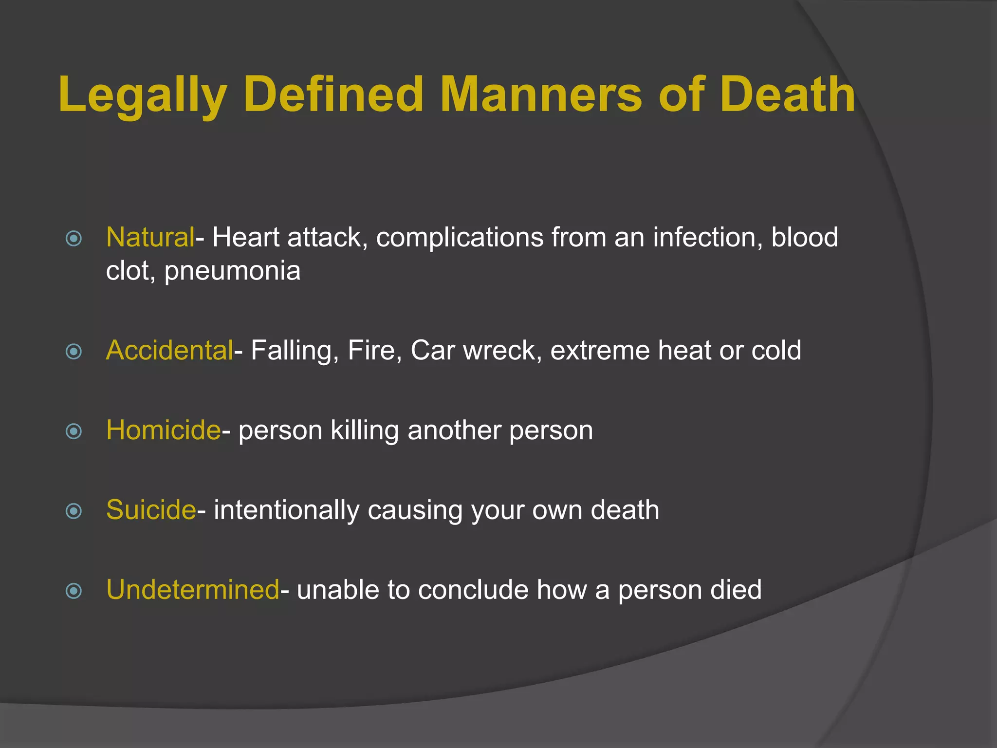
Legally Defined Mannersof Death
Natural- Heart attack, complications from an infection, blood
clot, pneumonia
Accidental- Falling, Fire, Car wreck, extreme heat or cold
Homicide- person killing another person
Suicide- intentionally causing your own death
Undetermined- unable to conclude how a person died
8.
Reason for autopsy
When an unexpected, or perhaps even suspicious, death
occurs.
When the cause of death needs to be determined.
To confirm a clinical diagnosis.
For academic purposes, such as research or teaching at
a medical school.
To gain insight into possible genetic traits or diseases in a
family.
To provide evidence in a crime investigation.
To provide closure for family members of the deceased.
On request by a family member or medical doctor.
In case of a public medical health concern, such as an
outbreak of sorts.
9.
Levels of autopsy
Complete: All body cavities are examined.
Limited: Which may exclude the head.
Selective: Where specific organs only are examined.
10.
Getting Started
The bodyis received in the morgue and is refrigerated/stored
until examination time. Autopsies are best if performed within 24
hours of death before organs deteriorate and before embalming
which can interfere with toxicology and blood cultures.
There are two types of mortuary cold chambers:
Morgue - Positive temperature
Morgue - Negative temperature
Morgue + (35.6 / 39.2 F) most usual for keeping bodies for a few
days or weeks.
Morgue – (5 / -13 F) used for keeping bodies which not have
been identified. Body is completely frozen.
11.
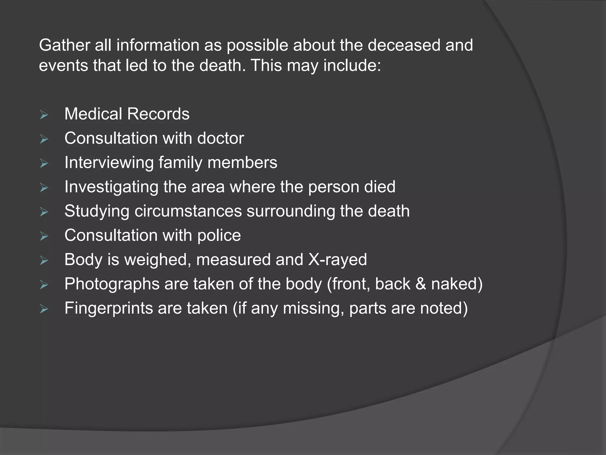
Gather all informationas possible about the deceased and
events that led to the death. This may include:
Medical Records
Consultation with doctor
Interviewing family members
Investigating the area where the person died
Studying circumstances surrounding the death
Consultation with police
Body is weighed, measured and X-rayed
Photographs are taken of the body (front, back & naked)
Fingerprints are taken (if any missing, parts are noted)
12.
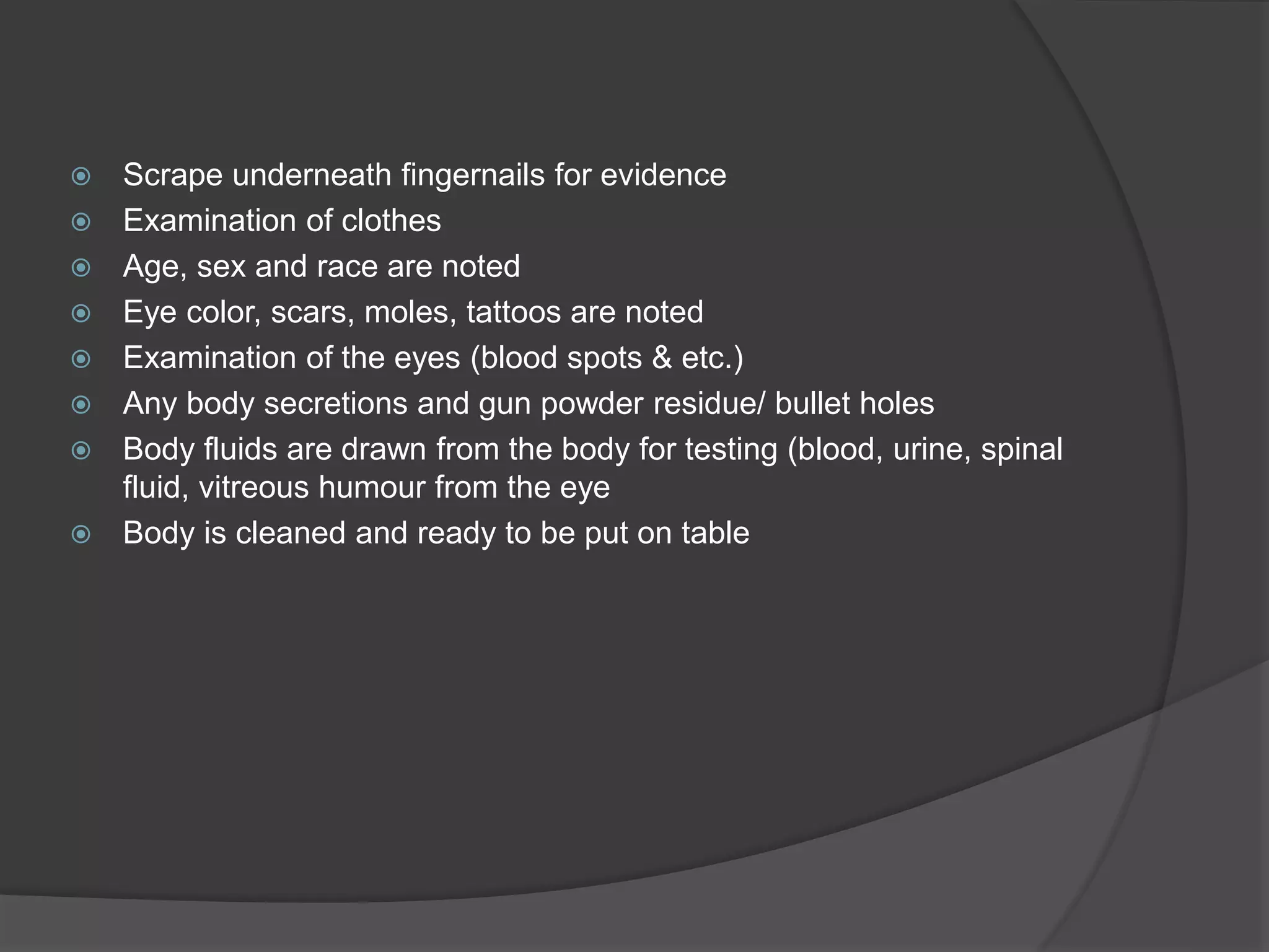
Scrape underneathfingernails for evidence
Examination of clothes
Age, sex and race are noted
Eye color, scars, moles, tattoos are noted
Examination of the eyes (blood spots & etc.)
Any body secretions and gun powder residue/ bullet holes
Body fluids are drawn from the body for testing (blood, urine, spinal
fluid, vitreous humour from the eye
Body is cleaned and ready to be put on table
13.
1. External examination
A pathologist starts an autopsy from the
outside of the body and works inwards.
Therefore, the first step in the procedure is an
external examination. The pathologist will first
look at the outer appearance, including clothes
and accessories.
They also note characteristics such as weight,
height, eye colour, hair colour, texture and
length, sex, and approximate age.
This information can help provide evidence, as
well as give clues to an identity if the body has
not been positively identified.
14.
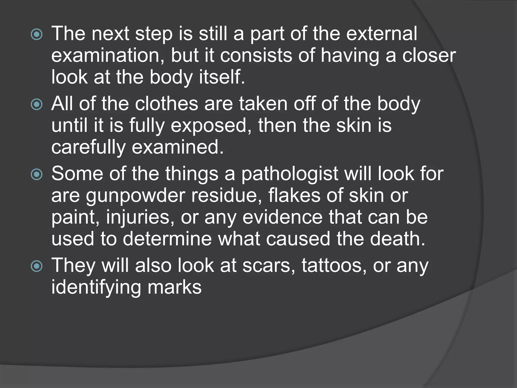
The nextstep is still a part of the external
examination, but it consists of having a closer
look at the body itself.
All of the clothes are taken off of the body
until it is fully exposed, then the skin is
carefully examined.
Some of the things a pathologist will look for
are gunpowder residue, flakes of skin or
paint, injuries, or any evidence that can be
used to determine what caused the death.
They will also look at scars, tattoos, or any
identifying marks
15.
2. X-rays
Finally,X-rays can be used to assess
whether there are any bone abnormalities
or foreign objects in the body, and
ultraviolet light can be used to detect
specific residues.
This is the part of the procedure where hair
or nail samples may be taken for further
examination.
This marks the end of the external
examination. Note that all of the
observations are written down, as well as
recorded.
16.
3. Internal examination
The internal examination includes the examining of the
chest and abdominal cavities, as well as the brain.
This is done by making careful incisions. The chest and
abdomen are accessed through Y-incisions, which start at
the shoulder, then meets at the sternum, and finally reach
the pubic bone.
The brain is reached by making an incision from ear to
ear in the back of the skull, or by a triangular incision
across the top part of the skull.
These incisions bleed minimally, as the heart is no longer
pumping blood. After performing the Y-incision, the
pathologist first examines all of the organs in place (by
removing the frontal part of the rib cage), then they can
remove all of the major organs (including the heart, lungs,
liver, stomach, and spleen).
17.
The removalcan be done using one of two techniques;
The Virchow technique, which consists of
removing each organ individually, or
The Rokitansky technique, during which all of
the organs are removed at once. The organs are then weighed
to detect the presence of certain illnesses.
Blood samples are also taken for further investigation. Small
pieces of tissue from each organ is examined under a
microscope.
The contents of the stomach are then examined. This is a good
indicator of time of death, as the last meal, as well as its level of
digestion, can be seen and used to determine a timeframe.
18.
4. Testing ofbody fluids
Body fluids are tested for anything from drugs, to
chemical and genetic composition, to infection, depending
on the type of autopsy.
1. Some of these fluids include
2. Blood,
3. Urine,
4. Bile,
5. Eye fluid.
Note that some poisons will only be observable in some
parts of the body, but not in others. The organs are then
either placed back in the body or preserved for teaching
or research purposes
19.
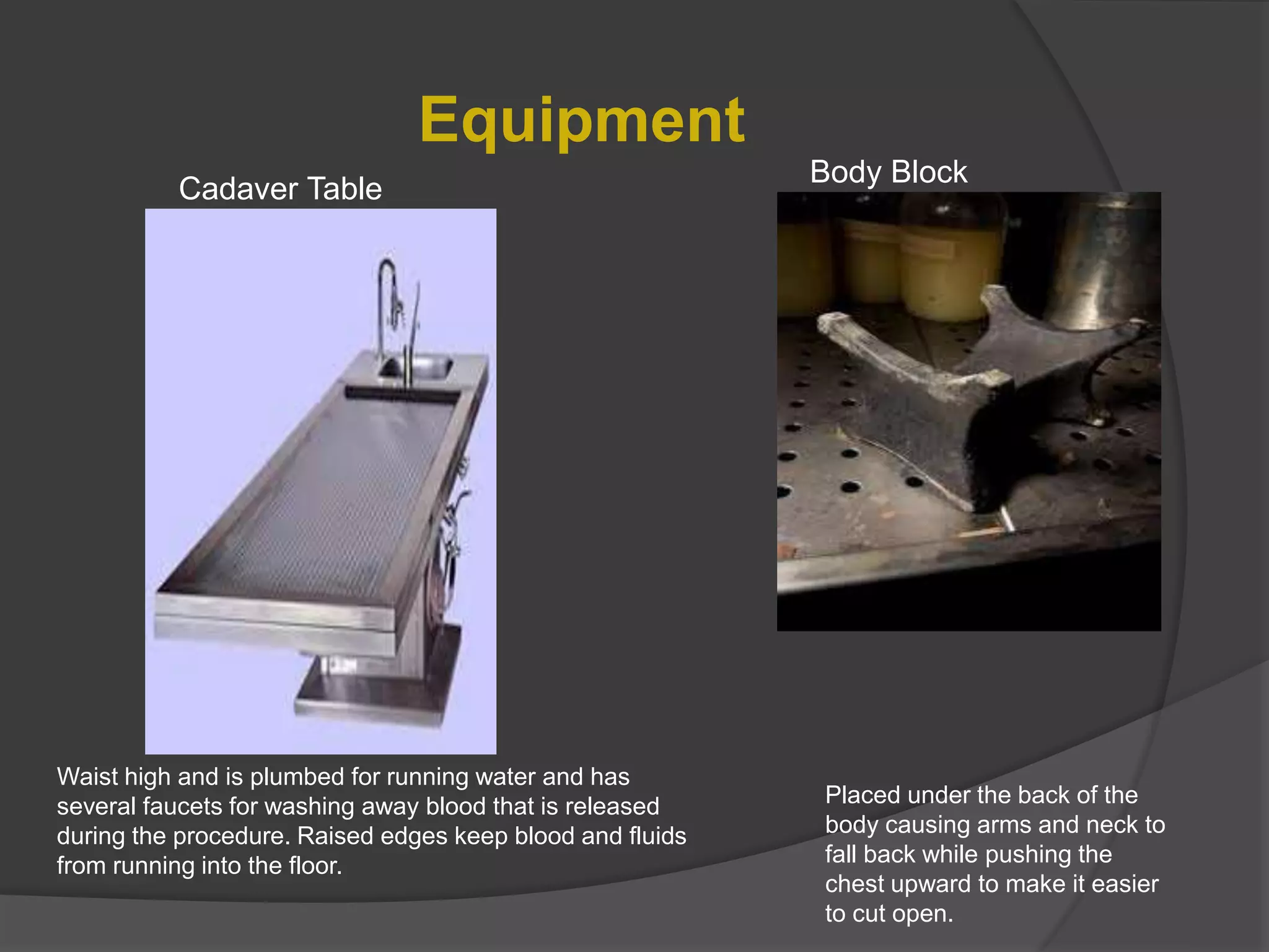
Equipment
Waist high andis plumbed for running water and has
several faucets for washing away blood that is released
during the procedure. Raised edges keep blood and fluids
from running into the floor.
Body Block
Placed under the back of the
body causing arms and neck to
fall back while pushing the
chest upward to make it easier
to cut open.
Cadaver Table
Procedure
Y-Incision: The Y-Incisionis the procedure used by the
pathologist or examiner to open up the breastplate of the
deceased and gain access to the body's major organs;
heart, lungs, liver, stomach, spleen etc.
22.
All theorgans are removed and weighed ( usually removed
in one unit but sometimes in sequence depending on the
trauma to the body).
Slices of each organ are taken and tested
Depending on type of death, stomach contents are removed,
examined and recorded
If gun shot was involved, then any bullets would be removed
and documented and saved for evidence
23.
After the mainorgans are examined the examiner proceeds to the
brain; (The body block is then moved to underneath the head)
Deep incision begins behind one ear, travels over the top of the head and
behind the opposite ear.
The scalp is pulled away from the skull in two flaps; front going over the face
and the rear going over the back of the neck so the skull is fully exposed.
Electric saw known as the “ Stryker saw” is used to cut and remove a wedge
shape portion of the skull which exposes the brain.
Brain is removed, weighed
and examined.
Any findings noted
24.
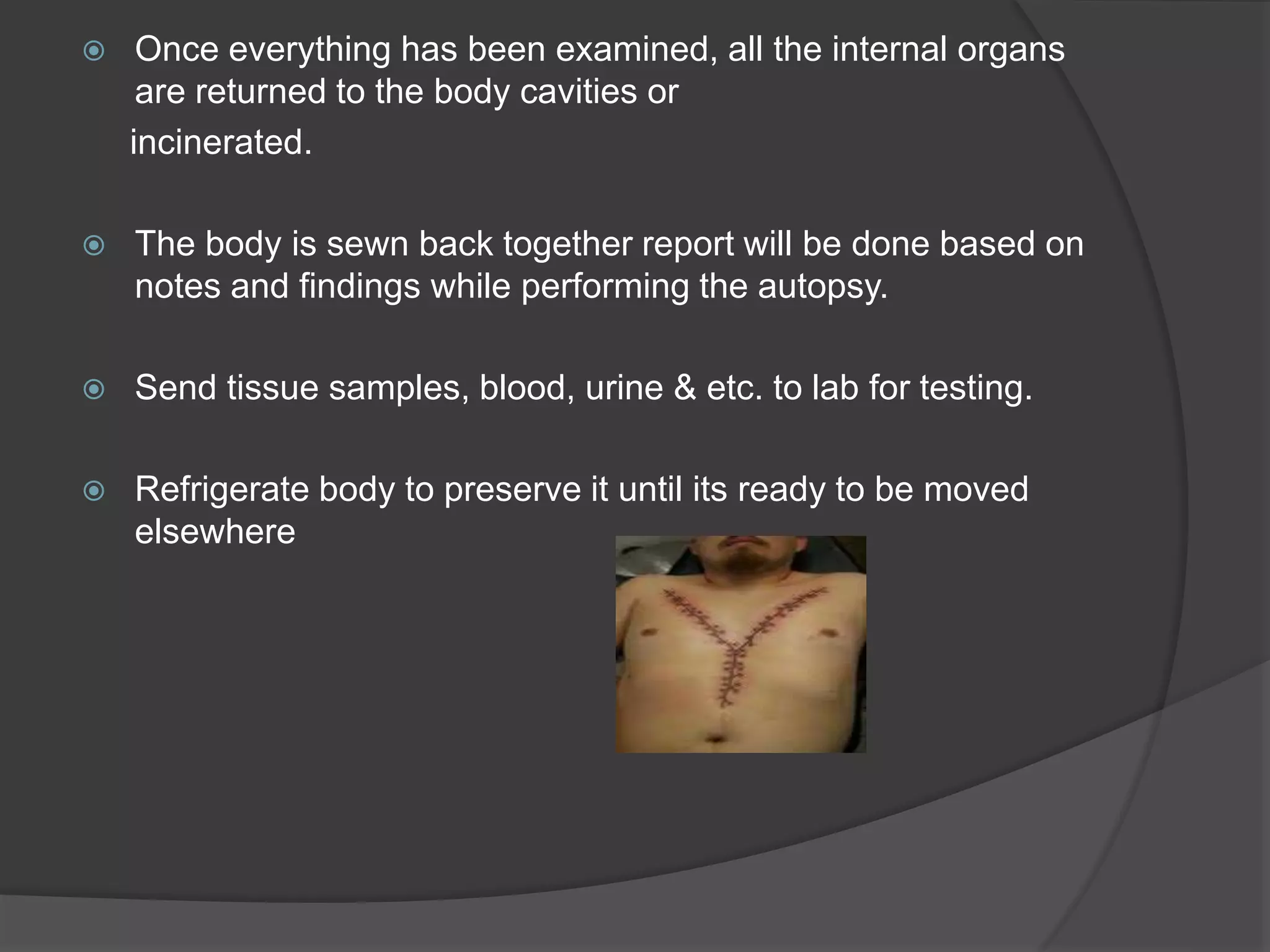
Once everythinghas been examined, all the internal organs
are returned to the body cavities or
incinerated.
The body is sewn back together report will be done based on
notes and findings while performing the autopsy.
Send tissue samples, blood, urine & etc. to lab for testing.
Refrigerate body to preserve it until its ready to be moved
elsewhere
25.
Once all testresults are back, a final report will be
provided giving the findings of the autopsy and the
cause of death.