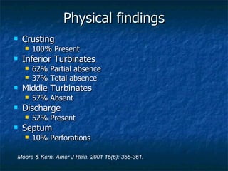
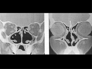
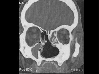
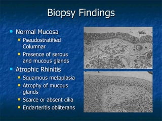
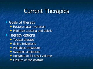
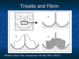
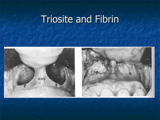
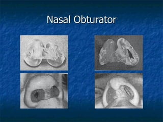
Atrophic Rhinitis UTMB Dept of Otolaryngology March 30, 2005 Alan L. Cowan, M.D. Matthew Ryan, M.D.
Atrophic Rhinitis Common Terms Ozena Dry Rhinitis Rhinitis Sicca
Atrophic Rhinitis Dr. Spencer Watson.  Diseases of the nose and its associated cavities . London, 1875. 1) Accidental or Simple Ozoena “ due to the retention of mucous.” “ easily dealt with by the frequent employment of the nasal douche …” 2) Idiopathic or constitutional “ commences in early childhood ... And remains during the early years or throughout the whole adult life.” “ The patient is generally anosmic … and he is, therefore, unaware of the offensive odor of his breath.” “ The nature of the inflammatory process is very probably allied to that of lupus erythematosus of the face.” 3) Syphilitic Ozoena “ the most common form” “ These ulcers may be preceded or followed by caries or necrosis of the bones, and the stench is then more horribly sickening than in any other form of this disgusting malady.”
Atrophic Rhinitis Described in 1876 by Dr. Bernhard Fraenkel as a triad of: Fetor Crusting Atrophy of nasal structures Dr. Francke Bosworth.  A Manual of Diseases of the Nose and Throat.  1881. “ the breath is often so penetrating as to render the near presence of the sufferer not only unpleasant but almost unendurable.”
Atrophic Rhinitis Clinical Features Anosmia Ozena, i.e. foul odor Extensive nasal crusting Subjective nasal congestion Enlargement of the nasal cavity Resorption or absence of turbinates Squamous metaplasia of nasal mucosa Depression
Atrophic rhinitis Primary History of prior sinus surgery, radiation, granulomatous disease, or nasal trauma are exclusions. Primary AR is rare in the US Most cases are reported in China, Egypt, and India Microbiology of primary AR is almost uniformly  Klebsiella ozenae . Radiographic and clinical features similar to secondary AR.
Atrophic rhinitis Secondary Complication of sinus surgery (89%) Complication of radiation (2.5%) Following nasal trauma (1%) Sequela of granulomatous diseases (1%) Sarcoid Leprosy Rhinoscleroma Sequlae of other infectious processes Tuberculosis Syphilis Moore & Kern. Amer J Rhin. 2001 15(6): 355-361.
Surgical causes Based on review of 242 cases from Mayo Clinic. Procedures per patient 2.3 Partial middle or inferior turbinectomy 56% Total middle and inferior turbinectomy 24% No turbinectomy 10% Partial maxillectomy 6% Moore & Kern. Amer J Rhin. 2001 15(6): 355-361.
Other suggested causes Infectious (Ssali) Case report of AR developed in 7 children of one family after contact with another known AR child. Dietary  (Bernat) Iron therapy found to benefit 50% of patients treated (Han-Sen) Hypocholesterolemia present in 50% of patients. (Han-Sen) Vitamin A therapy showed symptomatic improvement in 84%. Hereditary (Barton, Sibert) Proposed autosomal dominant disease due to father and 8 of 15 children contracting the disease. Hormonal  Symptoms known to worsen with menstraution or pregnancy. Developmental (Hagrass) Radiologic evidence of poor maxillary antrum pneumatization and short nasal lengths Vascular (Ruskin) Postulated overactivation of sympathetic activity. Environmental (Mickiewicz) Chronic exposure to phosphorite and apatide dust Autoimmune (Ricci)
Physical findings Crusting  100% Present Inferior Turbinates 62% Partial absence 37% Total absence Middle Turbinates 57% Absent Discharge  52% Present Septum 10% Perforations Moore & Kern. Amer J Rhin. 2001 15(6): 355-361.
Radiographic Findings Mucoperiosteal thickening of the paranasal sinuses. Loss of definition of the OMC secondary to resorption of the ethmoid bulla and uncinate process. Hypoplasia of the maxillary sinuses. Enlargement of the nasal cavities with erosion and bowing of the lateral nasal wall. Bony resorption and mucosal atrophy of the inferior and middle turbinates. Pace-Balzan, Shankar, Hawke. J Otolaryngol 1991; 20:428-32.


Biopsy Findings Normal Mucosa Pseudostratified Columnar Presence of serous and mucous glands Atrophic Rhinitis Squamous metaplasia Atrophy of mucous glands Scarce or absent cilia Endarteritis obliterans
Microbiology Klebsiella ozenae May be found in almost 100% of primary AR No predominance in secondary AR Staphylococcus aureus Proteus mirabilis Escherichia coli Corynebacterium diphtheriae
Current Therapies Goals of therapy Restore nasal hydration Minimize crusting and debris Therapy options Topical therapy Saline irrigations Antibiotic irrigations Systemic antibiotics Implants to fill nasal volume Closure of the nostrils
Local therapy Irrigations Saline Mixtures Sodium bicarbonate Shehata: Sodium Carbonate 25g, Sodium Biborate 25g, and Sodium Chloride 50g in 250ml water. Antibiotic solution Moore: Gentamycin solution 80mg/L Anti-drying agents Glycerine Mineral Oil Paraffin with 2% Menthol Other Acetylcholine Pilocarpine
Systemic therapy Oral antibiotics Tetracycline Ciprofloxacin Aminoglycosides Streptomycin injections Medication avoidance Vasoconstrictors Topical steroids * Other Vitamin A (12,500 to 15,000 Units daily) Potassium Iodide (Increases nasal secretions) Vasodilators Iron therapy Estrogen Corticosteroids * Vaccines Antibacterial (Pasturella, Bordetella) Autogenous
Surgical therapies Young procedure Modified Young procedure Turbinate reconstruction Volume reduction procedures Denervating operations
Nasal Closure Young’s procedure Circumferential flap elevation 1 cm cephalic to the alar rim. Sutures placed in center of elevated flap to close the nostril Staged second side in 3 months Advantages Often provided relief of symptoms Disadvantages Difficult to elevate circumferential flap Breakdown of central suture area common Does not allow for cleaning Did not allow for periodic examination Recurrence after flap takedown Young. “Closure of the nostril in atrophic rhinitis.” Journal of Laryngology and Otology, 81: 515-524.
Nasal Closure Modified Young’s Elevation of extended perichondrial flap through contralateral hemitransfixion incision. Short skin flap elevated from the intercartilaginous line on the ipsilateral side. Suture lateral and medial flaps with vicryl. Staged second side with first side takedown in 6 mon. Advantages Technically easier than Young procedure No suture line breakdown No vestibular stenosis on takedown Disadvantages Not possible with large septal defects Does not allow for cleaning Does not allow for periodic examination Recurrence after flap takedown El Kholy, Habib, Abdel-Monem, Safia. “Septal mucoperichondrial flap for closure of nostril in atrophic rhinitis.” Rhinology, 36, 202-203, 1998.
Modified Young
Volume reduction Plastipore implantation Porus material allows tissue ingrowth. Implants shaped then fenestrated for ingrowth. Implants placed submucosally along the septum and nasal floor. Advantages Easier than other surgical options (Young’s) Plastipore has low extrusion/complication rate May be done under local anesthesia Disadvantages Possibility of extrusion (occurred in 1/8 pts) Requires septal mucosa (not discussed) Goldenberg, Danino, Netzer, Joachims. Oto HNS, Vol. 122 (6). pp. 794-97.
Plastipore
Volume Reduction (cont) Triosite and Fibrin Triosite (60% hydroxyapetite, 40% calcium triphosphate) mixed with Fibrin 1:1. Deglove the labial vestibule Elevate periosteum of the floor posteriorly to the end of the hard palate, extend medially onto the septum. Insert Triosite & Fibrin mixture (~3.3g per side) Advantages Good to excellent result (7/9 patients) Material can be molded easily  Disadvantages Leakage of material (4/9 patients) Infection of material (3/9 patients) Potential damage to lacrimal system Bertrand, Doyen, Eloy. Laryngoscope 106: May 1996. p 652-57.
Triosite and Fibrin Bertrand, Doyen, Eloy. Laryngoscope 106: May 1996. p 652-57.
Triosite and Fibrin
Other Therapies Non-surgical nasal closure Nasal vestibule impressions taken similar to hearing aid moulds. Impressions are used to create a silastic obturator. Advantages Reversible Easily removed Allows for irrigations Allows for serial clinical exams Avoids surgical morbidity  Disadvantages May be uncomfortable May cause sore throat due to obligate mouth breathing. Lobo, Hartley, Farrington. J of Laryn and Oto. June 1998, Vol 112, p 543-46.
Nasal Obturator
Other Therapies Other Implants Acrylic Silicone Teflon Silastic Boplant Denervation Cervical sympathectomy (Bertein) Stellate ganglion block (Bahl) Sphenopalatine ganglion block (Girgis) Parasympathectomy, i.e. GSPN section (Krmptotic) Salivary Irrigation Involves reimplantation of parotid duct into the maxillary sinus Accupuncture Time Disease often resolves spontaneously after age 40
Bibliography Lobo, Hartley, Farrington. “Closure of the nasal vestibule in atrophic rhinitis – a new non-surgical technique.”  The Journal of Laryngology and Otology . June 1998, Vol. 112, pp. 543-46. Moore, Kern. “Atrophic Rhinitis: A Review of 242 cases.”  American Journal of Rhinology . November-December 2001, Vol. 15, No. 6, p 355-61. Shehata. “Atrophic Rhinitis.”  American Journal of Otolaryngology , Vol. 17, No. 2. March-April, 1996: pp 81-86. Chand, MacArthur. “Primary atrophic rhinitis: A summary of four cases and review of the literature.” Otolaryngology – Head and Neck Surgery. Vol. 116, No. 4: pp 554-57. Bertrand, Doyen, Eloy. “Triosite Implants and Fibrin Glue in the Treatment of Atrophic Rhinitis: Technique and Results.” Laryngoscope (106): May 1996: pp 652-57. Goldenberg, Danino, Netzer, Joachims. “Plastipore implants in the surgical treatment of atrophic rhinitis: Technique and results.” Otolaryngology Head and Neck Surgery. Vol 122 No 6: pp 794-97. Watson, Spencer.  Diseases of the nose and its accessory cavities . London: 1875. El Kholy, Habib, Abdel-Monem, Safia. “Septal mucoperichondrial flap for closure of nostril in atrophic rhinitis.”  Rhinology , 36, 202-203, 1998.

Atrophic Rhinitis Slides 050330

  • 1.
    Atrophic Rhinitis UTMBDept of Otolaryngology March 30, 2005 Alan L. Cowan, M.D. Matthew Ryan, M.D.
  • 2.
    Atrophic Rhinitis CommonTerms Ozena Dry Rhinitis Rhinitis Sicca
  • 3.
    Atrophic Rhinitis Dr.Spencer Watson. Diseases of the nose and its associated cavities . London, 1875. 1) Accidental or Simple Ozoena “ due to the retention of mucous.” “ easily dealt with by the frequent employment of the nasal douche …” 2) Idiopathic or constitutional “ commences in early childhood ... And remains during the early years or throughout the whole adult life.” “ The patient is generally anosmic … and he is, therefore, unaware of the offensive odor of his breath.” “ The nature of the inflammatory process is very probably allied to that of lupus erythematosus of the face.” 3) Syphilitic Ozoena “ the most common form” “ These ulcers may be preceded or followed by caries or necrosis of the bones, and the stench is then more horribly sickening than in any other form of this disgusting malady.”
  • 4.
    Atrophic Rhinitis Describedin 1876 by Dr. Bernhard Fraenkel as a triad of: Fetor Crusting Atrophy of nasal structures Dr. Francke Bosworth. A Manual of Diseases of the Nose and Throat. 1881. “ the breath is often so penetrating as to render the near presence of the sufferer not only unpleasant but almost unendurable.”
  • 5.
    Atrophic Rhinitis ClinicalFeatures Anosmia Ozena, i.e. foul odor Extensive nasal crusting Subjective nasal congestion Enlargement of the nasal cavity Resorption or absence of turbinates Squamous metaplasia of nasal mucosa Depression
  • 6.
    Atrophic rhinitis PrimaryHistory of prior sinus surgery, radiation, granulomatous disease, or nasal trauma are exclusions. Primary AR is rare in the US Most cases are reported in China, Egypt, and India Microbiology of primary AR is almost uniformly Klebsiella ozenae . Radiographic and clinical features similar to secondary AR.
  • 7.
    Atrophic rhinitis SecondaryComplication of sinus surgery (89%) Complication of radiation (2.5%) Following nasal trauma (1%) Sequela of granulomatous diseases (1%) Sarcoid Leprosy Rhinoscleroma Sequlae of other infectious processes Tuberculosis Syphilis Moore & Kern. Amer J Rhin. 2001 15(6): 355-361.
  • 8.
    Surgical causes Basedon review of 242 cases from Mayo Clinic. Procedures per patient 2.3 Partial middle or inferior turbinectomy 56% Total middle and inferior turbinectomy 24% No turbinectomy 10% Partial maxillectomy 6% Moore & Kern. Amer J Rhin. 2001 15(6): 355-361.
  • 9.
    Other suggested causesInfectious (Ssali) Case report of AR developed in 7 children of one family after contact with another known AR child. Dietary (Bernat) Iron therapy found to benefit 50% of patients treated (Han-Sen) Hypocholesterolemia present in 50% of patients. (Han-Sen) Vitamin A therapy showed symptomatic improvement in 84%. Hereditary (Barton, Sibert) Proposed autosomal dominant disease due to father and 8 of 15 children contracting the disease. Hormonal Symptoms known to worsen with menstraution or pregnancy. Developmental (Hagrass) Radiologic evidence of poor maxillary antrum pneumatization and short nasal lengths Vascular (Ruskin) Postulated overactivation of sympathetic activity. Environmental (Mickiewicz) Chronic exposure to phosphorite and apatide dust Autoimmune (Ricci)
  • 10.
    Physical findings Crusting 100% Present Inferior Turbinates 62% Partial absence 37% Total absence Middle Turbinates 57% Absent Discharge 52% Present Septum 10% Perforations Moore & Kern. Amer J Rhin. 2001 15(6): 355-361.
  • 11.
    Radiographic Findings Mucoperiostealthickening of the paranasal sinuses. Loss of definition of the OMC secondary to resorption of the ethmoid bulla and uncinate process. Hypoplasia of the maxillary sinuses. Enlargement of the nasal cavities with erosion and bowing of the lateral nasal wall. Bony resorption and mucosal atrophy of the inferior and middle turbinates. Pace-Balzan, Shankar, Hawke. J Otolaryngol 1991; 20:428-32.
  • 12.
  • 13.
  • 14.
  • 15.
    Biopsy Findings NormalMucosa Pseudostratified Columnar Presence of serous and mucous glands Atrophic Rhinitis Squamous metaplasia Atrophy of mucous glands Scarce or absent cilia Endarteritis obliterans
  • 16.
    Microbiology Klebsiella ozenaeMay be found in almost 100% of primary AR No predominance in secondary AR Staphylococcus aureus Proteus mirabilis Escherichia coli Corynebacterium diphtheriae
  • 17.
    Current Therapies Goalsof therapy Restore nasal hydration Minimize crusting and debris Therapy options Topical therapy Saline irrigations Antibiotic irrigations Systemic antibiotics Implants to fill nasal volume Closure of the nostrils
  • 18.
    Local therapy IrrigationsSaline Mixtures Sodium bicarbonate Shehata: Sodium Carbonate 25g, Sodium Biborate 25g, and Sodium Chloride 50g in 250ml water. Antibiotic solution Moore: Gentamycin solution 80mg/L Anti-drying agents Glycerine Mineral Oil Paraffin with 2% Menthol Other Acetylcholine Pilocarpine
  • 19.
    Systemic therapy Oralantibiotics Tetracycline Ciprofloxacin Aminoglycosides Streptomycin injections Medication avoidance Vasoconstrictors Topical steroids * Other Vitamin A (12,500 to 15,000 Units daily) Potassium Iodide (Increases nasal secretions) Vasodilators Iron therapy Estrogen Corticosteroids * Vaccines Antibacterial (Pasturella, Bordetella) Autogenous
  • 20.
    Surgical therapies Youngprocedure Modified Young procedure Turbinate reconstruction Volume reduction procedures Denervating operations
  • 21.
    Nasal Closure Young’sprocedure Circumferential flap elevation 1 cm cephalic to the alar rim. Sutures placed in center of elevated flap to close the nostril Staged second side in 3 months Advantages Often provided relief of symptoms Disadvantages Difficult to elevate circumferential flap Breakdown of central suture area common Does not allow for cleaning Did not allow for periodic examination Recurrence after flap takedown Young. “Closure of the nostril in atrophic rhinitis.” Journal of Laryngology and Otology, 81: 515-524.
  • 22.
    Nasal Closure ModifiedYoung’s Elevation of extended perichondrial flap through contralateral hemitransfixion incision. Short skin flap elevated from the intercartilaginous line on the ipsilateral side. Suture lateral and medial flaps with vicryl. Staged second side with first side takedown in 6 mon. Advantages Technically easier than Young procedure No suture line breakdown No vestibular stenosis on takedown Disadvantages Not possible with large septal defects Does not allow for cleaning Does not allow for periodic examination Recurrence after flap takedown El Kholy, Habib, Abdel-Monem, Safia. “Septal mucoperichondrial flap for closure of nostril in atrophic rhinitis.” Rhinology, 36, 202-203, 1998.
  • 23.
  • 24.
    Volume reduction Plastiporeimplantation Porus material allows tissue ingrowth. Implants shaped then fenestrated for ingrowth. Implants placed submucosally along the septum and nasal floor. Advantages Easier than other surgical options (Young’s) Plastipore has low extrusion/complication rate May be done under local anesthesia Disadvantages Possibility of extrusion (occurred in 1/8 pts) Requires septal mucosa (not discussed) Goldenberg, Danino, Netzer, Joachims. Oto HNS, Vol. 122 (6). pp. 794-97.
  • 25.
  • 26.
    Volume Reduction (cont)Triosite and Fibrin Triosite (60% hydroxyapetite, 40% calcium triphosphate) mixed with Fibrin 1:1. Deglove the labial vestibule Elevate periosteum of the floor posteriorly to the end of the hard palate, extend medially onto the septum. Insert Triosite & Fibrin mixture (~3.3g per side) Advantages Good to excellent result (7/9 patients) Material can be molded easily Disadvantages Leakage of material (4/9 patients) Infection of material (3/9 patients) Potential damage to lacrimal system Bertrand, Doyen, Eloy. Laryngoscope 106: May 1996. p 652-57.
  • 27.
    Triosite and FibrinBertrand, Doyen, Eloy. Laryngoscope 106: May 1996. p 652-57.
  • 28.
  • 29.
    Other Therapies Non-surgicalnasal closure Nasal vestibule impressions taken similar to hearing aid moulds. Impressions are used to create a silastic obturator. Advantages Reversible Easily removed Allows for irrigations Allows for serial clinical exams Avoids surgical morbidity Disadvantages May be uncomfortable May cause sore throat due to obligate mouth breathing. Lobo, Hartley, Farrington. J of Laryn and Oto. June 1998, Vol 112, p 543-46.
  • 30.
  • 31.
    Other Therapies OtherImplants Acrylic Silicone Teflon Silastic Boplant Denervation Cervical sympathectomy (Bertein) Stellate ganglion block (Bahl) Sphenopalatine ganglion block (Girgis) Parasympathectomy, i.e. GSPN section (Krmptotic) Salivary Irrigation Involves reimplantation of parotid duct into the maxillary sinus Accupuncture Time Disease often resolves spontaneously after age 40
  • 32.
    Bibliography Lobo, Hartley,Farrington. “Closure of the nasal vestibule in atrophic rhinitis – a new non-surgical technique.” The Journal of Laryngology and Otology . June 1998, Vol. 112, pp. 543-46. Moore, Kern. “Atrophic Rhinitis: A Review of 242 cases.” American Journal of Rhinology . November-December 2001, Vol. 15, No. 6, p 355-61. Shehata. “Atrophic Rhinitis.” American Journal of Otolaryngology , Vol. 17, No. 2. March-April, 1996: pp 81-86. Chand, MacArthur. “Primary atrophic rhinitis: A summary of four cases and review of the literature.” Otolaryngology – Head and Neck Surgery. Vol. 116, No. 4: pp 554-57. Bertrand, Doyen, Eloy. “Triosite Implants and Fibrin Glue in the Treatment of Atrophic Rhinitis: Technique and Results.” Laryngoscope (106): May 1996: pp 652-57. Goldenberg, Danino, Netzer, Joachims. “Plastipore implants in the surgical treatment of atrophic rhinitis: Technique and results.” Otolaryngology Head and Neck Surgery. Vol 122 No 6: pp 794-97. Watson, Spencer. Diseases of the nose and its accessory cavities . London: 1875. El Kholy, Habib, Abdel-Monem, Safia. “Septal mucoperichondrial flap for closure of nostril in atrophic rhinitis.” Rhinology , 36, 202-203, 1998.