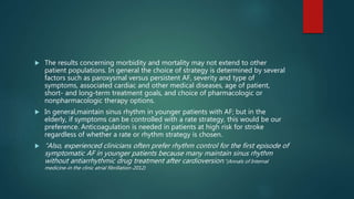
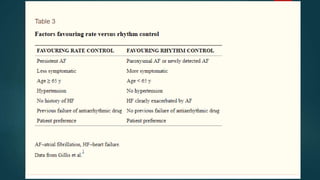
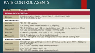
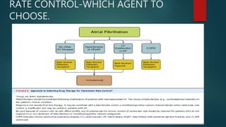
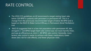
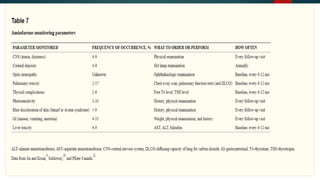
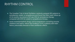
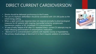
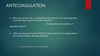
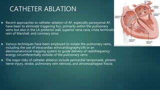
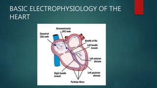
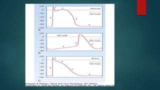
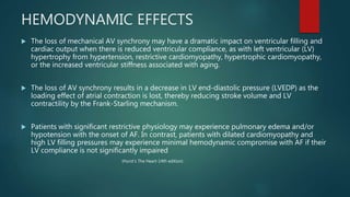
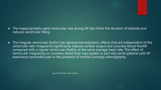
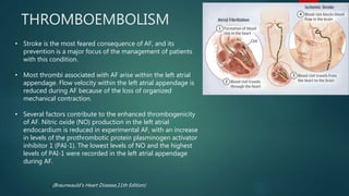
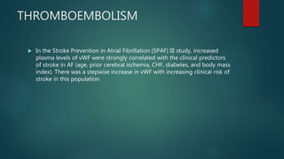
ATRIAL FIBRILLATION
PRESENTER-DR ANIRUDH
MODERATOR-DR NAGAPPA
PREVIOUS YEARS QUESTIONS
 Atrial Fibrillation
 Ennumerate causes of atrial firbrillation.Describe management of a patient
with newly diagnosed atrial fibrillation
 Newer anticoagulants
REFERENCES
 Harrison’s Principles of Internal Medicine-19th edition
 Hurst’s The Heart-14th edition
 Braunwauld’s Heart Disease,11th Edition
 Management of Atrial Fibrillation in Indian Scenario -Saumitra Ray,
Amitava Mazumdar
 2014 AHA/ACC/HRS Guideline for the Management of Patients With Atrial
Fibrillation
 New frontiers in Atrial fibrillation:Annals of Internal Medicine 2017
OUTLINE
 Basic Electrophysiology of the Heart
 Definition
 Classification
 Pathophysiology
 History and clinical examination
 Investigations
 Risk of stroke-scores and their comparison
 Management-AHA 2014 guidelines
 Newer OAC
BASIC ELECTROPHYSIOLOGY OF THE
HEART
ATRIAL FIBRILLATION-DEFINITION
 “Atrial fibrillation (AF) is characterized by disorganized, rapid, and
atrial activation with loss of atrial contraction and with an irregular
ventricular rate that is determined by AV nodal conduction”
(Harrison’s Principles of Internal Medicine-19th edition)
“Atrial fibrillation (AF) is characterized by disorganized atrial electrical
activation and uncoordinated atrial contraction. The surface
electrocardiogram characteristically demonstrates rapid fibrillatory
waves with changing morphology and rate and a ventricular rhythm that
is irregularly irregular “
(Hurst’s The Heart-14th edition)
CLASSIFICATION
Published guidelines from an American College of Cardiology
(ACC)/American Heart Association (AHA)/European Society of Cardiology
(ESC) committee of experts on the treatment of patients with atrial
fibrillation recommend classification of AF into the following 3 patterns :
1. Paroxysmal AF – Episodes of AF that terminate spontaneously within 7
days (most episodes last less than 24 hours)
2. Persistent AF - Episodes of AF that last more than 7 days and may require
either pharmacologic or electrical intervention to terminate
3. Permanent AF - AF that has persisted for more than 1 year, either
because cardioversion has failed or because cardioversion has not been
attempted
• This classification schema pertains to cases that are not related to a
reversible cause of AF (eg, thyrotoxicosis, electrolyte abnormalities, acute
ethanol intoxication).
(Hurst’s The Heart-14th edition)
 OTHER TERMS-CHRONIC AF
 -LONE AF
 -VALVULAR AND NON VALVULAR AF
EPIDEMIOLOGY
 Overall global prevalence of AF is 1-2% in the adult population with prevalence increasing
with increasing age.
 Most common sustained cardiac arrhythmia
 Incidence and prevalence increases with age
 The incidence <0.5% below 50Yrs
2% in age 60-69
4.6% in age 70-79
8.8% in age 80-89
 Men > Women
 Whites > Blacks
 In the absence of anticoagulation, the relative risk of stroke in patients with rheumatic AF
is increased approximately 17-fold.
 The Framingham Study demonstrated that the risk of stroke in AF is clearly related to
age, with an annual risk of stroke of 1.5 percent in patients aged 50 to 59 years, which
increased to 23.5 percent in patients aged 80 to 89 years.
 The risk of stroke in nonvalvular AF has been estimated to be approximately 7 percent
per year.
 The development of AF is a strong predictor of increased mortality in cardiac conditions
such as hypertrophic or restrictive cardiomyopathy.
EPIDEMIOLOGY
 The prevalences of paroxysmal AF in the RE-LY (Randomised Evaluation of
Long term anticoagulant therapY), REALISE-AF, and IHRSAF study were
38%, 43% and 19.5% respectively and prevalence rates of permanent AF
were 18.6%, 34.3% and 33.7% respectively
 REALIZE AF and IHRS AF study, the average age of patients with AF in India
was 60 years and 54 years, respectively (a decade younger than Western
counterparts). Thus, it increases the economic burden by more DALY’s lost
.
 RHD still remained the most common etiology (42%)
 Other etiologies which were a distant second to RHD were hypertension
(10.2%) and chronic obstructive pulmonary disease (10.2%).
CAUSES
PATHOGENESIS
 ATRAIL FIBRILLATION
INITIATING
TRIGGER
PROPOGATING
FACTORS
PATHOGENESIS-TRIGGERS
 Triggering foci of rapidly firing cells within the sleeve of atrial myocytes extending into the
pulmonary veins have been clearly shown to be the underlying mechanism of most paroxysmal
AF.
 The pulmonary veins of patients with paroxysmal AF demonstrate abnormal properties of
conduction such that there is a markedly reduced effective refractory period within the
pulmonary veins.
 Autopsy studies have identified pacemaker cells, transitional cells, and Purkinje cells within the
PVs. The molecular basis for PV triggers has been primarily attributed to abnormal calcium
Ca2+ handling
 Although most triggering foci that are mapped during electrophysiologic studies occur in the
pulmonary veins in patients with paroxysmal AF, foci within the superior vena cava,the
ligament of Marshall, and the musculature of the coronary sinus have been identified . Other
sites of initiating foci may be recorded in the left atrial wall or along the crista terminalis in the
right atrium.
 Role of vagal tone.
Circ Res. 2017;120:1501-1517
PROPOGATING FACTORS
 MULTIPLE WAVELET THEORY
HEMODYNAMIC EFFECTS
 The loss of mechanical AV synchrony may have a dramatic impact on ventricular filling and
cardiac output when there is reduced ventricular compliance, as with left ventricular (LV)
hypertrophy from hypertension, restrictive cardiomyopathy, hypertrophic cardiomyopathy,
or the increased ventricular stiffness associated with aging.
 The loss of AV synchrony results in a decrease in LV end-diastolic pressure (LVEDP) as the
loading effect of atrial contraction is lost, thereby reducing stroke volume and LV
contractility by the Frank-Starling mechanism.
 Patients with significant restrictive physiology may experience pulmonary edema and/or
hypotension with the onset of AF. In contrast, patients with dilated cardiomyopathy and
high LV filling pressures may experience minimal hemodynamic compromise with AF if their
LV compliance is not significantly impaired
(Hurst’s The Heart-14th edition)
 The inappropriately rapid ventricular rate during AF also limits the duration of diastole and
reduces ventricular filling.
 The irregular ventricular rhythm has adverse hemodynamic effects that are independent of the
ventricular rate. Irregularity significantly reduces cardiac output and coronary blood flow68
compared with a regular ventricular rhythm at the same average heart rate. The effect of
ventricular irregularity on coronary blood flow may explain in part why some patients with AF
experience precordial pain in the presence of normal coronary arteriography.
(Hurst’s The Heart-14th edition)
THROMBOEMBOLISM
• Stroke is the most feared consequence of AF, and its
prevention is a major focus of the management of patients
with this condition.
• Most thrombi associated with AF arise within the left atrial
appendage. Flow velocity within the left atrial appendage is
reduced during AF because of the loss of organized
mechanical contraction.
• Several factors contribute to the enhanced thrombogenicity
of AF. Nitric oxide (NO) production in the left atrial
endocardium is reduced in experimental AF, with an increase
in levels of the prothrombotic protein plasminogen activator
inhibitor 1 (PAI-1). The lowest levels of NO and the highest
levels of PAI-1 were recorded in the left atrial appendage
during AF.
(Braunwauld’s Heart Disease,11th Edition)
THROMBOEMBOLISM
 In the Stroke Prevention in Atrial Fibrillation (SPAF) III study, increased
plasma levels of vWF were strongly correlated with the clinical predictors
of stroke in AF (age, prior cerebral ischemia, CHF, diabetes, and body mass
index). There was a stepwise increase in vWF with increasing clinical risk of
stroke in this population
STROKE RISK ASSESSMENT SCORES
 How do we determine stroke risk ?
 0 points – low risk (1.2-3.0 strokes per 100 patient years)
 1-2 points – moderate risk (2.8-4.0 strokes per 100 patient years)
 > 3 points – high risk (5.9-18.2 strokes per 100 patient years)
BLEEDING RISK ASSESSMENT
 Assessment of bleeding risk should be part of the clinical assessment of AF
patients prior to starting anticoagulation
 Antithrombotic benefits and potential bleeding risks of long-term
coagulation should be explained and discussed with the patient
 Aim for a target INR of between 2.0 and 3.0
 HAS-BLED Score
HISTORY AND PHYSICAL EXAMINATION
 DEFINE SYMPTOMS-Presence and nature of symptoms associated with AF.
 While majority of AF episodes may not cause symptoms, many patients
experience a wide variety of symptoms, including palpitations, dyspnea,
fatigue, dizziness, angina, and decompensated heart failure
 DEFINE PATTERN-paroxysmal,persistent,permanent
 ONSET AND DATE OF DISCOVERY
 FREQUENCY AND DURATION OF EPISODES
 PRECIPITATING CAUSES AND MODES OF TERMINATION.
 Presence of underlying heart disease or other reversible factors (e.g.,
hyperthyroidism, alcohol consumption)
HISTORY AND PHYSICAL EXAMINATION
PHYSICAL EXAMINATION
 Patients will have an irregularly irregular pulse and will commonly be
tachycardic, with heart rates typically in the 110- to 140-range, but rarely
over 160-170.
 Apex pulse deficit of >10
 Absence of a waves in JVP
 Examination of the head and neck may reveal exophthalmos, thyromegaly,
elevated jugular venous pressures.
 Heart sounds are irregular with variable intensity of heart sounds.
INVESTIGATIONS
 ECG
 P waves is absent in all leads
 multiple oscillating baseline waves ‘ f ’ (fibrillation) of various amplitude
and shape are recorded instead of P waves (usually best seen in the leads
II, III, aVF, V1 and V2)
 RR intervals are of various duration (irregular ventricular rhythm );
 QRS complexes are not changed.
2D ECHO
 Valvular heart disease
 Left and right atrial size
 Left ventricular size and function
 Peak right ventricular pressure (pulmonary hypertension)
 Left atrial thrombus (low sensitivity)
 Pericardial disease
ADDITIONAL TESTING
 Six-minute walk test (if the adequacy of rate control is in question) Exercise
test (if the adequacy of rate control is in question [permanent atrial
fibrillation])
 To reproduce exercise-induced atrial fibrillation
 To exclude ischemia before treatment of selected patients with a type IC*
antiarrhythmic drug
 Holter monitor test or event recording (if diagnosis of the type of
arrhythmia is in question) to evaluate rate control
ATRIAL FIBRILLATION-MANAGEMENT
RATE V/S RHYTHM CONTROL
 The AFFIRM trial enrolled 4060 patients aged older than 65 years or with risk
factors for stroke, randomizing them to rate versus rhythm control.
 Over a mean follow-up period of 3.5 years, there was no significant difference
in overall mortality between the two groups .
 The RACE trial randomly assigned 522 patients with persistent AF after
electrical cardioversion to either a rate-control or a rhythm-control treatment.
There was no difference in the primary endpoint of the study (a composite of
cardiovascular death, CHF, thromboemboli, bleeding, need for pacemaker, or
serious drug side effects) between the two strategies.
 Thus, these studies indicate that both approaches have similar clinical
outcomes provided that appropriate anticoagulation is maintained.
 The results concerning morbidity and mortality may not extend to other
patient populations. In general the choice of strategy is determined by several
factors such as paroxysmal versus persistent AF, severity and type of
symptoms, associated cardiac and other medical diseases, age of patient,
short- and long-term treatment goals, and choice of pharmacologic or
nonpharmacologic therapy options.
 In general,maintain sinus rhythm in younger patients with AF; but in the
elderly, if symptoms can be controlled with a rate strategy, this would be our
preference. Anticoagulation is needed in patients at high risk for stroke
regardless of whether a rate or rhythm strategy is chosen.
 “Also, experienced clinicians often prefer rhythm control for the first episode of
symptomatic AF in younger patients because many maintain sinus rhythm
without antiarrhythmic drug treatment after cardioversion.”(Annals of Internal
medicine-in the clinic atrial fibrillation-2012)
RATE CONTROL AGENTS
RATE CONTROL-WHICH AGENT TO
CHOOSE.
RATE CONTROL
 The 2010 CCS guidelines on Af recommend a target resting heart rate
below 100 BPM in patients with persistent or permanent AF. This is a
change from the previously recommended target of below 80 BPM and is
based on the RACE-II (Rate Control Efficacy in Permanent Atrial Fibrillation)
trial.
 RACE-II suggested that in a low-risk population with permanent AF, less
stringent (< 110 BPM) target heart rate control was not more harmful than,
and was as efficacious as, strict (< 80 BPM) rate control. Generally, more
lenient rate control is easier to achieve with fewer medications, lower
doses (less risk for side effects), and fewer physician visits.
RHYTHM CONTROL
Due to more wide spread availability amiodarone is commonly used in India in all situations
http://www.apiindia.org/pdf/medicine_update_2017/mu_210.pdf
What strategies should clinicians consider for
rhythm control in patients with AF?
 Either direct electrical or antiarrhythmic drug cardioversion:
 AF >48h or undetermined duration: establish rate control &
anticoagulation before elective cardioversion
 INR 2.0 – 3.0 for > 3 weeks prior and > 4 weeks after cardioversion
 Alternative- Transesophageal Echo:
 No clot: heparin for 48 hr prior to cardioversion & warfarin for 4 wks
after
 Clot present: anticoagulation for 4 weeks; most confirm thrombus
resolution with repeat TEE before cardioversion
RHYTHM CONTROL
 The Canadian Trial of Atrial Fibrillation randomly assigned 403 patients to
amiodarone, sotalol, or propafenone and found that after mean follow-up
of 16 months, recurrence of AF was 35% for amiodarone therapy
compared with 63% for sotalol or propafenone therapy.
 Some nonantiarrhythmic drugs, such as angiotensin-converting enzyme
inhibitors and statins, reduce the incidence of AF in patients with heart
failure, presumably because of their antifibrotic effects .
DIRECT CURRENT CARDIOVERSION
• Shocks should be delivered synchronous to the R-wave.
• The use of a biphasic defibrillator should be considered with 150-200 joules as the
initial energy setting.
• When a rapid ventricular response does not respond promptly to pharmacological
measures for AF patients with ongoing myocardial ischemia, symptomatic
hypotension, angina, or HF, immediate CV is recommended.
• In case of early relapse of AF after CV, repeated direct- current CV attempts may be
made following administration of antiarrhythmic medication.
• Electrical CV is contraindicated in patients with digitalis toxicity or hypokalemia.
• The primary disadvantage of electrical CV is that it requires sedation or anesthesia
ANTICOAGULATION
 With prior stroke, TIA, or CHA2DS2-VASc score ≥2, oral anticoagulants
recommended. Options include: Warfarin
Dabigatran, rivaroxaban, or
apixaban (Class I)
 With nonvalvular AF and CHA2DS2-VASc score of 0, it is reasonable to
omit antithrombotic therapy (Class IIa)
(AHA 2014 GUIDELINES FOR ATRIAL FIBRILLATION)
 With CHA2DS2-VASc score ≥2 and end-stage CKD (CrCl <15 mL/min) or
on hemodialysis, it is reasonable to prescribe warfarin for oral
anticoagulation (Class IIa)
 With nonvalvular AF and a CHA2DS2-VASc score of 1, no antithrombotic
therapy or treatment with oral anticoagulant or aspirin may be considered
(Class IIb)
 After coronary revascularization in patients with CHA2DS2-VASc score ≥2,
it may be reasonable to use clopidogrel concurrently with oral
anticoagulants but without aspirin (Class IIb)
(AHA 2014 GUIDELINES FOR ATRIAL FIBRILLATION)
Warfarin: Advantages
 INR assesses anticoagulant level
 Multiple antidotes available
 Introduced in 1954. Has “stood the test of time.” No liver toxicity
 No anticoagulant has demonstrated superior efficacy or safety
 Inexpensive
Warfarin: Walking a Tightrope
 Excessive dose precipitates hemorrhage
 Inadequate dose predisposes to stroke and pulmonary embolism
 Dosing nomograms are cumbersome
 Dosing by trial and error predominates
Fuster et al. J Am Coll Cardiol. 2001;38:1231-1266
WARFARIN-DIET
THE IDEAL ANTICOAGULANT-wide
therapeutic margin.
NEWER ORAL ANTICOAGULANTS
 Dabigatran Oral direct thrombin inhibitor
Twice daily dosing
Renal clearance
 Rivaroxaban Direct factor Xainhibitor
Once daily (maintenance), twice daily (loading)
Renal clearance
 Apixaban Direct factor Xainhibitor
Twice daily dosing
Hepatic clearance
 Edoxaban Direct factor Xainhibitor
Once daily dosing
Hepatic clearance
AF IN ACUTE CORONARY SYNDROME
 Urgent cardioversion of new-onset AF in the setting of ACS is
recommended for patients with hemodynamic compromise, ongoing
ischemia, or inadequate rate control (Class I)
 IV beta blockers are recommended to slow RVR with ACS and no HF,
hemodynamic instability, or bronchospasm (Class I)
 With ACS and AF with CHA2DS2-VASc score ≥2, anticoagulation with
warfarin is recommended unless contraindicated (Class I)
 Amiodarone or digoxin may be considered to slow RVR with ACS and AF
and severe LV dysfunction and HF or hemodynamic instability (Class IIb)
 Non-DHP CCBs might be considered to slow RVR with ACS and AF only in
the absence of significant HF or hemodynamic instability (Class IIb)
AF IN HYPERTHYROIDISM
 Beta blockers are recommended to control ventricular rate with AF
complicating thyrotoxicosis unless contraindicated (Class I)
 When beta blockers cannot be used, a Non-DHP CCBs is recommended to
control ventricular rate (Class I)
AF IN PULMONARY DISEASE
 Non-DHP CCBs is recommended to control ventricular rate with AF and
COPD (Class I)
 Cardioversion should be attempted for patients with pulmonary disease
who become hemodynamically unstable with new-onset AF (Class I)
AF WITH WPW SYNDROME
 Cardioversion is recommended for patients with AF, WPW syndrome who
are hemodynamically compromised (Class I)
 IV procainamide or ibutilide to restore sinus rhythm or slow ventricular
rate is (Class I) recommended for patients with pre-excited AF who are not
hemodynamically compromised (Class I)
 IV amiodarone, adenosine, digoxin, or non-DHP CCBs in patients with
WPW syndrome who have pre-excited AF is potentially harmful (Class III)
AF IN PREGNANCY
 Digoxin, a beta blocker, or a nondihydropyridine calcium channel
antagonists are recommended for rate control.
 Direct cardioversion if there is hemodynamic instability
 Except in patients with low risk profile, either aspirin or an anticoagulant is
recommended for prevention of thromboembolic complications.
 Unfractionated or LMWH in 1st & last trimester, oral anticoagulant in 2nd
trimester for high risk group.
 Quinidine or procainamide for pharmacologic cardioversion in stable
patients.
CATHETER ABLATION
 Recent approaches to catheter ablation of AF, especially paroxysmal AF,
have been to eliminate triggering foci, primarily within the pulmonary
veins but also in the LA posterior wall, superior vena cava, crista terminalis,
vein of Marshall, and coronary sinus
 Various techniques have been employed to isolate the pulmonary veins,
including the use of intracardiac echocardiography146 or an
electroanatomical mapping system to guide delivery of radiofrequency
energy circumferentially outside of the pulmonary veins
 The major risks of catheter ablation include pericardial tamponade, phrenic
nerve injury, stroke, pulmonary vein stenosis, and atrioesophageal fistula.
ATRIAL FIBRILLATION.pptx

ATRIAL FIBRILLATION.pptx

  • 1.
  • 2.
    PREVIOUS YEARS QUESTIONS Atrial Fibrillation  Ennumerate causes of atrial firbrillation.Describe management of a patient with newly diagnosed atrial fibrillation  Newer anticoagulants
  • 3.
    REFERENCES  Harrison’s Principlesof Internal Medicine-19th edition  Hurst’s The Heart-14th edition  Braunwauld’s Heart Disease,11th Edition  Management of Atrial Fibrillation in Indian Scenario -Saumitra Ray, Amitava Mazumdar  2014 AHA/ACC/HRS Guideline for the Management of Patients With Atrial Fibrillation  New frontiers in Atrial fibrillation:Annals of Internal Medicine 2017
  • 4.
    OUTLINE  Basic Electrophysiologyof the Heart  Definition  Classification  Pathophysiology  History and clinical examination  Investigations  Risk of stroke-scores and their comparison  Management-AHA 2014 guidelines  Newer OAC
  • 5.
  • 7.
    ATRIAL FIBRILLATION-DEFINITION  “Atrialfibrillation (AF) is characterized by disorganized, rapid, and atrial activation with loss of atrial contraction and with an irregular ventricular rate that is determined by AV nodal conduction” (Harrison’s Principles of Internal Medicine-19th edition) “Atrial fibrillation (AF) is characterized by disorganized atrial electrical activation and uncoordinated atrial contraction. The surface electrocardiogram characteristically demonstrates rapid fibrillatory waves with changing morphology and rate and a ventricular rhythm that is irregularly irregular “ (Hurst’s The Heart-14th edition)
  • 8.
    CLASSIFICATION Published guidelines froman American College of Cardiology (ACC)/American Heart Association (AHA)/European Society of Cardiology (ESC) committee of experts on the treatment of patients with atrial fibrillation recommend classification of AF into the following 3 patterns : 1. Paroxysmal AF – Episodes of AF that terminate spontaneously within 7 days (most episodes last less than 24 hours) 2. Persistent AF - Episodes of AF that last more than 7 days and may require either pharmacologic or electrical intervention to terminate 3. Permanent AF - AF that has persisted for more than 1 year, either because cardioversion has failed or because cardioversion has not been attempted • This classification schema pertains to cases that are not related to a reversible cause of AF (eg, thyrotoxicosis, electrolyte abnormalities, acute ethanol intoxication). (Hurst’s The Heart-14th edition)
  • 9.
     OTHER TERMS-CHRONICAF  -LONE AF  -VALVULAR AND NON VALVULAR AF
  • 10.
    EPIDEMIOLOGY  Overall globalprevalence of AF is 1-2% in the adult population with prevalence increasing with increasing age.  Most common sustained cardiac arrhythmia  Incidence and prevalence increases with age  The incidence <0.5% below 50Yrs 2% in age 60-69 4.6% in age 70-79 8.8% in age 80-89  Men > Women  Whites > Blacks
  • 11.
     In theabsence of anticoagulation, the relative risk of stroke in patients with rheumatic AF is increased approximately 17-fold.  The Framingham Study demonstrated that the risk of stroke in AF is clearly related to age, with an annual risk of stroke of 1.5 percent in patients aged 50 to 59 years, which increased to 23.5 percent in patients aged 80 to 89 years.  The risk of stroke in nonvalvular AF has been estimated to be approximately 7 percent per year.  The development of AF is a strong predictor of increased mortality in cardiac conditions such as hypertrophic or restrictive cardiomyopathy.
  • 13.
    EPIDEMIOLOGY  The prevalencesof paroxysmal AF in the RE-LY (Randomised Evaluation of Long term anticoagulant therapY), REALISE-AF, and IHRSAF study were 38%, 43% and 19.5% respectively and prevalence rates of permanent AF were 18.6%, 34.3% and 33.7% respectively  REALIZE AF and IHRS AF study, the average age of patients with AF in India was 60 years and 54 years, respectively (a decade younger than Western counterparts). Thus, it increases the economic burden by more DALY’s lost .  RHD still remained the most common etiology (42%)  Other etiologies which were a distant second to RHD were hypertension (10.2%) and chronic obstructive pulmonary disease (10.2%).
  • 14.
  • 15.
  • 16.
    PATHOGENESIS-TRIGGERS  Triggering fociof rapidly firing cells within the sleeve of atrial myocytes extending into the pulmonary veins have been clearly shown to be the underlying mechanism of most paroxysmal AF.  The pulmonary veins of patients with paroxysmal AF demonstrate abnormal properties of conduction such that there is a markedly reduced effective refractory period within the pulmonary veins.  Autopsy studies have identified pacemaker cells, transitional cells, and Purkinje cells within the PVs. The molecular basis for PV triggers has been primarily attributed to abnormal calcium Ca2+ handling  Although most triggering foci that are mapped during electrophysiologic studies occur in the pulmonary veins in patients with paroxysmal AF, foci within the superior vena cava,the ligament of Marshall, and the musculature of the coronary sinus have been identified . Other sites of initiating foci may be recorded in the left atrial wall or along the crista terminalis in the right atrium.  Role of vagal tone. Circ Res. 2017;120:1501-1517
  • 17.
  • 19.
    HEMODYNAMIC EFFECTS  Theloss of mechanical AV synchrony may have a dramatic impact on ventricular filling and cardiac output when there is reduced ventricular compliance, as with left ventricular (LV) hypertrophy from hypertension, restrictive cardiomyopathy, hypertrophic cardiomyopathy, or the increased ventricular stiffness associated with aging.  The loss of AV synchrony results in a decrease in LV end-diastolic pressure (LVEDP) as the loading effect of atrial contraction is lost, thereby reducing stroke volume and LV contractility by the Frank-Starling mechanism.  Patients with significant restrictive physiology may experience pulmonary edema and/or hypotension with the onset of AF. In contrast, patients with dilated cardiomyopathy and high LV filling pressures may experience minimal hemodynamic compromise with AF if their LV compliance is not significantly impaired (Hurst’s The Heart-14th edition)
  • 20.
     The inappropriatelyrapid ventricular rate during AF also limits the duration of diastole and reduces ventricular filling.  The irregular ventricular rhythm has adverse hemodynamic effects that are independent of the ventricular rate. Irregularity significantly reduces cardiac output and coronary blood flow68 compared with a regular ventricular rhythm at the same average heart rate. The effect of ventricular irregularity on coronary blood flow may explain in part why some patients with AF experience precordial pain in the presence of normal coronary arteriography. (Hurst’s The Heart-14th edition)
  • 21.
    THROMBOEMBOLISM • Stroke isthe most feared consequence of AF, and its prevention is a major focus of the management of patients with this condition. • Most thrombi associated with AF arise within the left atrial appendage. Flow velocity within the left atrial appendage is reduced during AF because of the loss of organized mechanical contraction. • Several factors contribute to the enhanced thrombogenicity of AF. Nitric oxide (NO) production in the left atrial endocardium is reduced in experimental AF, with an increase in levels of the prothrombotic protein plasminogen activator inhibitor 1 (PAI-1). The lowest levels of NO and the highest levels of PAI-1 were recorded in the left atrial appendage during AF. (Braunwauld’s Heart Disease,11th Edition)
  • 22.
    THROMBOEMBOLISM  In theStroke Prevention in Atrial Fibrillation (SPAF) III study, increased plasma levels of vWF were strongly correlated with the clinical predictors of stroke in AF (age, prior cerebral ischemia, CHF, diabetes, and body mass index). There was a stepwise increase in vWF with increasing clinical risk of stroke in this population
  • 23.
  • 24.
     How dowe determine stroke risk ?  0 points – low risk (1.2-3.0 strokes per 100 patient years)  1-2 points – moderate risk (2.8-4.0 strokes per 100 patient years)  > 3 points – high risk (5.9-18.2 strokes per 100 patient years)
  • 25.
    BLEEDING RISK ASSESSMENT Assessment of bleeding risk should be part of the clinical assessment of AF patients prior to starting anticoagulation  Antithrombotic benefits and potential bleeding risks of long-term coagulation should be explained and discussed with the patient  Aim for a target INR of between 2.0 and 3.0  HAS-BLED Score
  • 27.
    HISTORY AND PHYSICALEXAMINATION  DEFINE SYMPTOMS-Presence and nature of symptoms associated with AF.  While majority of AF episodes may not cause symptoms, many patients experience a wide variety of symptoms, including palpitations, dyspnea, fatigue, dizziness, angina, and decompensated heart failure  DEFINE PATTERN-paroxysmal,persistent,permanent  ONSET AND DATE OF DISCOVERY  FREQUENCY AND DURATION OF EPISODES  PRECIPITATING CAUSES AND MODES OF TERMINATION.  Presence of underlying heart disease or other reversible factors (e.g., hyperthyroidism, alcohol consumption)
  • 28.
  • 29.
    PHYSICAL EXAMINATION  Patientswill have an irregularly irregular pulse and will commonly be tachycardic, with heart rates typically in the 110- to 140-range, but rarely over 160-170.  Apex pulse deficit of >10  Absence of a waves in JVP  Examination of the head and neck may reveal exophthalmos, thyromegaly, elevated jugular venous pressures.  Heart sounds are irregular with variable intensity of heart sounds.
  • 30.
  • 31.
     P wavesis absent in all leads  multiple oscillating baseline waves ‘ f ’ (fibrillation) of various amplitude and shape are recorded instead of P waves (usually best seen in the leads II, III, aVF, V1 and V2)  RR intervals are of various duration (irregular ventricular rhythm );  QRS complexes are not changed.
  • 34.
    2D ECHO  Valvularheart disease  Left and right atrial size  Left ventricular size and function  Peak right ventricular pressure (pulmonary hypertension)  Left atrial thrombus (low sensitivity)  Pericardial disease
  • 35.
    ADDITIONAL TESTING  Six-minutewalk test (if the adequacy of rate control is in question) Exercise test (if the adequacy of rate control is in question [permanent atrial fibrillation])  To reproduce exercise-induced atrial fibrillation  To exclude ischemia before treatment of selected patients with a type IC* antiarrhythmic drug  Holter monitor test or event recording (if diagnosis of the type of arrhythmia is in question) to evaluate rate control
  • 36.
  • 38.
    RATE V/S RHYTHMCONTROL  The AFFIRM trial enrolled 4060 patients aged older than 65 years or with risk factors for stroke, randomizing them to rate versus rhythm control.  Over a mean follow-up period of 3.5 years, there was no significant difference in overall mortality between the two groups .  The RACE trial randomly assigned 522 patients with persistent AF after electrical cardioversion to either a rate-control or a rhythm-control treatment. There was no difference in the primary endpoint of the study (a composite of cardiovascular death, CHF, thromboemboli, bleeding, need for pacemaker, or serious drug side effects) between the two strategies.  Thus, these studies indicate that both approaches have similar clinical outcomes provided that appropriate anticoagulation is maintained.
  • 40.
     The resultsconcerning morbidity and mortality may not extend to other patient populations. In general the choice of strategy is determined by several factors such as paroxysmal versus persistent AF, severity and type of symptoms, associated cardiac and other medical diseases, age of patient, short- and long-term treatment goals, and choice of pharmacologic or nonpharmacologic therapy options.  In general,maintain sinus rhythm in younger patients with AF; but in the elderly, if symptoms can be controlled with a rate strategy, this would be our preference. Anticoagulation is needed in patients at high risk for stroke regardless of whether a rate or rhythm strategy is chosen.  “Also, experienced clinicians often prefer rhythm control for the first episode of symptomatic AF in younger patients because many maintain sinus rhythm without antiarrhythmic drug treatment after cardioversion.”(Annals of Internal medicine-in the clinic atrial fibrillation-2012)
  • 42.
  • 43.
  • 44.
    RATE CONTROL  The2010 CCS guidelines on Af recommend a target resting heart rate below 100 BPM in patients with persistent or permanent AF. This is a change from the previously recommended target of below 80 BPM and is based on the RACE-II (Rate Control Efficacy in Permanent Atrial Fibrillation) trial.  RACE-II suggested that in a low-risk population with permanent AF, less stringent (< 110 BPM) target heart rate control was not more harmful than, and was as efficacious as, strict (< 80 BPM) rate control. Generally, more lenient rate control is easier to achieve with fewer medications, lower doses (less risk for side effects), and fewer physician visits.
  • 45.
  • 46.
    Due to morewide spread availability amiodarone is commonly used in India in all situations http://www.apiindia.org/pdf/medicine_update_2017/mu_210.pdf
  • 47.
    What strategies shouldclinicians consider for rhythm control in patients with AF?  Either direct electrical or antiarrhythmic drug cardioversion:  AF >48h or undetermined duration: establish rate control & anticoagulation before elective cardioversion  INR 2.0 – 3.0 for > 3 weeks prior and > 4 weeks after cardioversion  Alternative- Transesophageal Echo:  No clot: heparin for 48 hr prior to cardioversion & warfarin for 4 wks after  Clot present: anticoagulation for 4 weeks; most confirm thrombus resolution with repeat TEE before cardioversion
  • 49.
    RHYTHM CONTROL  TheCanadian Trial of Atrial Fibrillation randomly assigned 403 patients to amiodarone, sotalol, or propafenone and found that after mean follow-up of 16 months, recurrence of AF was 35% for amiodarone therapy compared with 63% for sotalol or propafenone therapy.  Some nonantiarrhythmic drugs, such as angiotensin-converting enzyme inhibitors and statins, reduce the incidence of AF in patients with heart failure, presumably because of their antifibrotic effects .
  • 50.
    DIRECT CURRENT CARDIOVERSION •Shocks should be delivered synchronous to the R-wave. • The use of a biphasic defibrillator should be considered with 150-200 joules as the initial energy setting. • When a rapid ventricular response does not respond promptly to pharmacological measures for AF patients with ongoing myocardial ischemia, symptomatic hypotension, angina, or HF, immediate CV is recommended. • In case of early relapse of AF after CV, repeated direct- current CV attempts may be made following administration of antiarrhythmic medication. • Electrical CV is contraindicated in patients with digitalis toxicity or hypokalemia. • The primary disadvantage of electrical CV is that it requires sedation or anesthesia
  • 51.
    ANTICOAGULATION  With priorstroke, TIA, or CHA2DS2-VASc score ≥2, oral anticoagulants recommended. Options include: Warfarin Dabigatran, rivaroxaban, or apixaban (Class I)  With nonvalvular AF and CHA2DS2-VASc score of 0, it is reasonable to omit antithrombotic therapy (Class IIa) (AHA 2014 GUIDELINES FOR ATRIAL FIBRILLATION)
  • 52.
     With CHA2DS2-VAScscore ≥2 and end-stage CKD (CrCl <15 mL/min) or on hemodialysis, it is reasonable to prescribe warfarin for oral anticoagulation (Class IIa)  With nonvalvular AF and a CHA2DS2-VASc score of 1, no antithrombotic therapy or treatment with oral anticoagulant or aspirin may be considered (Class IIb)  After coronary revascularization in patients with CHA2DS2-VASc score ≥2, it may be reasonable to use clopidogrel concurrently with oral anticoagulants but without aspirin (Class IIb) (AHA 2014 GUIDELINES FOR ATRIAL FIBRILLATION)
  • 53.
    Warfarin: Advantages  INRassesses anticoagulant level  Multiple antidotes available  Introduced in 1954. Has “stood the test of time.” No liver toxicity  No anticoagulant has demonstrated superior efficacy or safety  Inexpensive
  • 54.
    Warfarin: Walking aTightrope  Excessive dose precipitates hemorrhage  Inadequate dose predisposes to stroke and pulmonary embolism  Dosing nomograms are cumbersome  Dosing by trial and error predominates
  • 55.
    Fuster et al.J Am Coll Cardiol. 2001;38:1231-1266
  • 56.
  • 57.
  • 58.
    NEWER ORAL ANTICOAGULANTS Dabigatran Oral direct thrombin inhibitor Twice daily dosing Renal clearance  Rivaroxaban Direct factor Xainhibitor Once daily (maintenance), twice daily (loading) Renal clearance  Apixaban Direct factor Xainhibitor Twice daily dosing Hepatic clearance  Edoxaban Direct factor Xainhibitor Once daily dosing Hepatic clearance
  • 60.
    AF IN ACUTECORONARY SYNDROME  Urgent cardioversion of new-onset AF in the setting of ACS is recommended for patients with hemodynamic compromise, ongoing ischemia, or inadequate rate control (Class I)  IV beta blockers are recommended to slow RVR with ACS and no HF, hemodynamic instability, or bronchospasm (Class I)  With ACS and AF with CHA2DS2-VASc score ≥2, anticoagulation with warfarin is recommended unless contraindicated (Class I)  Amiodarone or digoxin may be considered to slow RVR with ACS and AF and severe LV dysfunction and HF or hemodynamic instability (Class IIb)  Non-DHP CCBs might be considered to slow RVR with ACS and AF only in the absence of significant HF or hemodynamic instability (Class IIb)
  • 61.
    AF IN HYPERTHYROIDISM Beta blockers are recommended to control ventricular rate with AF complicating thyrotoxicosis unless contraindicated (Class I)  When beta blockers cannot be used, a Non-DHP CCBs is recommended to control ventricular rate (Class I)
  • 62.
    AF IN PULMONARYDISEASE  Non-DHP CCBs is recommended to control ventricular rate with AF and COPD (Class I)  Cardioversion should be attempted for patients with pulmonary disease who become hemodynamically unstable with new-onset AF (Class I)
  • 63.
    AF WITH WPWSYNDROME  Cardioversion is recommended for patients with AF, WPW syndrome who are hemodynamically compromised (Class I)  IV procainamide or ibutilide to restore sinus rhythm or slow ventricular rate is (Class I) recommended for patients with pre-excited AF who are not hemodynamically compromised (Class I)  IV amiodarone, adenosine, digoxin, or non-DHP CCBs in patients with WPW syndrome who have pre-excited AF is potentially harmful (Class III)
  • 64.
    AF IN PREGNANCY Digoxin, a beta blocker, or a nondihydropyridine calcium channel antagonists are recommended for rate control.  Direct cardioversion if there is hemodynamic instability  Except in patients with low risk profile, either aspirin or an anticoagulant is recommended for prevention of thromboembolic complications.  Unfractionated or LMWH in 1st & last trimester, oral anticoagulant in 2nd trimester for high risk group.  Quinidine or procainamide for pharmacologic cardioversion in stable patients.
  • 65.
    CATHETER ABLATION  Recentapproaches to catheter ablation of AF, especially paroxysmal AF, have been to eliminate triggering foci, primarily within the pulmonary veins but also in the LA posterior wall, superior vena cava, crista terminalis, vein of Marshall, and coronary sinus  Various techniques have been employed to isolate the pulmonary veins, including the use of intracardiac echocardiography146 or an electroanatomical mapping system to guide delivery of radiofrequency energy circumferentially outside of the pulmonary veins  The major risks of catheter ablation include pericardial tamponade, phrenic nerve injury, stroke, pulmonary vein stenosis, and atrioesophageal fistula.