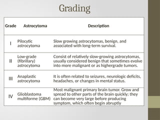
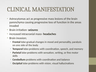
Astrocytoma
Tharun ponnuvel kumar
• Glioma- most common group of primary brain tumors
• Glial cells- support and protection for neurons. They are
thus known as the "supporting cells" of the nervous
system
Four main functions of glial cells:
1. To surround neurons and hold them in place
2. To supply nutrients and oxygen to neurons
3. To insulate one neuron from another
4. To destroy pathogens and remove dead neurons.
ETIOLOGY
• The exact cause is unknown
• Astrocytomas of all grades have been associated with
cranial therapeutic irradiation
• People who have certain rare genetic conditions such as
neurofibromatosis or Li-Fraumeni syndrome
• People over the age of 65 are diagnosed with brain
cancer at a rate four times higher than younger people.
Etiology
• mutations affecting p53
• overexpression of platelet-derived growth
factor α (PDGF-A) and its receptor
• Disruption of tumor suppressor genes
• RB and p16/CDKNaA
• tumor suppressor on chromosome 19q
most common in
the low-grade
astrocytomas
transition to higher
grade astrocytoma
• Mostly it is supratentorial in adults: Cerebral cortex (MC
site)
• Mostly it is infratentorial in children: Cerebellum (MC
site)
• MC type of astrocytoma in children: Low grade-Pilocytic
astrocytoma (Best prognosis)
• MC type in adults; High grade Glioblastoma Multiforme
[GBM: most malignant (Worst prognosis)]
Grading
Grade Astrocytoma Description
I Pilocytic
astrocytoma
Slow growing astrocytomas, benign, and
associated with long-term survival.
II
Low-grade
(fibrillary)
astrocytoma
Consist of relatively slow-growing astrocytomas,
usually considered benign that sometimes evolve
into more malignant or as highergrade tumors.
III Anaplastic
astrocytoma
It is often related to seizures, neurologic deficits,
headaches, or changes in mental status.
IV Glioblastoma
multiforme (GBM)
Most malignant primary brain tumor. Grow and
spread to other parts of the brain quickly; they
can become very large before producing
symptom, which often begin abruptly
Pilocytic astrocytoma
• Slow growing, sometimes very slow growing
• Most common type Of astrocytoma in childrens
• Relatively contained (have well-defined edges), which can
make it easier to remove completely
• Know to typically carry a mutation in the BRAF-1 gene
• The presence of eosinophilic Rosenthal fibers is a
characteristic feature
Radiological finding
• They are cystic tumor with mural nodule
Low grade astrocytoma
Slow growing
diffuse (don’t have well-defined edges), which can make it
more difficult to remove completely
• can sometimes return following treatment (known as a
recurrence), as a grade 3 tumour.
• They occur most often in adults between the ages of 20 and
45.
• They’re more common in males than females.
Anaplastic astrocytoma
Fast growing
often referred to as malignant or cancerous
• often come back following treatment (known as a
recurrence) in a more advanced form.
• Grade 3 astrocytomas are a type of high grade glioma.
• They’re more common in adults between the ages of 30
and 70 and are more common in males.
Glioblastoma multiforme
• The most common and dangerous
cancerous brain tumor, glioblastoma
multiforme (GMB) makes up more
than 50% of all astrocytomas
• Aggressive type of tumor is known
to spread quickly and typically
affects adults between the ages of
50 and 70.
• Know to typically carry a mutation in
the IDH gene
• Unique Feature of GBM: Necrosis
Morphology
Serpentine necrosis
Glomerulai tumor cells
Radiological finding
• Butterfly glioma-are high-grade astrocytomas, IDH-mutant or
glioblastomas, IDH-wildtype that cross the midline via the
corpus callosum
CLINICAL MANIFESTATION
• Astrocytomas act as progressive mass lesions of the brain
parenchyma causing progressive loss of function in the areas
invaded
• Brain irritation- seizures
• increased intracranial mass- headaches
• Brain invasion;
• Frontal lobe-gradual changes in mood and personality, paralysis
on one side of the body
• Temporal lobe-problems with coordination, speech, and memory
• Parietal lobe-problems with sensation, writing, or fine motor
skills
• Cerebellum-problems with coordination and balance
• Occipital lobe-problems with vision, visual hallucinations
Treatment
• The treatment for a brain tumour depends on its grade, size
and location
• GRADE 1
Radical resection is curable
• GRADE 2
Radiotherapy+Chemotherapy+Surgery
• GRADE 3
Cytoreductive surgery followed by EBRT (external beam
radiotherapy)
• GRADE 4
• Cytoreductive sx followed by EBRT(external beam
radiotherapy)
• Two agents used in Malignant glioma:
1.Carmustine ,2.Cisplatin
• New agents (used for newly diagnosed GBM and Recurrent
GBM):Temozolomide
CASE
• 34 year old male
• Has recurrent severe headache of 8 weeks
• Initially relieved by Mefenamic Acid.
• 1 day PTA, the patient noted weakness of the left upper
extremity.
• Few minutes PTA, he had seizures thus consulted and
subsequently admitted.
MRI features of a mixed solid and cystic tumor of the right
cerebellum suggestive of a pilocytic astrocytoma (pathologically
proven).
• The patient went to have a resection of the tumor with
histopathological exam which confirmed the diagnosis of a
pilocytic astrocytoma grade I
astrocytoma ppt.pptx.                     .

astrocytoma ppt.pptx. .

  • 1.
  • 2.
    • Glioma- mostcommon group of primary brain tumors • Glial cells- support and protection for neurons. They are thus known as the "supporting cells" of the nervous system Four main functions of glial cells: 1. To surround neurons and hold them in place 2. To supply nutrients and oxygen to neurons 3. To insulate one neuron from another 4. To destroy pathogens and remove dead neurons.
  • 3.
    ETIOLOGY • The exactcause is unknown • Astrocytomas of all grades have been associated with cranial therapeutic irradiation • People who have certain rare genetic conditions such as neurofibromatosis or Li-Fraumeni syndrome • People over the age of 65 are diagnosed with brain cancer at a rate four times higher than younger people.
  • 4.
    Etiology • mutations affectingp53 • overexpression of platelet-derived growth factor α (PDGF-A) and its receptor • Disruption of tumor suppressor genes • RB and p16/CDKNaA • tumor suppressor on chromosome 19q most common in the low-grade astrocytomas transition to higher grade astrocytoma
  • 5.
    • Mostly itis supratentorial in adults: Cerebral cortex (MC site) • Mostly it is infratentorial in children: Cerebellum (MC site) • MC type of astrocytoma in children: Low grade-Pilocytic astrocytoma (Best prognosis) • MC type in adults; High grade Glioblastoma Multiforme [GBM: most malignant (Worst prognosis)]
  • 6.
    Grading Grade Astrocytoma Description IPilocytic astrocytoma Slow growing astrocytomas, benign, and associated with long-term survival. II Low-grade (fibrillary) astrocytoma Consist of relatively slow-growing astrocytomas, usually considered benign that sometimes evolve into more malignant or as highergrade tumors. III Anaplastic astrocytoma It is often related to seizures, neurologic deficits, headaches, or changes in mental status. IV Glioblastoma multiforme (GBM) Most malignant primary brain tumor. Grow and spread to other parts of the brain quickly; they can become very large before producing symptom, which often begin abruptly
  • 7.
    Pilocytic astrocytoma • Slowgrowing, sometimes very slow growing • Most common type Of astrocytoma in childrens • Relatively contained (have well-defined edges), which can make it easier to remove completely • Know to typically carry a mutation in the BRAF-1 gene • The presence of eosinophilic Rosenthal fibers is a characteristic feature
  • 8.
    Radiological finding • Theyare cystic tumor with mural nodule
  • 9.
    Low grade astrocytoma Slowgrowing diffuse (don’t have well-defined edges), which can make it more difficult to remove completely • can sometimes return following treatment (known as a recurrence), as a grade 3 tumour. • They occur most often in adults between the ages of 20 and 45. • They’re more common in males than females.
  • 10.
    Anaplastic astrocytoma Fast growing oftenreferred to as malignant or cancerous • often come back following treatment (known as a recurrence) in a more advanced form. • Grade 3 astrocytomas are a type of high grade glioma. • They’re more common in adults between the ages of 30 and 70 and are more common in males.
  • 11.
    Glioblastoma multiforme • Themost common and dangerous cancerous brain tumor, glioblastoma multiforme (GMB) makes up more than 50% of all astrocytomas • Aggressive type of tumor is known to spread quickly and typically affects adults between the ages of 50 and 70. • Know to typically carry a mutation in the IDH gene • Unique Feature of GBM: Necrosis
  • 12.
  • 13.
    Radiological finding • Butterflyglioma-are high-grade astrocytomas, IDH-mutant or glioblastomas, IDH-wildtype that cross the midline via the corpus callosum
  • 14.
    CLINICAL MANIFESTATION • Astrocytomasact as progressive mass lesions of the brain parenchyma causing progressive loss of function in the areas invaded • Brain irritation- seizures • increased intracranial mass- headaches • Brain invasion; • Frontal lobe-gradual changes in mood and personality, paralysis on one side of the body • Temporal lobe-problems with coordination, speech, and memory • Parietal lobe-problems with sensation, writing, or fine motor skills • Cerebellum-problems with coordination and balance • Occipital lobe-problems with vision, visual hallucinations
  • 15.
    Treatment • The treatmentfor a brain tumour depends on its grade, size and location • GRADE 1 Radical resection is curable • GRADE 2 Radiotherapy+Chemotherapy+Surgery • GRADE 3 Cytoreductive surgery followed by EBRT (external beam radiotherapy)
  • 16.
    • GRADE 4 •Cytoreductive sx followed by EBRT(external beam radiotherapy) • Two agents used in Malignant glioma: 1.Carmustine ,2.Cisplatin • New agents (used for newly diagnosed GBM and Recurrent GBM):Temozolomide
  • 17.
    CASE • 34 yearold male • Has recurrent severe headache of 8 weeks • Initially relieved by Mefenamic Acid. • 1 day PTA, the patient noted weakness of the left upper extremity. • Few minutes PTA, he had seizures thus consulted and subsequently admitted.
  • 19.
    MRI features ofa mixed solid and cystic tumor of the right cerebellum suggestive of a pilocytic astrocytoma (pathologically proven). • The patient went to have a resection of the tumor with histopathological exam which confirmed the diagnosis of a pilocytic astrocytoma grade I

Editor's Notes

  • #4 amplification of MDM2, a gene that encodes an inhibitor of p53. Similarly, while secondary glioblastomas have increased signaling through the PDGF-A receptor, primary glioblastomas often have amplified, mutated epidermal growth factor receptor (EGFR) genes, which encode aberrant forms of EGFR known as EGFRvIII. Both types of mutations lead to increased receptor tyrosine kinase activity and the activation of the RAS and PI-3 kinase pathways, whichstimulate the growth and survival of tumor cells ( Chapter 7 ). Based on whole genome sequencing, it is estimated that combinations of mutations that activate RAS and PI-3 kinase and inactivate p53 and RB are present in 80% to 90% of primary glioblastomas
  • #6 I- Slow growing astrocytomas, benign, and associated with long-term survival. Individuals with very slow growing tumors where complete surgical removal is possible may experience total remission. II-Consist of relatively slow-growing astrocytomas, usually considered benign that sometimes evolve into more malignant or as highergrade tumors. They are prevalent in younger people who are often present with seizures. III-It is often related to seizures, neurologic deficits, headaches, or changes in mental status. The standard initial treatment is to remove as much of the tumor as possible without worsening neurologic deficits IV-Most malignant primary brain tumor. Grow and spread to other parts of the brain quickly; they can become very large before producing symptom, which often begin abruptly
  • #14 progression from lower to higher grades is associated with a concomitant lower incidence of seizures and a higher incidence of focal neurologic deficits.